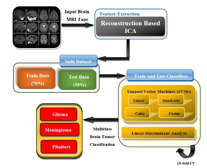
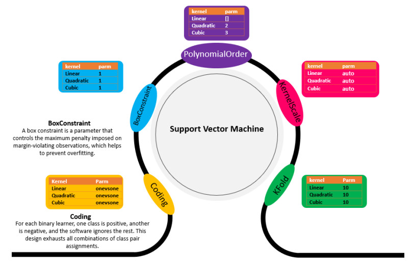
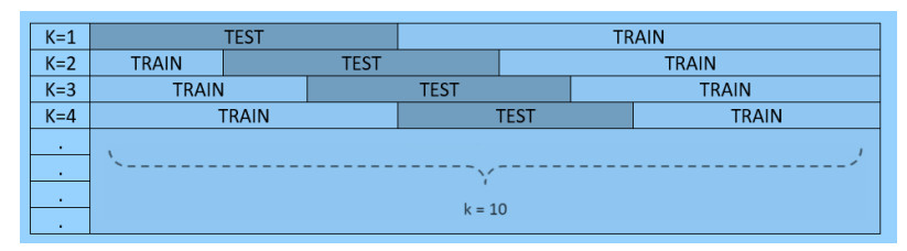
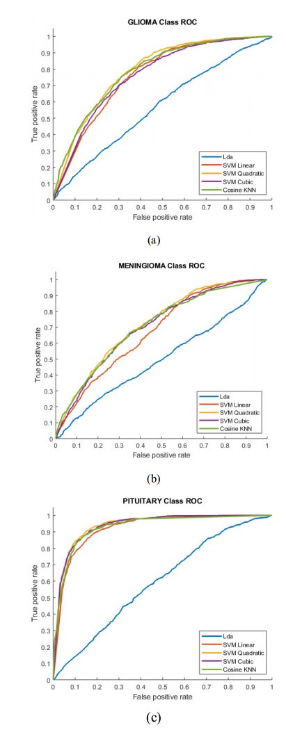
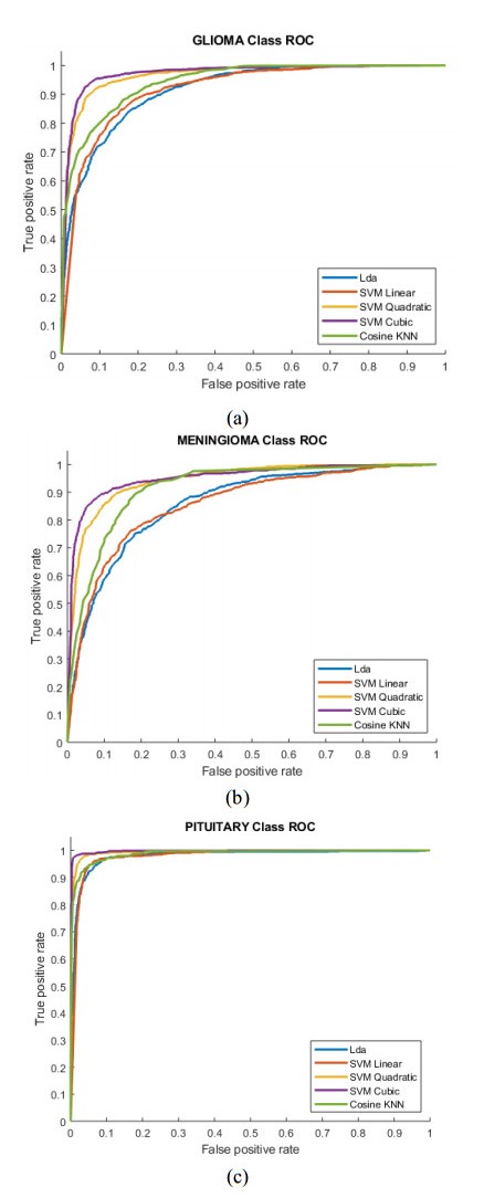
Among the other cancer types, the brain tumor is one the leading cause of cancer across globe. If the tumor is properly identified at an earlier stage, then the chances of the survival can be increased. To categorize the brain tumor there are several factors including texture, type and location of brain tumor. We proposed a novel reconstruction independent component analysis (RICA) feature extraction method to detect multi-class brain tumor types (pituitary, meningioma, and glioma). We then employed the robust machine learning techniques as support vector machine (SVM) with quadratic and linear kernels and linear discriminant analysis (LDA). For training and testing of the data validation, a 10-fold cross validation was employed. For the multi-class classification, the sensitivity, specificity, positive predictive value (PPV), negative predictive value (NPV), accuracy and AUC were, respectively, 97.78%, 100%, 100%, 99.07, 99.34% and 0.9892 to detect pituitary using SVM Cubic followed by meningioma with accuracy (96.96%0, AUC (0.9348) and glioma with accuracy (95.88%), AUC (0.9635). The findings indicates that RICA feature based proposed methodology has more potential to detect the multiclass brain tumor types for improving diagnostic efficiency and can further improve the prediction accuracy to achieve the clinical outcomes.
Citation: Sadia Anjum, Lal Hussain, Mushtaq Ali, Adeel Ahmed Abbasi, Tim Q. Duong. Automated multi-class brain tumor types detection by extracting RICA based features and employing machine learning techniques[J]. Mathematical Biosciences and Engineering, 2021, 18(3): 2882-2908. doi: 10.3934/mbe.2021146
Among the other cancer types, the brain tumor is one the leading cause of cancer across globe. If the tumor is properly identified at an earlier stage, then the chances of the survival can be increased. To categorize the brain tumor there are several factors including texture, type and location of brain tumor. We proposed a novel reconstruction independent component analysis (RICA) feature extraction method to detect multi-class brain tumor types (pituitary, meningioma, and glioma). We then employed the robust machine learning techniques as support vector machine (SVM) with quadratic and linear kernels and linear discriminant analysis (LDA). For training and testing of the data validation, a 10-fold cross validation was employed. For the multi-class classification, the sensitivity, specificity, positive predictive value (PPV), negative predictive value (NPV), accuracy and AUC were, respectively, 97.78%, 100%, 100%, 99.07, 99.34% and 0.9892 to detect pituitary using SVM Cubic followed by meningioma with accuracy (96.96%0, AUC (0.9348) and glioma with accuracy (95.88%), AUC (0.9635). The findings indicates that RICA feature based proposed methodology has more potential to detect the multiclass brain tumor types for improving diagnostic efficiency and can further improve the prediction accuracy to achieve the clinical outcomes.

| [1] | S. B. Gaikwad, M. S. Joshi, Brain tumor classification using principal component analysis and probabilistic neural network, Int. J. Comput. Appl., 120 (2015), 5-9. |
| [2] |
Q. T. Ostrom, G. Cioffi, H. Gittleman, N. Patil, K. Waite, C. Kruchko, et al., CBTRUS statistical report: primary brain and other central nervous system tumors diagnosed in the United States in 2012-2016, Neuro. Oncol., 21 (2019), v1-v100. doi: 10.1093/neuonc/noz150

|
| [3] |
D. N. Louis, A. Perry, G. Reifenberger, A. von Deimling, D. Figarella-Branger, W. K. Cavenee, et al., The 2016 World Health Organization Classification of tumors of the central nervous system: A summary, Acta Neuropathol., 131 (2016), 803-820. doi: 10.1007/s00401-016-1545-1
|
| [4] |
Z. N. K. Swati, Q. Zhao, M. Kabir, F. Ali, Z. Ali, S. Ahmed, et al., Content-Based brain tumor retrieval for MR images using transfer learning, IEEE Access, 7 (2019), 17809-17822. doi: 10.1109/ACCESS.2019.2892455
|
| [5] | S. Pereira, R. Meier, V. Alves, M. Reyes, C. A. Silva, Automatic brain tumor grading from mri data using convolutional neural networks and quality assessment, in: Understanding and interpreting machine learning in medical image computing applications, Springer, Cham., 2018,106-114. |
| [6] |
S. Deepak, P. M. Ameer, Brain tumor classification using deep CNN features via transfer learning, Comput. Biol. Med., 111 (2019), 103345. doi: 10.1016/j.compbiomed.2019.103345
|
| [7] | K. Machhale, H. B. Nandpuru, V. Kapur, L. Kosta, MRI brain cancer classification using hybrid classifier (SVM-KNN), in: 2015 Int. Conf. Ind. Instrum. Control, IEEE, 2015, 60-65. |
| [8] |
S. Rathore, M. Hussain, M. Aksam Iftikhar, A. Jalil, Ensemble classification of colon biopsy images based on information rich hybrid features, Comput. Biol. Med., 47 (2014), 76-92. doi: 10.1016/j.compbiomed.2013.12.010
|
| [9] |
S. Rathore, M. Hussain, A. Khan, Automated colon cancer detection using hybrid of novel geometric features and some traditional features, Comput. Biol. Med., 65 (2015) 279-296. doi: 10.1016/j.compbiomed.2015.03.004
|
| [10] |
L. Hussain, A. Ahmed, S. Saeed, S. Rathore, I. A. Awan, S. A. Shah, et al., Prostate cancer detection using machine learning techniques by employing combination of features extracting strategies, Cancer Biomarkers, 21 (2018), 393-413. doi: 10.3233/CBM-170643
|
| [11] |
Y. Asim, B. Raza, A. Kamran, M. Saima, A.K. Malik, S. Rathore, et al., A multi-modal, multi-atlas-based approach for Alzheimer detection via machine learning, Int. J. Imag. Sys. Tech., 28 (2018), 113-123. doi: 10.1002/ima.22263
|
| [12] | A. Islam, M. F. Hossain, C. Saha, A new hybrid approach for brain tumor classification using BWT-KSVM, in: 2017 4th Int. Conf. Adv. Electr. Eng., IEEE, 2017,241-246. |
| [13] |
P. P. Rebouças Filho, E. de S. Rebouças, L. B. Marinho, R. M. Sarmento, J. M. R. S. Tavares, V. H. C. de Albuquerque, Analysis of human tissue densities: A new approach to extract features from medical images, Pattern Recognit. Lett., 94 (2017), 211-218. doi: 10.1016/j.patrec.2017.02.005
|
| [14] |
B. Dhruv, N. Mittal, M. Modi, Study of Haralick's and GLCM texture analysis on 3D medical images, Int. J. Neurosci., 129 (2019), 350-362. doi: 10.1080/00207454.2018.1536052
|
| [15] |
Q. Zheng, H. Li, B. Fan, S. Wu, J. Xu, Integrating support vector machine and graph cuts for medical image segmentation, J. Vis. Commun. Image Represent, 55 (2018), 157-165. doi: 10.1016/j.jvcir.2018.06.005
|
| [16] | S. A. Taie, W. Ghonaim, Title CSO-based algorithm with support vector machine for brain tumor's disease diagnosis, in: 2017 IEEE Int. Conf. Pervasive Comput. Commun. Work. (PerCom Work.), IEEE, 2017,183-187. |
| [17] | M. K. Abd-Ellah, A. I. Awad, A. A. M. Khalaf, H. F. A. Hamed, Classification of brain tumor MRIs using a kernel support vector machine, in: International Conference on Well-Being in the Information Society, Springer, Cham. 2016,151-160. |
| [18] | H. Alquran, I. A. Qasmieh, A. M. Alqudah, S. Alhammouri, E. Alawneh, A. Abughazaleh, et al., The melanoma skin cancer detection and classification using support vector machine, in: 2017 IEEE Jordan Conf. Appl. Electr. Eng. Comput. Technol., IEEE, 2017, 1-5. |
| [19] |
S. Wang, S. Du, A. Atangana, A. Liu, Z. Lu, Application of stationary wavelet entropy in pathological brain detection, Multimed. Tools Appl., 77 (2018), 3701-3714. doi: 10.1007/s11042-017-4476-5
|
| [20] |
Y. Zhang, J. Yang, S. Wang, Z. Dong, P. Phillips, Pathological brain detection in MRI scanning via Hu moment invariants and machine learning, J. Exp. Theor. Artif. Intell., 29 (2017), 299-312. doi: 10.1080/0952813X.2015.1132274
|
| [21] |
J. Cheng, W. Yang, M. Huang, W. Huang, J. Jiang, Y. Zhou, et al., Retrieval of brain tumors by adaptive spatial pooling and fisher vector representation, PLoS One, 11 (2016), e0157112. doi: 10.1371/journal.pone.0157112
|
| [22] |
J. Cheng, W. Huang, S. Cao, R. Yang, W. Yang, Z. Yun, et al., Enhanced performance of brain tumor classification via tumor region augmentation and partition, PLoS One., 10 (2015), e0140381. doi: 10.1371/journal.pone.0140381
|
| [23] | N. Abiwinanda, M. Hanif, S. T. Hesaputra, A. Handayani, T. R. Mengko, Brain tumor classification using convolutional neural network, in: World Congr. Med. Phys. Biomed. Eng. 2018, Springer, Singapore, 2019,183-189. |
| [24] |
M. Sajjad, S. Khan, K. Muhammad, W. Wu, A. Ullah, S. W. Baik, Multi-grade brain tumor classification using deep CNN with extensive data augmentation, J. Comput. Sci., 30 (2019), 174-182. doi: 10.1016/j.jocs.2018.12.003
|
| [25] |
R. Zia, P. Akhtar, A. Aziz, A new rectangular window based image cropping method for generalization of brain neoplasm classification systems, Int. J. Imag. Syst. Technol., 28 (2018), 153-162. doi: 10.1002/ima.22266
|
| [26] |
M. M. Badža, M. Č. Barjaktarović, Classification of brain tumors from MRI images using a convolutional neural network, Appl. Sci., 10 (2020), 1999. doi: 10.3390/app10061999
|
| [27] |
A. Gumaei, M. M. Hassan, M. R. Hassan, A. Alelaiwi, G. Fortino, A hybrid feature extraction method with regularized extreme learning machine for brain tumor classification, IEEE Access, 7 (2019), 36266-36273. doi: 10.1109/ACCESS.2019.2904145
|
| [28] |
Z. Huang, X. Du, L. Chen, Y. Li, M. Liu, Y. Chou, et al., Convolutional neural network based on complex networks for brain tumor image classification with a modified activation function, IEEE Access, 8 (2020), 89281-89290. doi: 10.1109/ACCESS.2020.2993618
|
| [29] | L. Hussain, I. A. Awan, W. Aziz, S. Saeed, A. Ali, F. Zeeshan, et al., Detecting congestive heart failure by extracting multimodal features and employing machine learning techniques, Biomed. Res. Int., 2020 (2020), 1-19. |
| [30] |
L. Hussain, W. Aziz, S. Saeed, I. A. Awan, A. A. Abbasi, N. Maroof, Arrhythmia detection by extracting hybrid features based on refined Fuzzy entropy (FuzEn) approach and employing machine learning techniques, Waves Rand. Complex Media., 30 (2020), 656-686. doi: 10.1080/17455030.2018.1554926
|
| [31] | D. S. Guru, Y. H. Sharath, S. Manjunath, Texture features and KNN in classification of flower images, Int. J. Comput. Appl., (2010), 21-29. |
| [32] |
A. N. Esgiar, R. N. Naguib, B. S. Sharif, M. K. Bennett, A. Murray, Microscopic image analysis for quantitative measurement and feature identification of normal and cancerous colonic mucosa, IEEE Trans. Inf. Technol. Biomed., 2 (1998), 197-203. doi: 10.1109/4233.735785
|
| [33] |
A. N. Esgiar, R. N. G. Naguib, B. S. Sharif, M. K. Bennett, A. Murray, Fractal analysis in the detection of colonic cancer images, IEEE Trans. Inf. Technol. Biomed., 6 (2002), 54-58. doi: 10.1109/4233.992163
|
| [34] | M. Masseroli, A. Bollea, G. Forloni, Quantitative morphology and shape classification of neurons by computerized image analysis, Comput. Methods Programs Biomed., (1993), 89-99. |
| [35] |
Y. M. Li, X. P. Zeng, A new strategy for urinary sediment segmentation based on wavelet, morphology and combination method, Comput. Methods Programs Biomed., 84 (2006), 162-173. doi: 10.1016/j.cmpb.2006.07.010
|
| [36] |
A. Hyvä;rinen, E. Oja, Independent component analysis: Algorithms and applications, Neural Networks, 13 (2000), 411-430. doi: 10.1016/S0893-6080(00)00026-5
|
| [37] |
Y. Xiao, Z. Zhu, Y. Zhao, Kernel reconstruction ICA for sparse representation, IEEE Trans. Neural Networks Learn. Syst., 26 (2015), 1222-1232. doi: 10.1109/TNNLS.2014.2334711
|
| [38] | J. Hurri, P. O. Hoyer, Natural Image Statistics, A probabilistic approach to early computational vision, Springer Science & Business Media, 39 (2009). |
| [39] | Q. V. Le, A. Karpenko, J. Ngiam, A. Y. Ng, ICA with reconstruction cost for efficient overcomplete feature learning, Adv. Neural. Inform. Process Syst., 24 (2011), 1017-1025. |
| [40] | Q. V. Le, M. A. Ranzato, M. Devin, G. S. Corrado, A. Y. Ng, Building high-level features using large scale unsupervised learning, In 2013 IEEE international conference on acoustics, speech and signal processing, IEEE, (2013), 8595-8598 |
| [41] | Y. Boureau, A theoretical analysis of feature pooling in visual recognition, In Proceedings of the 27th international conference on machine learning (ICML-10), (2010), 111-118. |
| [42] | Y. Lecun, Learning invariant feature hierarchies, In European conference on computer vision, Springer, Berlin, Heidelberg, (2012), 496-505 |
| [43] |
A. P. Dobrowolski, M. Wierzbowski, K. Tomczykiewicz, Multiresolution MUAPs decomposition and SVM-based analysis in the classification of neuromuscular disorders, Comput. Methods Programs Biomed., 107 (2012), 393-403. doi: 10.1016/j.cmpb.2010.12.006
|
| [44] |
J. Schmidhuber, Deep learning in neural networks: An overview, Neural Networks, 61 (2015), 85-117. doi: 10.1016/j.neunet.2014.09.003
|
| [45] |
H. Papadopoulos, V. Vovk, A. Gammerman, Guest editors' preface to the special issue on conformal prediction and its applications, Ann. Math. Artif. Intell., 74 (2015), 1-7. doi: 10.1007/s10472-014-9429-3
|
| [46] |
N. Kambhatla, T. K. Leen, Dimension reduction by local principal component analysis, Neural Comput., 9 (1997), 1493-1516. doi: 10.1162/neco.1997.9.7.1493
|
| [47] | A. Pathak, B. Vohra, K. Gupta, Supervised learning approach towards class separability-linear discriminant analysis, in: 2019 Int. Conf. Intell. Comput. Control Syst., IEEE, (2019), 1088-1093. |
| [48] |
W. Yang, Q. Feng, M. Yu, Z. Lu, Y. Gao, Y. Xu, et al., Content-based retrieval of brain tumor in contrast-enhanced MRI images using tumor margin information and learned distance metric, Med. Phys., 39 (2012), 6929-6942. doi: 10.1118/1.4754305
|
| [49] | M. Huang, W. Yang, M. Yu, Z. Lu, Q. Feng, W. Chen, Retrieval of brain tumors with region-specific bag-of-visual-words representations in contrast-enhanced MRI images, Comput. Math. Methods Med., 2012 (2012), 1-17. |
| [50] |
M. Huang, W. Yang, Y. Wu, J. Jiang, Y. Gao, Y. Chen, et al., Content-based image retrieval using spatial layout information in brain tumor T1-weighted contrast-enhanced mr images, PLoS One, 9 (2014), e102754. doi: 10.1371/journal.pone.0102754
|
| [51] | P. Afshar, K. N. Plataniotis, A. Mohammadi, Capsule networks for brain tumor classification based on mri images and coarse tumor boundaries, in: ICASSP 2019 - 2019 IEEE Int. Conf. Acoust. Speech Signal Process., IEEE, (2019), 1368-1372. |
| [52] | P. Afshar, A. Mohammadi, K. N. Plataniotis, Brain tumor type classification via capsule networks, in: 2018 25th IEEE Int. Conf. Image Process., IEEE, (2018), 3129-3133. |
| [53] | J. S. Paul, A. J. Plassard, B. A. Landman, D. Fabbri, Deep learning for brain tumor classification, in: Medical Imaging 2017: Biomedical Applications in Molecular, Structural, and Functional Imaging., International Society for Optics and Photonics, 10137 (2017), 1013710. |
| [54] |
E. I. Zacharaki, S. Wang, S. Chawla, D. Soo, R. Yoo, E. R. Wolf, et al., Classification of brain tumor type and grade using MRI texture and shape in a machine learning scheme, Magn. Reson. Med., 62 (2009), 1609-1618. doi: 10.1002/mrm.22147
|
| [55] |
A. Kabir Anaraki, M. Ayati, F. Kazemi, Magnetic resonance imaging-based brain tumor grades classification and grading via convolutional neural networks and genetic algorithms, Biocybern. Biomed. Eng., 39 (2019), 63-74. doi: 10.1016/j.bbe.2018.10.004
|
| [56] |
J. Sachdeva, V. Kumar, I. Gupta, N. Khandelwal, C. K. Ahuja, Segmentation, feature extraction, and multiclass brain tumor classification, J. Digit. Imag., 26 (2013), 1141-1150. doi: 10.1007/s10278-013-9600-0
|
| [57] |
R. A. Lerski, K. Straughan, L. R. Schad, D. Boyce, S. Blüml, I. Zuna, VⅢ. MR image texture analysis-An approach to tissue characterization, Magn. Reson. Imaging., 11 (1993), 873-887. doi: 10.1016/0730-725X(93)90205-R
|
| [58] |
S. Herlidou-Même, J. Constans, B. Carsin, D. Olivie, P. Eliat, L. Nadal-Desbarats, et al., MRI texture analysis on texture test objects, normal brain and intracranial tumors, Magn. Reson. Imaging., 21 (2003), 989-993. doi: 10.1016/S0730-725X(03)00212-1
|
| [59] |
L. R. Schad, S. Blüml, I. Zuna, IX. MR tissue characterization of intracranial tumors by means of texture analysis, Magn. Reson. Imaging., 11 (1993), 889-896. doi: 10.1016/0730-725X(93)90206-S
|
| [60] |
A. Devos, A.W. Simonetti, M. van der Graaf, L. Lukas, J. A. K. Suykens, L. Vanhamme, et al., The use of multivariate MR imaging intensities versus metabolic data from MR spectroscopic imaging for brain tumour classification, J. Magn. Reson., 173 (2005), 218-228. doi: 10.1016/j.jmr.2004.12.007
|
| [61] |
L. Lukas, A. Devos, J. A. K. Suykens, L. Vanhamme, F. A. Howe, C. Majós, et al., Brain tumor classification based on long echo proton MRS signals, Artif. Intell. Med., 31 (2004), 73-89. doi: 10.1016/j.artmed.2004.01.001
|
| [62] |
A. Devos, L. Lukas, J. A. K. Suykens, L. Vanhamme, A. R. Tate, F. A. Howe, et al., Classification of brain tumours using short echo time 1H MR spectra, J. Magn. Reson., 170 (2004), 164-175. doi: 10.1016/j.jmr.2004.06.010
|
| [63] |
Y. Huang, P. J. G. Lisboa, W. El-Deredy, Tumour grading from magnetic resonance spectroscopy: A comparison of feature extraction with variable selection, Stat. Med., 22 (2003), 147-164. doi: 10.1002/sim.1321
|
| [64] |
A. R. Tate, C. Majós, A. Moreno, F. A. Howe, J. R. Griffiths, C. Arús, Automated classification of short echo time in in vivo 1 H brain tumor spectra: A multicenter study, Magn. Reson. Med., 49 (2003), 29-36. doi: 10.1002/mrm.10315
|
| [65] |
Y. D. Cho, G. H. Choi, S. P. Lee, J. K. Kim, 1H-MRS metabolic patterns for distinguishing between meningiomas and other brain tumors, Magn. Reson. Imaging., 21 (2003), 663-672. doi: 10.1016/S0730-725X(03)00097-3
|
| [66] | Y. Pan, W. Huang, Z. Lin, W. Zhu, J. Zhou, J. Wong, et al., Brain tumor grading based on Neural Networks and Convolutional Neural Networks, in: 2015 37th Annu. Int. Conf. IEEE Eng. Med. Biol. Soc., IEEE, (2015), 699-702. |

Figures(9) / Tables(6)

Sadia Anjum, Lal Hussain, Mushtaq Ali, Adeel Ahmed Abbasi, Tim Q. Duong. Automated multi-class brain tumor types detection by extracting RICA based features and employing machine learning techniques[J]. Mathematical Biosciences and Engineering, 2021, 18(3): 2882-2908. doi: 10.3934/mbe.2021146


 DownLoad: