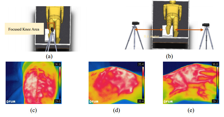
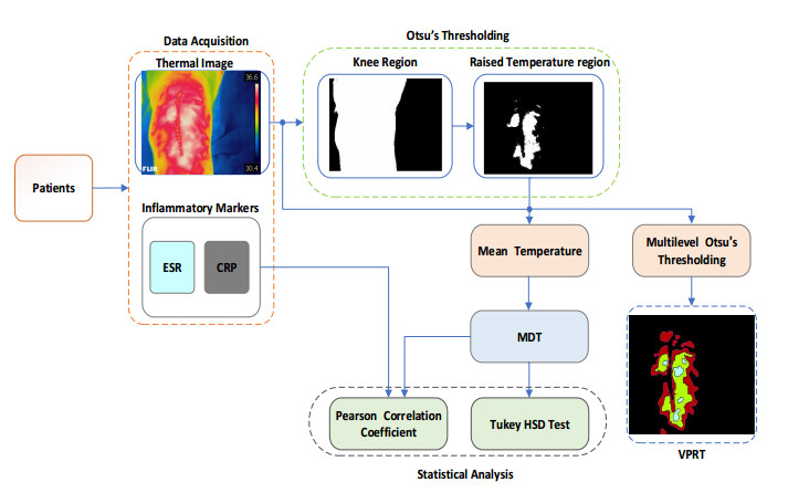
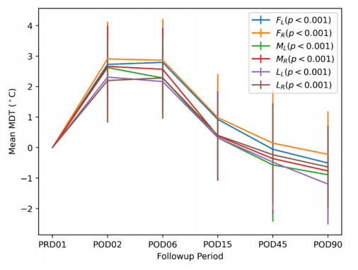
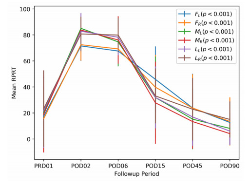
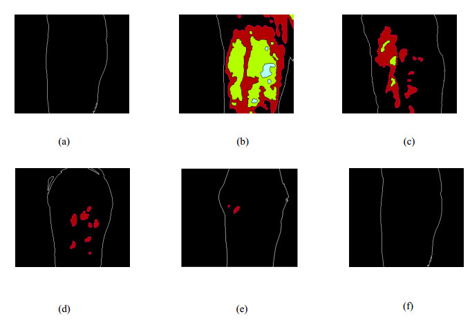
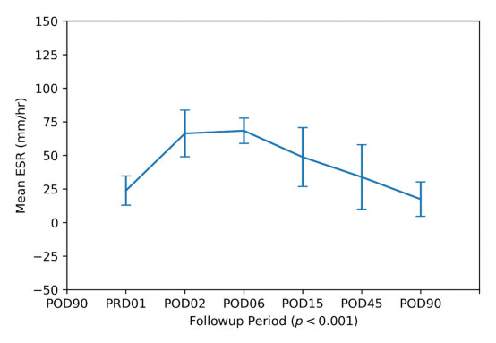
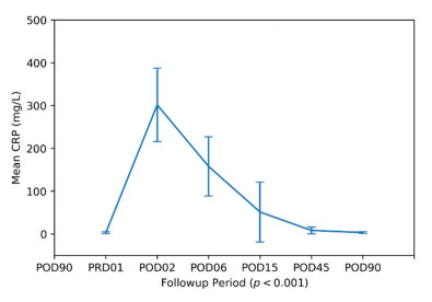
Total knee replacement is an end-stage surgical treatment of osteoarthritis patients to improve their quality of life. The study presents a thermal imaging-based approach to assess the recovery of operated-knees. The study focuses on the potential of thermal imaging for total knee replacement and its relation with clinical inflammatory markers. A total of 20 patients with bilateral knee replacement were included for thermal imaging and serology, where data was acquired on pre-operative day and five post-operative days. To quantify the inflammation, the temperature-based parameters (like mean differential temperature, relative percentage of raised temperature) were evaluated from thermal images, while the clinically proven inflammation markers were obtained from blood samples for clinical validation. Initially, the knee region was segmented by applying the automatic method, subsequently, the mean skin temperature was calculated and investigated for a statistical relevant relationship with inflammatory markers. After surgery, the mean skin temperature was first increased (>2.15 ℃ for different views) then settled to pre-operative level by 90th day. Consequently, the mean differential temperature showed a strong correlation with erythrocyte sedimentation rate (r > 0.893) and C-reactive protein (r > 0.955). Also, the visual profile and relative percentage of raised temperature showed promising results in quantifying the temperature changes both qualitatively and quantitatively. This study provides an automatic and non-invasive way of screening the patients for raised levels of skin temperature, which can be a sign of inflammation. Hence, the proposed temperature-based technique can help the clinicians for visual assessment of post-operative recovery of patients.
Citation: Viney Lohchab, Jaspreet Singh, Prasant Mahapatra, Vikas Bachhal, Aman Hooda, Karan Jindal, MS Dhillon. Thermal imaging in total knee replacement and its relation with inflammation markers[J]. Mathematical Biosciences and Engineering, 2021, 18(6): 7759-7773. doi: 10.3934/mbe.2021385
Total knee replacement is an end-stage surgical treatment of osteoarthritis patients to improve their quality of life. The study presents a thermal imaging-based approach to assess the recovery of operated-knees. The study focuses on the potential of thermal imaging for total knee replacement and its relation with clinical inflammatory markers. A total of 20 patients with bilateral knee replacement were included for thermal imaging and serology, where data was acquired on pre-operative day and five post-operative days. To quantify the inflammation, the temperature-based parameters (like mean differential temperature, relative percentage of raised temperature) were evaluated from thermal images, while the clinically proven inflammation markers were obtained from blood samples for clinical validation. Initially, the knee region was segmented by applying the automatic method, subsequently, the mean skin temperature was calculated and investigated for a statistical relevant relationship with inflammatory markers. After surgery, the mean skin temperature was first increased (>2.15 ℃ for different views) then settled to pre-operative level by 90th day. Consequently, the mean differential temperature showed a strong correlation with erythrocyte sedimentation rate (r > 0.893) and C-reactive protein (r > 0.955). Also, the visual profile and relative percentage of raised temperature showed promising results in quantifying the temperature changes both qualitatively and quantitatively. This study provides an automatic and non-invasive way of screening the patients for raised levels of skin temperature, which can be a sign of inflammation. Hence, the proposed temperature-based technique can help the clinicians for visual assessment of post-operative recovery of patients.

| [1] |
B. A. Kohl, C. S. Deutschman, The inflammatory response to surgery and trauma, Curr. Opin. Crit. Care, 12 (2006), 325-332. doi: 10.1097/01.ccx.0000235210.85073.fc

|
| [2] |
N. V. Kalore, T. J. Gioe, J. A. Singh, Diagnosis and management of infected total knee arthroplasty, Open Orthop. J., 5 (2011), 86. doi: 10.2174/1874325001105010086
|
| [3] |
J. C. Martínez-Pastor, F. Maculé-Beneyto, S. Suso-Vergara, Suppl 2: acute infection in total knee arthroplasty: diagnosis and treatment, Open Orthop. J., 7 (2013), 197. doi: 10.2174/1874325001307010197
|
| [4] |
E. Ghanem, Jr. V. Antoci, L. Pulido, A. Joshi, W. Hozack, J. Parvizi, The use of receiver operating characteristics analysis in determining erythrocyte sedimentation rate and C-reactive protein levels in diagnosing periprosthetic infection prior to revision total hip arthroplasty, Int. J. Infect. Dis., 13 (2009), e444-e449. doi: 10.1016/j.ijid.2009.02.017
|
| [5] |
J. Parvizi, B. Zmistowski, EF. Berbari, T. W. Bauer, B. D. Springer, C. J. D. Valle, et al., New definition for periprosthetic joint infection: from the workgroup of the musculoskeletal infection society, Clin. Orthop. Relat. Res., 469 (2011), 2992-2994. doi: 10.1007/s11999-011-2102-9
|
| [6] |
N. V. Greidanus, B. A. Masri, D. S. Garbuz, S. D. Wilson, M. G. Mcalinden, M. Xu, et al., Use of erythrocyte sedimentation rate and C-reactive protein level to diagnose infection before revision total knee arthroplasty: a prospective evaluation, J. Bone Joint. Surg., 89 (2007), 1409-1416. doi: 10.2106/00004623-200707000-00001
|
| [7] |
P. E. Di Cesare, E. Chang, C.F. Preston, C. J. Liu, Serum interleukin-6 as a marker of periprosthetic infection following total hip and knee arthroplasty, J. Bone Joint. Surg., 87 (2005), 1921-1927. doi: 10.2106/00004623-200509000-00003
|
| [8] |
A. Mehra, V. Langkamer, A. Day, S. Harris, R. F. Spencer, C reactive protein and skin temperature post total knee replacement, Knee, 12 (2005), 297-300. doi: 10.1016/j.knee.2004.09.005
|
| [9] |
S. Honsawek, B. Deepaisarnsakul, A. Tanavalee, M. Sakdinakiattikoon, S. Ngarmukos, K. Preativatanyou, et al., Relationship of serum IL-6, C-reactive protein, erythrocyte sedimentation rate, and knee skin temperature after total knee arthroplasty: a prospective study, Int. Orthop., 35 (2011), 31-35. doi: 10.1007/s00264-010-0973-0
|
| [10] |
Y. Zeng, W. Feng, X. Qi, J. Li, J. Chen, L. Lu, et al., Differential knee skin temperature following total knee arthroplasty and its relationship with serum indices and outcome: a prospective study, J. Int. Med. Res., 44 (2016), 1023-1033. doi: 10.1177/0300060516655237
|
| [11] | M. Yishake, Z. Xindie, H. Rongxin, Value of knee skin temperature measured by infrared thermography and soluble intercellular adhesion molecule-1 in the diagnosis of periprosthetic knee infection in Chinese individuals following total knee arthroplasty, Chin. Med. J., 127 (2014), 3105-3109. |
| [12] |
C. L. Romano, D. Romano, F. Dell'Oro, N. Logoluso, L. Drago, Healing of surgical site after total hip and knee replacements show similar telethermographic patterns, J. Orthop. Traumatol., 12 (2011), 81-86. doi: 10.1007/s10195-011-0135-1
|
| [13] |
J. I. Arias, M. A. Aller, J. Arias, Surgical inflammation: a pathophysiological rainbow, J. Transl. Intern. Med., 7 (2009), 1-15. doi: 10.1186/1479-5876-7-1
|
| [14] | E. Lambiris, H. Stoboy, Thermography in osteosyntheses and total endoprostheses of the knee joint with and without infection, Z. Orthop. Grenzgeb., 119 (1981), 521-524. |
| [15] |
S.T. Larsen, L. Larsen, Temperature elevation during knee arthroplasty, Acta Orthop. Scand., 60 (1989), 439-442. doi: 10.3109/17453678909149314
|
| [16] |
S. G. Haidar, R. M. Charity, R. S. Bassi, P. Nicolai, B. K. Singh, Knee skin temperature following uncomplicated total knee replacement, Knee, 13 (2006), 422-426. doi: 10.1016/j.knee.2006.08.003
|
| [17] | Y. He, B. Deng, H. Wang, K. Zhou, S. Cai, F. Ciampa, Infrared machine vision and infrared thermography with deep learning: a review, Infrared Phys. Techn., (2021), 103754. |
| [18] | World Medical Association, World Medical Association Declaration of Helsinki, Ethical Principles for Medical Research Involving Human Subjects, (2008). Available form: https://pubmed.ncbi.nlm.nih.gov/24141714/. |
| [19] | D. G. Moreira, J. T. Costello, C. J. Brito, J. G. Adamczyk, K. Ammer, A. J. Bach, et al., Thermographic imaging in sports and exercise medicine: a Delphi study and consensus statement on the measurement of human skin temperature, J. Therm. Miol., 69 (2017), 155-162. |
| [20] |
N. Otsu, A threshold selection method from gray-level histograms, IEEE Trans. Syst. Man Cybern., 9 (1979), 62-66. doi: 10.1109/TSMC.1979.4310076
|
| [21] | M. M. Mukaka, A guide to appropriate use of correlation coefficient in medical research, Malawi. Med. J., 24 (2012), 69-71. |
| [22] |
F. Feldman, E. L. Nickoloff, Normal thermographic standards for the cervical spine and upper extremities, Skeletal Radiol., 12 (1984), 235-249. doi: 10.1007/BF00349505
|
| [23] |
S. Ariyaratnam, J. Rood, Measurement of facial skin temperature, J. Dent., 18 (1990), 250-253. doi: 10.1016/0300-5712(90)90022-7
|
| [24] | B. M. Graft, E.A. Sickles, V. J. Shetty, Thermography for the clinical assessment of inferior alveolar nerve deficit: a pilot study, J. Orofac. Pain., 8 (1994). |
| [25] |
S. Uematsu, D. H. Edwin, W. R. Jankel, J. Kozikowski, M. Trattner, Quantification of thermal asymmetry: part 1: normal values and reproducibility, J. Neurosurge., 69 (1988), 552-555. doi: 10.3171/jns.1988.69.4.0552
|
| [26] |
J. Christensen, M. Væth, A. Wenzel, Thermographic imaging of facial skin-gender differences and temperature changes over time in healthy subjects, Dentomaxillofac. Rad., 41 (2012), 662-667. doi: 10.1259/dmfr/55922484
|
| [27] | C. L. Romanò, N. Logoluso, F. Dell'Oro, A. Elia, L. Drago, Telethermographic findings after uncomplicated and septic total knee replacement, Knee, 19 (2011), 193-197. |
| [28] | S. J. Lin, F. C. Chang, T. W. Huang, K. T. Peng, H. N. Shin, M. S. Lee, Temporal change of interleukin-6, c-reactive protein, and skin temperature after total knee arthroplasty using triclosan-coated sutures, Biomed Res. Int., 2018. |
| [29] | K. Ammer, Temperature of the human knee-a review, Therm. Int., 22 (2012), 137-151. |
| [30] |
C. Windisch, S. Brodt, E. Roehner, G. Matziolis, Regional differences in temperature course after knee arthroplasty, Knee Surg. Sport. Traumatol. Arthroscopy, 24 (2016), 2686-2691. doi: 10.1007/s00167-015-3809-z
|
Figures(7) / Tables(6)

Viney Lohchab, Jaspreet Singh, Prasant Mahapatra, Vikas Bachhal, Aman Hooda, Karan Jindal, MS Dhillon. Thermal imaging in total knee replacement and its relation with inflammation markers[J]. Mathematical Biosciences and Engineering, 2021, 18(6): 7759-7773. doi: 10.3934/mbe.2021385


 DownLoad: