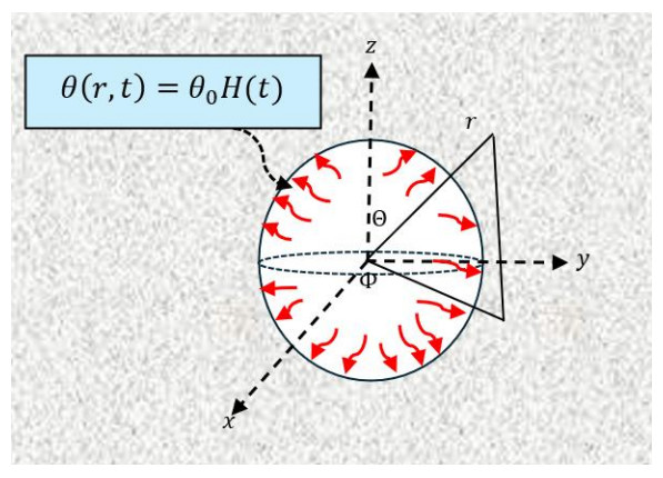
In this work, we present the nonlocal Moore-Gibson-Thompson photothermal (NMGTPT) theory, a novel framework that integrates spatial and temporal nonlocality to address limitations in both traditional and advanced thermoelastic models. Specifically tailored for semiconductor materials with microstructural features, memory effects, and photo-excited phenomena, the NMGTPT theory unifies nonlocal elasticity, MGT thermal relaxation, and photothermal effects to model the complex interplay between heat, deformation, and photo-induced processes. Unlike prior models, the NMGTPT framework incorporates spatial and temporal nonlocalities, enabling the accurate representation of long-range interactions and memory effects. Additionally, the Atangana-Baleanu (AB) fractional operator is integrated into the NMGTPT model to further enhance its ability to describe nonlocal and memory-dependent behavior, making it particularly suitable for advanced material systems. By incorporating a thermal relaxation coefficient, the framework ensures finite-speed thermal wave propagation, effectively addressing the unrealistic prediction of infinite heat speed found in classical models. The theory also integrates photo-excited free carriers, thermal waves, and acoustic waves, proving highly effective in photothermal and photoacoustic studies involving semiconductors. With the inclusion of an internal length scale, the NMGTPT theory successfully captures size-dependent behaviors, which are essential for accurately modeling nanostructured materials, thin films, and composites. This innovation provides a robust platform for investigating the complex dynamics of photothermal and thermoelastic phenomena in advanced material systems.
Citation: Mofareh Alhazmi, Ahmed E. Abouelregal. Nonlocal fractional MGT-non-Fourier photothermal model with spatial and temporal nonlocality for controlling the behavior of semiconductor materials with spherical cavities[J]. AIMS Mathematics, 2025, 10(3): 7559-7590. doi: 10.3934/math.2025348
In this work, we present the nonlocal Moore-Gibson-Thompson photothermal (NMGTPT) theory, a novel framework that integrates spatial and temporal nonlocality to address limitations in both traditional and advanced thermoelastic models. Specifically tailored for semiconductor materials with microstructural features, memory effects, and photo-excited phenomena, the NMGTPT theory unifies nonlocal elasticity, MGT thermal relaxation, and photothermal effects to model the complex interplay between heat, deformation, and photo-induced processes. Unlike prior models, the NMGTPT framework incorporates spatial and temporal nonlocalities, enabling the accurate representation of long-range interactions and memory effects. Additionally, the Atangana-Baleanu (AB) fractional operator is integrated into the NMGTPT model to further enhance its ability to describe nonlocal and memory-dependent behavior, making it particularly suitable for advanced material systems. By incorporating a thermal relaxation coefficient, the framework ensures finite-speed thermal wave propagation, effectively addressing the unrealistic prediction of infinite heat speed found in classical models. The theory also integrates photo-excited free carriers, thermal waves, and acoustic waves, proving highly effective in photothermal and photoacoustic studies involving semiconductors. With the inclusion of an internal length scale, the NMGTPT theory successfully captures size-dependent behaviors, which are essential for accurately modeling nanostructured materials, thin films, and composites. This innovation provides a robust platform for investigating the complex dynamics of photothermal and thermoelastic phenomena in advanced material systems.

| [1] |
E. Gutiérrez-Reyes, C. García-Segundo, A. García-valenzuela, R. Ortega, C. Buj, F. Filbir, Heat transport considerations in the mathematical analysis of the photoacoustic and photothermal effects, J. Phys. Commun., 3 (2019), 085007. https://doi.org/10.1088/2399-6528/ab376d doi: 10.1088/2399-6528/ab376d

|
| [2] |
J. Zakrzewski, M. Pawlak, O. Matsuda, D. Todorovic, J. Liu, Semiconductor physics: Plasma, thermal, elastic, and acoustic phenomena, J. Appl. Phys., 136 (2024), 120401. https://doi.org/10.1063/5.0234837 doi: 10.1063/5.0234837
|
| [3] |
K. Lotfy, I. S. Elshazly, B. Halouani, P. Ailawalia, A. A. El-Bary, Photo-thermo-acoustic waves interaction for nanostructured rotational semiconductor material subjected to laser pulse, Eur. Phys. J. B, 97 (2024), 189. https://doi.org/10.1140/epjb/s10051-024-00830-0 doi: 10.1140/epjb/s10051-024-00830-0
|
| [4] | A. R. Warrier, K. P. Vijayakumar, Photothermal studies in semiconductor materials, In: Photoacoustic Photothermal Spectrosc, Elsevier, 2023,245–26. |
| [5] |
S. Galovic, K. Djordjevic, M. Dragas, D. Milicevic, E. Suljovrujic, Dynamics of photoinduced charge carrier and photothermal effect in pulse-illuminated narrow gap and moderate doped semiconductors, Mathematics, 13 (2025), 258. https://doi.org/10.3390/math13020258 doi: 10.3390/math13020258
|
| [6] |
A. E. Abouelregal, H. M. Sedighi, A. H. Sofiyev, Modeling photoexcited carrier interactions in a solid sphere of a semiconductor material based on the photothermal Moore–Gibson–Thompson model, Appl. Phys. A, 127 (2021), 845. https://doi.org/10.1007/s00339-021-04971-2 doi: 10.1007/s00339-021-04971-2
|
| [7] |
B. Feng, A. Mo, W. Dong, W. Fan, J. Ren, Z. Li, et al., Investigation into the carrier recombination in Sb2Se3: Photo thermal effect, trapped carrier absorption and hot carrier cooling, Chem. Phys., 588 (2025), 112448. https://doi.org/10.1016/j.chemphys.2024.112448 doi: 10.1016/j.chemphys.2024.112448
|
| [8] | V. D. Kupradze, Three-dimensional problems of elasticity and thermoelasticity, Elsevier, 2012. |
| [9] | C. Truesdell, Linear theories of elasticity and thermoelasticity: Linear and nonlinear theories of rods, plates, and shells, Springer, 2013. |
| [10] |
S. L. Guo, Y. X. Zhang, K. F. Wang, B. L. Wang, C. W. Zhang, Effects of non-Fourier heat conduction and surface heating rate on thermoelastic waves in semi-infinite ceramics subject to thermal shock, Ceram. Int., 47 (2021), 17494–17501. https://doi.org/10.1016/j.ceramint.2021.03.067 doi: 10.1016/j.ceramint.2021.03.067
|
| [11] |
G. A. Maugin, A. Berezovski, Material formulation of finite-strain thermoelasticity and applications, J. Therm. Stresses, 22 (1999), 421–449. https://doi.org/10.1080/014957399280823 doi: 10.1080/014957399280823
|
| [12] |
J. Gawinecki, Initial-boundary value problems in linear and nonlinear hyperbolic thermoelasticity theory, Z. Angew. Math. Mech. 78 (1998), 911–912. https://doi.org/10.1002/zamm.19980781528 doi: 10.1002/zamm.19980781528
|
| [13] |
R. F. Silva, P. G. Coelho, F. M. Conde, C. J. Almeida, A. L. Custódio, Topology optimization of thermoelastic structures with single and functionally graded materials exploring energy and stress-based formulations, Struct. Multidisc. Optim., 68 (2025), 11. https://doi.org/10.1007/s00158-024-03929-1 doi: 10.1007/s00158-024-03929-1
|
| [14] |
A. E. Green, P. Naghdi, A re-examination of the basic postulates of thermomechanics, Proc. R. Soc. Lond. A, 432 (1991), 171–194. https://doi.org/10.1098/rspa.1991.0012 doi: 10.1098/rspa.1991.0012
|
| [15] |
A. E. Green, P. Naghdi, On undamped heat waves in an elastic solid, J. Therm. Stresses, 15 (1992), 253–264. https://doi.org/10.1080/01495739208946136 doi: 10.1080/01495739208946136
|
| [16] |
A. E. Green, P. Naghdi, Thermoelasticity without energy dissipation, J. Elast., 31 (1993), 189–208. https://doi.org/10.1007/BF00044969 doi: 10.1007/BF00044969
|
| [17] |
R. Quintanilla, Moore–Gibson–Thompson thermoelasticity, Math. Mech. Solids, 24 (2019), 4020–4031. https://doi.org/10.1177/1081286519862007 doi: 10.1177/1081286519862007
|
| [18] |
R. Quintanilla, Moore-Gibson-Thompson thermoelasticity with two temperatures, Appl. Eng. Sci., 1 (2020), 100006. https://doi.org/10.1016/j.apples.2020.100006 doi: 10.1016/j.apples.2020.100006
|
| [19] | C. Cattaneo, A form of heat-conduction equations which eliminates the paradox of instantaneous propagation, C. R. Acad. Sci., 247 (1958), 431. https://ci.nii.ac.jp/naid/10018112216 |
| [20] | P. Vernotte, Some possible complications in the phenomena of thermal conduction, C. R. Acad. Sci., 252 (1961), 2190–2191. |
| [21] |
A. E. Abouelregal, M. Marin, A. Foul, S. S. Askar, Thermoviscoelastic responses in Kirchhoff circular micro-plate via MGT thermoelastic model and modified couple stress theory, Mech. Solids, 59 (2024), 2269–2291. https://doi.org/10.1134/S002565442460449X doi: 10.1134/S002565442460449X
|
| [22] |
A. E. Abouelregal, Generalized thermoelastic MGT model for a functionally graded heterogeneous unbounded medium containing a spherical hole, Eur. Phys. J. Plus, 137 (2022), 953. https://doi.org/10.1140/epjp/s13360-022-03160-1 doi: 10.1140/epjp/s13360-022-03160-1
|
| [23] |
A. E. Abouelregal, M. Alesemi, Evaluation of the thermal and mechanical waves in anisotropic fiber-reinforced magnetic viscoelastic solid with temperature-dependent properties using the MGT thermoelastic model, Case Stud. Therm. Eng., 36 (2022), 102187. https://doi.org/10.1016/j.csite.2022.102187 doi: 10.1016/j.csite.2022.102187
|
| [24] |
A. E. Abouelregal, H. M. Sedighi, S. F. Megahid, Photothermal-induced interactions in a semiconductor solid with a cylindrical gap due to laser pulse duration using a fractional MGT heat conduction model, Arch. Appl. Mech., 93 (2023), 2287–2305. https://doi.org/10.1007/s00419-023-02383-7 doi: 10.1007/s00419-023-02383-7
|
| [25] |
F. K. Moore, W. E. Gibson, Propagation of weak disturbances in a gas subject to relaxation effects, J. Aerosp. Sci., 27 (1960), 117–127. https://doi.org/10.2514/8.8418 doi: 10.2514/8.8418
|
| [26] |
P. A. Thompson, A. A. Sonin, Compressible-fluid dynamics, Phys. Today, 26 (1973), 65–69. https://doi.org/10.1063/1.3127987 doi: 10.1063/1.3127987
|
| [27] |
R. Barretta, F. Marotti de Sciarra, M. Concurrent S. Vaccaro, Nonlocal elasticity for nanostructures: A review of recent achievements, Encyclopedia, 3 (2023), 279–310. https://doi.org/10.3390/encyclopedia3010018 doi: 10.3390/encyclopedia3010018
|
| [28] |
E. Kröner, Elasticity theory of materials with long range cohesive forces, Int. J. Solids Struct., 3 (1967), 731–742. https://doi.org/10.1016/0020-7683(67)90049-2 doi: 10.1016/0020-7683(67)90049-2
|
| [29] |
K. Duan, L. Li, Y. Hu, X. Wang, Enhanced interfacial strength of carbon nanotube/copper nanocomposites via Ni-coating: Molecular-dynamics insights, Physica E, 88 (2017), 259–264. https://doi.org/10.1016/j.physe.2017.01.015 doi: 10.1016/j.physe.2017.01.015
|
| [30] |
A. C. Eringen, Linear theory of nonlocal elasticity and dispersion of plane waves, Int. J. Eng. Sci., 10 (1972), 425–435. https://doi.org/10.1016/0020-7225(72)90050-X doi: 10.1016/0020-7225(72)90050-X
|
| [31] |
A. C. Eringen, On differential equations of nonlocal elasticity and solutions of screw dislocation and surface waves, J. Appl. Phys., 54 (1983), 4703–4710. https://doi.org/10.1063/1.332803 doi: 10.1063/1.332803
|
| [32] |
G. Romano, R. Barretta, Stress-driven versus strain-driven nonlocal integral model for elastic nano-beams, Compos. B Eng., 114 (2017), 184–188. https://doi.org/10.1016/j.compositesb.2017.01.008 doi: 10.1016/j.compositesb.2017.01.008
|
| [33] |
S. Li, W. Zheng, L. Li, Spatiotemporally nonlocal homogenization method for viscoelastic porous metamaterial structures, Int. J. Mech. Sci., 282 (2024), 109572. https://doi.org/10.1016/j.ijmecsci.2024.109572 doi: 10.1016/j.ijmecsci.2024.109572
|
| [34] |
Y. Jiang, L. Li, Y. Hu, A spatiotemporally-nonlocal continuum field theory of polymer networks, Sci. China Phys. Mech. Astron., 66 (2023), 254611. https://doi.org/10.1007/s11433-022-2053-1 doi: 10.1007/s11433-022-2053-1
|
| [35] |
A. E. Abouelregal, M. Marin, A. Öchsner, A modified spatiotemporal nonlocal thermoelasticity theory with higher-order phase delays for a viscoelastic micropolar medium exposed to short-pulse laser excitation, Continuum Mech. Thermodyn., 37 (2025), 15. https://doi.org/10.1007/s00161-024-01342-z doi: 10.1007/s00161-024-01342-z
|
| [36] | A. E. Abouelregal, M. Marin, Y. Alhassan, D. Atta, A novel space–time nonlocal thermo-viscoelastic model with two-phase lags for analyzing heat diffusion in a half-space subjected to a heat source, Iran. J. Sci. Technol. Trans. Mech. Eng., 2025. https://doi.org/10.1007/s40997-025-00835-9 |
| [37] |
S. El-Sapa, K. Lotfy, E. S. Elidy, A. El-Bary, R. S. Tantawi, Photothermal excitation process in semiconductor materials under the effect moisture diffusivity, Silicon, 15 (2023), 4171–4182. https://doi.org/10.1007/s12633-023-02311-y doi: 10.1007/s12633-023-02311-y
|
| [38] |
S. El-Sapa, A. A. Almoneef, K. Lotfy, A. A. El-Bary, A. M. Saeed, Moore-Gibson-Thompson theory of a non-local excited semiconductor medium with stability studies, Alex. Eng. J., 61 (2022), 11753–11764. https://doi.org/10.1016/j.aej.2022.05.036 doi: 10.1016/j.aej.2022.05.036
|
| [39] |
S. El-Sapa, N. Becheikh, H. Chtioui, K. Lotfy, M. A. Seddeek, A. A. El-Bary, et al., Moore–Gibson–Thompson model with the influence of moisture diffusivity of semiconductor materials during photothermal excitation, Front. Phys., 11 (2023), 1224326. https://doi.org/10.3389/fphy.2023.1224326 doi: 10.3389/fphy.2023.1224326
|
| [40] |
Y. Q. Song, J. T. Bai, Z. Y. Ren, Study on the reflection of photothermal waves in a semiconducting medium under generalized thermoelastic theory, Acta Mech., 223 (2012), 1545–1557. https://doi.org/10.1007/s00707-012-0677-1 doi: 10.1007/s00707-012-0677-1
|
| [41] |
D. M. Todorović, Plasma, thermal, and elastic waves in semiconductors, Rev. Sci. Instrum., 74 (2003), 582–585. https://doi.org/10.1063/1.1523133 doi: 10.1063/1.1523133
|
| [42] | W. Nowacki, Thermoelasticity, Elsevier, 2013. |
| [43] |
B. Audoin, H. Meri, C. Rossignol, Two-dimensional diffraction of plasma, thermal, and elastic waves generated by an infrared laser pulse in semiconductors, Phys. Rev. B, 74 (2006), 214304. https://doi.org/10.1103/PhysRevB.74.214304 doi: 10.1103/PhysRevB.74.214304
|
| [44] |
R. B. Hetnarski, J. Ignaczak, Generalized thermoelasticity, J. Therm. Stresses, 22 (1999), 451–476. https://doi.org/10.1080/014957399280832 doi: 10.1080/014957399280832
|
| [45] |
A. E. Abouelregal, M. A. Fahmy, Generalized Moore-Gibson-Thompson thermoelastic fractional derivative model without singular kernels for an infinite orthotropic thermoelastic body with temperature-dependent properties, ZAMM-J. Appl. Math. Mech., 102 (2022), e202100533. https://doi.org/10.1002/zamm.202100533 doi: 10.1002/zamm.202100533
|
| [46] |
A. E. Abouelregal, Y. Alhassan, S. S. Alsaeed, M. Marin, M. E. Elzayady, MGT photothermal model incorporating a generalized Caputo fractional derivative with a tempering parameter: Application to an unbounded semiconductor medium, Contemp. Math., 5 (2024), 6556–6581. https://doi.org/10.37256/cm.5420245963 doi: 10.37256/cm.5420245963
|
| [47] |
J. A. Tenreiro Machado, M. F. Silva, R. S. Barbosa, I. S. Jesus, C. M. Reis, M. G. Marcos, et al., Some applications of fractional calculus in engineering, Math. Probl. Eng., 2010 (2010), 639801, https://doi.org/10.1155/2010/639801 doi: 10.1155/2010/639801
|
| [48] |
M. Caputo, F. Mainardi, A new dissipation model based on memory mechanism, Pure Appl. Geophys., 91 (1971), 134–147. https://doi.org/10.1007/BF00879562 doi: 10.1007/BF00879562
|
| [49] |
M. Caputo, M. Fabrizio, A new definition of fractional derivative without singular kernel, Prog. Fract. Differ. Appl., 1 (2015), 73–85. http://doi.org/10.12785/pfda/010201 doi: 10.12785/pfda/010201
|
| [50] |
A. Atangana, D. Baleanu, New fractional derivatives with nonlocal and non-singular kernel: Theory and application to heat transfer model, Therm. Sci., 20 (2016), 763–769. https://doi.org/10.2298/TSCI160111018A doi: 10.2298/TSCI160111018A
|
| [51] |
M. Lazar, E. Agiasofitou, Nonlocal elasticity of Klein–Gordon type: Fundamentals and wave propagation, Wave Motion, 114 (2022), 103038. https://doi.org/10.1016/j.wavemoti.2022.103038 doi: 10.1016/j.wavemoti.2022.103038
|
| [52] |
E. Agiasofitou, M. Lazar, Nonlocal elasticity of Klein-Gordon type with internal length and time scales: Constitutive modelling and dispersion relations, PAMM, 23 (2023), e202300065. https://doi.org/10.1002/pamm.202300065 doi: 10.1002/pamm.202300065
|
| [53] |
F. Ebrahimi, K. Khosravi, A. Dabbagh, Wave dispersion in viscoelastic FG nanobeams via a novel spatial–temporal nonlocal strain gradient framework, Waves Random Complex Media, 34 (2024), 2962–2984. https://doi.org/10.1080/17455030.2021.1970282 doi: 10.1080/17455030.2021.1970282
|
| [54] |
F. Ebrahimi, K. Khosravi, A. Dabbagh, A novel spatial–temporal nonlocal strain gradient theorem for wave dispersion characteristics of FGM nanoplates, Waves Random Complex Media, 34 (2024), 3490–3509. https://doi.org/10.1080/17455030.2021.1979272 doi: 10.1080/17455030.2021.1979272
|
| [55] | R. G. Jacquot, J. W. Steadman, C. N. Rhodine, The Gaver-Stehfest algorithm for approximate inversion of Laplace transforms, IEEE Circuits Syst. Mag., 5 (1983), 4–8. |
| [56] |
A. Kuznetsov, On the convergence of the Gaver-Stehfest algorithm, SIAM J. Numer. Anal., 51 (2013), 2984–2998. https://doi.org/10.1137/13091974X doi: 10.1137/13091974X
|
| [57] |
A. Sur, Photo-thermoelastic interaction in a semiconductor with cylindrical cavity due to memory-effect, Mech. Time-Depend. Mater., 28 (2024), 1219–1243. https://doi.org/10.1007/s11043-023-09637-5 doi: 10.1007/s11043-023-09637-5
|


Figures(10)

Mofareh Alhazmi, Ahmed E. Abouelregal. Nonlocal fractional MGT-non-Fourier photothermal model with spatial and temporal nonlocality for controlling the behavior of semiconductor materials with spherical cavities[J]. AIMS Mathematics, 2025, 10(3): 7559-7590. doi: 10.3934/math.2025348


 DownLoad: