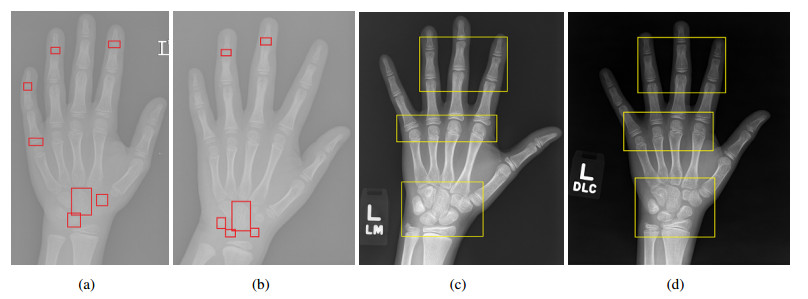
Bone age assessment plays a vital role in monitoring the growth and development of adolescents. However, it is still challenging to obtain precise bone age from hand radiography due to these problems: 1) Hand bone varies greatly and is always masked by the background; 2) the hand bone radiographs with successive ages offer high similarity. To solve such issues, a region fine-grained attention network (RFGA-Net) was proposed for bone age assessment, where the region aware attention (RAA) module was developed to distinguish the skeletal regions from the background by modeling global spatial dependency; then the fine-grained feature attention (FFA) module was devised to identify similar bone radiographs by recognizing critical fine-grained feature regions. The experimental results demonstrate that the proposed RFGA-Net shows the best performance on the Radiological Society of North America (RSNA) pediatric bone dataset, achieving the mean absolute error (MAE) of 3.34 and the root mean square error (RMSE) of 4.02, respectively.
Citation: Yamei Deng, Ting Song, Xu Wang, Yonglu Chen, Jianwei Huang. Region fine-grained attention network for accurate bone age assessment[J]. Mathematical Biosciences and Engineering, 2024, 21(2): 1857-1871. doi: 10.3934/mbe.2024081
Bone age assessment plays a vital role in monitoring the growth and development of adolescents. However, it is still challenging to obtain precise bone age from hand radiography due to these problems: 1) Hand bone varies greatly and is always masked by the background; 2) the hand bone radiographs with successive ages offer high similarity. To solve such issues, a region fine-grained attention network (RFGA-Net) was proposed for bone age assessment, where the region aware attention (RAA) module was developed to distinguish the skeletal regions from the background by modeling global spatial dependency; then the fine-grained feature attention (FFA) module was devised to identify similar bone radiographs by recognizing critical fine-grained feature regions. The experimental results demonstrate that the proposed RFGA-Net shows the best performance on the Radiological Society of North America (RSNA) pediatric bone dataset, achieving the mean absolute error (MAE) of 3.34 and the root mean square error (RMSE) of 4.02, respectively.

| [1] |
M. L. Danielson, R. H. Bitsko, R. M. Ghandour, J. R. Holbrook, M. D. Kogan, S. J. Blumberg, Prevalence of parent-reported ADHD diagnosis and associated treatment among US children and adolescents, 2016, J. Clin. Child Adolesc. Psychol., 47 (2018), 199–212. https://doi.org/10.1080/15374416.2017.1417860 doi: 10.1080/15374416.2017.1417860

|
| [2] |
S. E. Barlow, Expert committee recommendations regarding the prevention, assessment, and treatment of child and adolescent overweight and obesity: Summary report, Pediatrics, 120 (2007), 164–192. https://doi.org/10.1542/peds.2007-2329C doi: 10.1542/peds.2007-2329C
|
| [3] |
S. L Truesdell, M. M Saunders, Bone remodeling platforms: Understanding the need for multicellular lab-on-a-chip systems and predictive agent-based models, Math. Biosci. Eng., 17 (2020), 1233–1252. https://doi.org/10.3934/mbe.2020063 doi: 10.3934/mbe.2020063
|
| [4] |
P. Hao, S. Chokuwa, X. Xie, F. Wu, J. Wu, C. Bai, Skeletal bone age assessments for young children based on regression convolutional neural networks, Math. Biosci. Eng., 16 (2019), 6454–6466. https://doi.org/10.3934/mbe.2019323 doi: 10.3934/mbe.2019323
|
| [5] |
K. C. Lee, K. H. Lee, C. H. Kang, K. S. Ahn, L. Y. Chung, J. J. Lee, Clinical validation of a deep learning-based hybrid (Greulich-Pyle and modified Tanner-Whitehouse) method for bone age assessment, Korean J. Radiol., 22 (2021), 2017–2025. https://doi.org/10.3348/kjr.2020.1468 doi: 10.3348/kjr.2020.1468
|
| [6] |
P. Lv, C. Zhang, Tanner–Whitehouse skeletal maturity score derived from ultrasound images to evaluate bone age, Eur. Radiol., 2022 (2022), 1–8. https://doi.org/10.1007/s00330-022-09285-2 doi: 10.1007/s00330-022-09285-2
|
| [7] | S. Zhang, L. Liu, The skeletal development standards of hand and wrist for Chinese Children¡ªChina 05 I. TW3-C RUS, TW3-C Carpal, and RUS-CHN methods, Chin. J. Sports Med., 2023 (2023), 6–13. |
| [8] |
Z. Ling, S. Yang, F. Gou, Z. Dai, J. Wu, Intelligent assistant diagnosis system of osteosarcoma MRI image based on transformer and convolution in developing countries, IEEE J. Biomed. Health Inf., 26 (2022), 5563–5574. https://doi.org/10.1109/JBHI.2022.3196043 doi: 10.1109/JBHI.2022.3196043
|
| [9] |
Y. Deng, X. Wang, Y. Liao, ASA-Net: Adaptive sparse attention network for robust electric load forecasting, IEEE Internet Things J., (2023), 1–13. https://doi.org/10.1109/JIOT.2023.3300695 doi: 10.1109/JIOT.2023.3300695
|
| [10] | Q. H. Nguyen, R. Muthuraman, L. Singh, G. Sen, G. Sen, B. P. Nguyen, et al., Diabetic retinopathy detection using deep learning, in Proceedings of the International Conference on Machine Learning and Soft Computing, (2020), 103–107. https://doi.org/10.1145/3380688.3380709 |
| [11] | Q. H. Nguyen, B. P. Nguyen, S. Dao, B. Unnikrishnan, R. Dhingra, S. R. Ravichandran, et al., Deep learning models for tuberculosis detection from chest X-ray images, in Proceedings of the International Conference on Telecommunications (ICT), (2019), 381–385. https://doi.org/10.1109/ICT.2019.8798798 |
| [12] | H. N. Pham, R. J. Tan, Y. T. Cai, S. Mustafa, N. C. Yeo, H. J. Lim, Automated grading in diabetic retinopathy using image processing and modified efficientnet, in Proceedings of the International Conference on Computational Collective Intelligence (ICCCI), (2020), 505–515. |
| [13] |
Q. H. Nguyen, B. P. Nguyen, M. T. Nguyen, M. C. Chua, T. T. Do, N. Nghiem, Bone age assessment and sex determination using transfer learning, Expert Syst. Appl., 200 (2022), 1–11. https://doi.org/10.1016/j.eswa.2022.116926 doi: 10.1016/j.eswa.2022.116926
|
| [14] |
X. Wang, W. Fan, M. Hu, Y. Wang, F. Ren, CFJLNet: Coarse and fine feature joint learning network for bone age assessment, IEEE Trans. Instrum. Measure., 71 (2022), 1–11. https://doi.org/10.1109/TIM.2022.3193711 doi: 10.1109/TIM.2022.3193711
|
| [15] |
C. Liu, H. Xie, Y. Zhang, Self-supervised attention mechanism for pediatric bone age assessment with efficient weak annotation, IEEE Trans. Med. Imaging, 40 (2020), 2685–2697. https://doi.org/10.1109/TMI.2020.3046672 doi: 10.1109/TMI.2020.3046672
|
| [16] |
X. Ren, T. Li, X. Yang, S. Wang, S. Ahmad, L. Xiang, et al., Regression convolutional neural network for automated pediatric bone age assessment from hand radiograph, IEEE J. Biomed. Health Inf., 23 (2018), 2030–2038. https://doi.org/10.1109/JBHI.2018.2876916 doi: 10.1109/JBHI.2018.2876916
|
| [17] |
C. Chen, Z. Chen, X. Jin, L. Li, W. Speier, C. W. Arnold, Attention-guided discriminative region localization and label distribution learning for bone age assessment, IEEE J. Biomed. Health Inf., 26 (2021), 1208–1218. https://doi.org/10.1109/JBHI.2021.3095128 doi: 10.1109/JBHI.2021.3095128
|
| [18] |
N. Li, B. Cheng, J. Zhang, A cascade model with prior knowledge for bone age assessment, Appl. Sci., 12 (2022), 1–18. https://doi.org/10.55708/js0108002 doi: 10.55708/js0108002
|
| [19] |
S. Li, B. Liu, S. Li, X. Zhu, Y. Yan, D. Zhang, A deep learning-based computer-aided diagnosis method of X-ray images for bone age assessment, Complex Intell. Syst., 8 (2021), 1929–1939. https://doi.org/10.1007/s40747-021-00376-z doi: 10.1007/s40747-021-00376-z
|
| [20] |
A. A. Kasani, H. Sajedi, Hand bone age estimation using divide and conquer strategy and lightweight convolutional neural networks, Eng. Appl. Artif. Intell., 120 (2023), 1–12. https://doi.org/10.1016/j.engappai.2023.105935 doi: 10.1016/j.engappai.2023.105935
|
| [21] |
F. Cavallo, A. Mohn, F. Chiarelli, C. Giannini, Evaluation of bone age in children: A mini-review, Front. Pediatr., 9 (2021), 1–11. https://doi.org/10.3389/fped.2021.580314 doi: 10.3389/fped.2021.580314
|
| [22] |
H. N. Tuan, N. D. Hai, N. T. Thinh, Shape prediction of nasal bones by digital 2D-photogrammetry of the nose based on convolution and back-propagation neural network, Comput. Math. Methods Med., 2022 (2022), 1–18. https://doi.org/10.1155/2022/5938493 doi: 10.1155/2022/5938493
|
| [23] |
B. Dalisson, B. Charbonnier, A. Aoude, M. Gilardino, E. Harvey, N. Makhoul, Skeletal regeneration for segmental bone loss: Vascularised grafts, analogues and surrogates, Acta Biomater., 136 (2021), 37–55. https://doi.org/10.1016/j.actbio.2021.09.053 doi: 10.1016/j.actbio.2021.09.053
|
| [24] | J. Chen, X. Wang, Z. Guo, X. Zhang, J. Sun, Dynamic region-aware convolution, in Proceedings of the IEEE Conference on Computer Vision and Pattern Recognition (CVPR), (2021), 8064–8073. |
| [25] |
L. Wang, Y. Mao, J. Xu, J. Wu, K. Wu, K. Mao, A ROI extraction method for wrist imaging applied in smart bone-age assessment system, IEEE J. Biomed. Health Inf., (2023), 1–11. https://doi.org/10.1109/JBHI.2023.3284060 doi: 10.1109/JBHI.2023.3284060
|
| [26] |
R. Alexander, S. Waite, M. Bruno, E. Krupinski, L. Berlin, Mandating limits on workload, duty, and speed in radiology, Radiology, 304 (2022), 274–282. https://doi.org/10.1148/radiol.212631 doi: 10.1148/radiol.212631
|
| [27] |
P. Agarwal, A. Jagati, S. Rathod, K. Kalra, S. Patel, Clinical features of mycetoma and the appropriate treatment options, Res. Rep. Trop. Med., (2021), 173–179. https://doi.org/10.2147/RRTM.S282266 doi: 10.2147/RRTM.S282266
|
| [28] | Y. Chen, Y. Bai, W. Zhang, T. Mei, Destruction and construction learning for fine-grained image recognition, in Proceedings of the IEEE Conference on Computer Vision and Pattern Recognition (CVPR), (2019), 5157–5166. |
| [29] | G. Zhang, X. Lu, J. Tan, J. Li, Refinemask: Towards high-quality instance segmentation with fine-grained features, in Proceedings of the IEEE Conference on Computer Vision and Pattern Recognition (CVPR), (2021), 6861–6869. |
| [30] |
S. S. Halabi, L. M. Prevedello, J. Kalpathy-Cramer, A. B. Mamonov, A. Bilbily, M. Ciceroet, et al., The RSNA pediatric bone age machine learning challenge, Radiology, 290 (2019), 498–503. https://doi.org/10.1148/radiol.2018180736 doi: 10.1148/radiol.2018180736
|
| [31] | A. Paszke, S. Gross, F. Massa, A. Lerer, J. Bradbury, G. Chanan, Pytorch: An imperative style, high-performance deep learning library, in Proceedings of the Advances in Neural Information Processing Systems (NIPS), 2019, 8026–8037. |
| [32] |
L. Bottou, F. E. Curtis, J. Nocedal, Optimization methods for large-scale machine learning, Siam Review, 60 (2018), 223–311. https://doi.org/10.1137/16M1080173 doi: 10.1137/16M1080173
|
| [33] |
C. Spampinato, S. Palazzo, D. Giordano, M. Aldinucci, R. Leonardi, Deep learning for automated skeletal bone age assessment in X-ray images, Med. Image Anal., 36 (2017), 41–51. https://doi.org/10.1016/j.media.2016.10.010 doi: 10.1016/j.media.2016.10.010
|
| [34] |
Y. Deng, Y. Chen, Q. He, X. Wang, Y. Liao, J. Liu, et al., Bone age assessment from articular surface and epiphysis using deep neural networks, Math. Biosci. Eng., 20 (2023), 13133–13148. https://doi.org/10.3934/mbe.2023585 doi: 10.3934/mbe.2023585
|
| [35] |
T. D. Bui, J. Lee, J. Shin, Incorporated region detection and classification using deep convolutional networks for bone age assessment, Artif. Intell. Med., 97 (2019), 1–8. https://doi.org/10.1016/j.artmed.2019.04.005 doi: 10.1016/j.artmed.2019.04.005
|
| [36] |
A. Wibisono, P. Mursanto, Multi Region-Based Feature Connected Layer (RB-FCL) of deep learning models for bone age assessment, J. Big Data, 7 (2020), 1–17. https://doi.org/10.1186/s40537-019-0278-0 doi: 10.1186/s40537-019-0278-0
|
| [37] |
X. Zhou, E. Wang, Q. Lin, G. Lin, W. Wu, K. Huang, Diagnostic performance of convolutional neural network-based Tanner-Whitehouse 3 bone age assessment system, Quant. Imaging Med. Surg., 10 (2020), 657–667. https://doi.org/10.21037/qims.2020.02.20 doi: 10.21037/qims.2020.02.20
|
| [38] |
X. Li, Y. Jiang, Y. Liu, J. Zhang, S. Yin, H. Luo, RAGCN: Region aggregation graph convolutional network for bone age assessment from X-ray images, IEEE Trans. Instrum. Measure., 71 (2022), 1–12. https://doi.org/10.1109/TIM.2022.3190025 doi: 10.1109/TIM.2022.3190025
|
| [39] | C. Szegedy, V. Vanhoucke, S. Ioffe, J. Shlens, Z. Wojna, Rethinking the inception architecture for computer vision, in Proceedings of the IEEE Conference on Computer Vision and Pattern Recognition (CVPR), (2016), 2818–2826. |


Figures(5) / Tables(3)

Yamei Deng, Ting Song, Xu Wang, Yonglu Chen, Jianwei Huang. Region fine-grained attention network for accurate bone age assessment[J]. Mathematical Biosciences and Engineering, 2024, 21(2): 1857-1871. doi: 10.3934/mbe.2024081


 DownLoad: