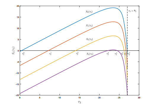
In this paper, we propose a delayed viral infection model with mitosis of uninfected target cells, two infection modes (virus-to-cell transmission and cell-to-cell transmission), and immune response. The model involves intracellular delays during the processes of viral infection, viral production, and CTLs recruitment. We verify that the threshold dynamics are determined by the basic reproduction number $ R_0 $ for infection and the basic reproduction number $ R_{IM} $ for immune response. The model dynamics become very rich when $ R_{IM} > 1 $. In this case, we use the CTLs recruitment delay $ \tau_3 $ as the bifurcation parameter to obtain stability switches on the positive equilibrium and global Hopf bifurcation diagrams for the model system. This allows us to show that $ \tau_3 $ can lead to multiple stability switches, the coexistence of multiple stable periodic solutions, and even chaos. A brief simulation of two-parameter bifurcation analysis indicates that both the CTLs recruitment delay $ \tau_3 $ and the mitosis rate $ r $ have a strong impact on the viral dynamics, but they do behave differently.
Citation: Jiawei Deng, Ping Jiang, Hongying Shu. Viral infection dynamics with mitosis, intracellular delays and immune response[J]. Mathematical Biosciences and Engineering, 2023, 20(2): 2937-2963. doi: 10.3934/mbe.2023139
In this paper, we propose a delayed viral infection model with mitosis of uninfected target cells, two infection modes (virus-to-cell transmission and cell-to-cell transmission), and immune response. The model involves intracellular delays during the processes of viral infection, viral production, and CTLs recruitment. We verify that the threshold dynamics are determined by the basic reproduction number $ R_0 $ for infection and the basic reproduction number $ R_{IM} $ for immune response. The model dynamics become very rich when $ R_{IM} > 1 $. In this case, we use the CTLs recruitment delay $ \tau_3 $ as the bifurcation parameter to obtain stability switches on the positive equilibrium and global Hopf bifurcation diagrams for the model system. This allows us to show that $ \tau_3 $ can lead to multiple stability switches, the coexistence of multiple stable periodic solutions, and even chaos. A brief simulation of two-parameter bifurcation analysis indicates that both the CTLs recruitment delay $ \tau_3 $ and the mitosis rate $ r $ have a strong impact on the viral dynamics, but they do behave differently.

| [1] |
M. A. Nowak, S. Bonhoeffer, G. M. Shaw, R. M. May, Anti-viral drug treatment: dynamics of resistance in free virus and infected cell populations, J. Theor. Biol., 184 (1997), 203–217. https://doi.org/10.1006/jtbi.1996.0307 doi: 10.1006/jtbi.1996.0307

|
| [2] |
A. S. Perelson, P. W. Nelson, Mathematical analysis of HIV-1 dynamics in vivo, SIAM Rev., 41 (1999), 3–44. https://doi.org/10.1137/S0036144598335107 doi: 10.1137/S0036144598335107
|
| [3] |
R. V. Culshaw, S. Ruan, G. Webb, A mathematical model of cell-to-cell spread of HIV-1 that includes a time delay, J. Math. Biol., 46 (2003), 425–444. https://doi.org/10.1007/s00285-002-0191-5 doi: 10.1007/s00285-002-0191-5
|
| [4] |
Y. Wang, Y. Zhou, J. Wu, J. Heffernan, Oscillatory viral dynamics in a delayed HIV pathogenesis model, Math. Biosci., 219 (2009), 104–112. https://doi.org/10.1016/j.mbs.2009.03.003 doi: 10.1016/j.mbs.2009.03.003
|
| [5] |
H. Shu, L. Wang, J. Watmough, Global stability of a nonlinear viral infection model with infinitely distributed intracellular delays and CTL immune responses, SIAM J. Appl. Math., 73 (2013), 1280–1302. https://doi.org/10.1137/120896463 doi: 10.1137/120896463
|
| [6] |
Y. Wang, J. Liu, J. M. Heffernan, Viral dynamics of an HTLV-I infection model with intracellular delay and CTL immune response delay, J. Math. Anal. Appl., 459 (2018), 506–527. https://doi.org/10.1016/j.jmaa.2017.10.027 doi: 10.1016/j.jmaa.2017.10.027
|
| [7] |
J. Ren, R. Xu, L. Li, Global stability of an HIV infection model with saturated CTL immune response and intracellular delay, Math. Biosci. Eng., 18 (2021), 57–68. https://doi.org/10.3934/mbe.2021003 doi: 10.3934/mbe.2021003
|
| [8] |
A. Sigal, J. T. Kim, A. B. Balazs, E. Dekel, A. Mayo, R. Milo, et al., Cell-to-cell spread of HIV permits ongoing replication despite antiretroviral therapy, Nature, 477 (2011), 95–98. https://doi.org/10.1038/nature10347 doi: 10.1038/nature10347
|
| [9] |
S. Iwami, J. S. Takeuchi, S. Nakaoka, F. Mammano, F. Clavel, H. Inaba, et al., Cell-to-cell infection by HIV contributes over half of virus infection, eLife, 4 (2015), e08150. https://doi.org/10.7554/eLife.08150 doi: 10.7554/eLife.08150
|
| [10] |
N. L. K. Galloway, G. Doitsh, K. M. Monroe, Z. Yang, I. Muñoz-Arias, D. N. Levy, et al., Cell-to-cell transmission of HIV-1 is required to trigger pyroptotic death of lymphoid-tissue-derived CD4 T cells, Cell Rep., 12 (2015), 1555–1563. https://doi.org/10.1016/j.celrep.2015.08.011 doi: 10.1016/j.celrep.2015.08.011
|
| [11] |
W. Hübner, G. P. McNerney, P. Chen, B. M. Dale, R. E. Gordan, F. Y. S. Chuang, et al., Quantitative 3D video microscopy of HIV transfer across T cell virological synapses, Science, 323 (2009), 1743–1747. https://doi.org/10.1126/science.1167525 doi: 10.1126/science.1167525
|
| [12] |
P. De Leenheer, H. L. Smith, Virus dynamics: a global analysis, SIAM J. Appl. Math., 63 (2003), 1313–1327. https://doi.org/10.1137/S0036139902406905 doi: 10.1137/S0036139902406905
|
| [13] |
L. Wang, M. Y. Li, Mathematical analysis of the global dynamics of a model for HIV infection of CD4+ T cells, Math. Biosci., 200 (2006), 44–57. https://doi.org/10.1016/j.mbs.2005.12.026 doi: 10.1016/j.mbs.2005.12.026
|
| [14] |
M. Tsiang, J. F. Rooney, J. J. Toole, C. S. Gibbs, Biphasic clearance kinetics of hepatitis B virus from patients during adefovir dipivoxil therapy, Hepatology, 29 (1999), 1863–1869. https://doi.org/10.1002/hep.510290626 doi: 10.1002/hep.510290626
|
| [15] |
X. Lai, X. Zou, Modeling cell-to-cell spread of HIV-1 with logistic target cell growth, J. Math. Anal. Appl., 426 (2015), 563–584. https://doi.org/10.1016/j.jmaa.2014.10.086 doi: 10.1016/j.jmaa.2014.10.086
|
| [16] |
Y. Yang, L. Zou, S. Ruan, Global dynamics of a delayed within-host viral infection model with both virus-to-cell and cell-to-cell transmissions, Math. Biosci., 270 (2015), 183–191. https://doi.org/10.1016/j.mbs.2015.05.001 doi: 10.1016/j.mbs.2015.05.001
|
| [17] |
J. E. Mittler, B. Sulzer, A. U. Neumann, A. S. Perelson, Influence of delayed viral production on viral dynamics in HIV-1 infected patients, Math. Biosci., 152 (1998), 143–163. https://doi.org/10.1016/S0025-5564(98)10027-5 doi: 10.1016/S0025-5564(98)10027-5
|
| [18] |
P. W. Nelson, A. S. Perelson, Mathematical analysis of delay differential equation models of HIV-1 infection, Math. Biosci., 179 (2002), 73–94. https://doi.org/10.1016/S0025-5564(02)00099-8 doi: 10.1016/S0025-5564(02)00099-8
|
| [19] |
K. Wang, W. Wang, H. Pang, X. Liu, Complex dynamic behavior in a viral model with delayed immune response, Physica D, 226 (2007), 197–208. https://doi.org/10.1016/j.physd.2006.12.001 doi: 10.1016/j.physd.2006.12.001
|
| [20] |
S. S. Chen, C. Y. Cheng, Y. Takeuchi, Stability analysis in delayed within-host viral dynamics with both viral and cellular infections, J. Math. Anal. Appl., 442 (2016), 642–672. https://doi.org/10.1016/j.jmaa.2016.05.003 doi: 10.1016/j.jmaa.2016.05.003
|
| [21] |
H. Shu, Y. Chen, L. Wang, Impacts of the cell-free and cell-to-cell infection modes on viral dynamics, J. Dyn. Differ. Equations, 30 (2018), 1817–1836. https://doi.org/10.1007/s10884-017-9622-2 doi: 10.1007/s10884-017-9622-2
|
| [22] |
T. Wang, Z. Hu, F. Liao, Stability and Hopf bifurcation for a virus infection model with delayed humoral immunity response, J. Math. Anal. Appl., 411 (2014), 63–74. https://doi.org/10.1016/j.jmaa.2013.09.035 doi: 10.1016/j.jmaa.2013.09.035
|
| [23] |
Y. Yang, T. Zhang, Y. Xu, J. Zhou, A delayed virus infection model with cell-to-cell transmission and CTL immune response, Int. J. Bifurcation Chaos, 27 (2017), 1750150. https://doi.org/10.1142/S0218127417501504 doi: 10.1142/S0218127417501504
|
| [24] | J. K. Hale, S. M. V. Lunel, Introduction to Functional Differential Equations, Springer-Verlag, New York, 1993. |
| [25] |
H. Shu, M. Y. Li, Joint effects of mitosis and intracellular delay on viral dynamics: two-parameter bifurcation analysis, J. Math. Biol., 64 (2012), 1005–1020. https://doi.org/10.1007/s00285-011-0436-2 doi: 10.1007/s00285-011-0436-2
|
| [26] |
H. R. Thieme, Spectral bound and reproduction number for infinite-dimensional population structure and time heterogeneity, SIAM J. Appl. Math., 70 (2009), 188–211. https://doi.org/10.1137/080732870 doi: 10.1137/080732870
|
| [27] |
J. K. Hale, P. Waltman, Persistence in infinite-dimensional systems, SIAM J. Math. Anal., 20 (1989), 388–395. https://doi.org/10.1137/0520025 doi: 10.1137/0520025
|
| [28] |
H. Shu, X. Hu, L. Wang, J. Watmough, Delay induced stability switch, multitype bistability and chaos in an intraguild predation model, J. Math. Biol., 71 (2015), 1269–1298. https://doi.org/10.1007/s00285-015-0857-4 doi: 10.1007/s00285-015-0857-4
|
| [29] |
E. Beretta, Y. Kuang, Geometric stability switch criteria in delay differential systems with delay dependent parameters, SIAM J. Math. Anal., 33 (2002), 1144–1165. https://doi.org/10.1137/S0036141000376086 doi: 10.1137/S0036141000376086
|
| [30] |
J. Xu, Y. Zhou, Bifurcation analysis of HIV-1 infection model with cell-to-cell transmission and immune response delay, Math. Biosci. Eng., 13 (2016), 343–367. https://doi.org/10.3934/mbe.2015006 doi: 10.3934/mbe.2015006
|
| [31] |
J. Wu, Symmetric functional differential equations and neural networks with memory, Trans. Am. Math. Soc., 350 (1998), 4799–4838. https://doi.org/10.1090/S0002-9947-98-02083-2 doi: 10.1090/S0002-9947-98-02083-2
|
| [32] |
H. Shu, W. Xu, X. S. Wang, J. Wu, Complex dynamics in a delay differential equation with two delays in tick growth with diapause, J. Differ. Equations, 269 (2020), 10937–10963. https://doi.org/10.1016/j.jde.2020.07.029 doi: 10.1016/j.jde.2020.07.029
|


Figures(7)

Jiawei Deng, Ping Jiang, Hongying Shu. Viral infection dynamics with mitosis, intracellular delays and immune response[J]. Mathematical Biosciences and Engineering, 2023, 20(2): 2937-2963. doi: 10.3934/mbe.2023139


 DownLoad: