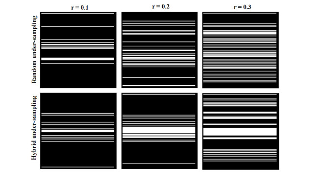
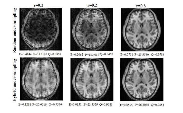
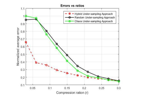
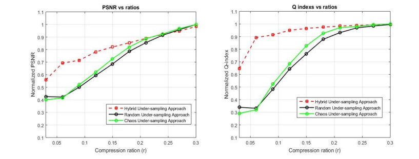
In magnetic resonance imaging (MRI), the scan time for acquiring an image is relatively long, resulting in patient uncomfortable and error artifacts. Fortunately, the compressed sensing (CS) and parallel magnetic resonance imaging (pMRI) can reduce the scan time of the MRI without significantly compromising the quality of the images. It has been found that the combination of pMRI and CS can better improve the image reconstruction, which will accelerate the speed of MRI acquisition because the number of measurements is much smaller than that by pMRI. In this paper, we propose combining a combined CS method and pMRI for better accelerating the MRI acquisition. In the combined CS method, the under-sampled data of the K-space is performed by taking both regular sampling and traditional random under-sampling approaches. MRI image reconstruction is then performed by using nonlinear conjugate gradient optimization. The performance of the proposed method is simulated and evaluated using the reconstruction error measure, the universal image quality Q-index, and the peak signal-to-noise ratio (PSNR). The numerical simulations confirmed that, the average error, the Q index, and the PSNR ratio of the appointed scheme are remarkably improved up to 59, 63, and 39% respectively as compared to the traditional scheme. For the first time, instead of using highly computational approaches, a simple and efficient combination of CS and pMRI is proposed for the better MRI reconstruction. These findings are very meaningful for reducing the imaging time of MRI systems.
Citation: Anh Quang Tran, Tien-Anh Nguyen, Phuc Thinh Doan, Duc-Nghia Tran, Duc-Tan Tran. Parallel magnetic resonance imaging acceleration with a hybrid sensing approach[J]. Mathematical Biosciences and Engineering, 2021, 18(3): 2288-2302. doi: 10.3934/mbe.2021116
In magnetic resonance imaging (MRI), the scan time for acquiring an image is relatively long, resulting in patient uncomfortable and error artifacts. Fortunately, the compressed sensing (CS) and parallel magnetic resonance imaging (pMRI) can reduce the scan time of the MRI without significantly compromising the quality of the images. It has been found that the combination of pMRI and CS can better improve the image reconstruction, which will accelerate the speed of MRI acquisition because the number of measurements is much smaller than that by pMRI. In this paper, we propose combining a combined CS method and pMRI for better accelerating the MRI acquisition. In the combined CS method, the under-sampled data of the K-space is performed by taking both regular sampling and traditional random under-sampling approaches. MRI image reconstruction is then performed by using nonlinear conjugate gradient optimization. The performance of the proposed method is simulated and evaluated using the reconstruction error measure, the universal image quality Q-index, and the peak signal-to-noise ratio (PSNR). The numerical simulations confirmed that, the average error, the Q index, and the PSNR ratio of the appointed scheme are remarkably improved up to 59, 63, and 39% respectively as compared to the traditional scheme. For the first time, instead of using highly computational approaches, a simple and efficient combination of CS and pMRI is proposed for the better MRI reconstruction. These findings are very meaningful for reducing the imaging time of MRI systems.

| [1] |
D. J. Larkman, R. G. Nunes, Parallel magnetic resonance imaging, Phys. Med. Biol., 52 (2007), R15-R55. doi: 10.1088/0031-9155/52/7/R01

|
| [2] |
J. Hamilton, D. Franson, N. Seiberlich, Recent advances in parallel imaging for MRI, Prog. Nucl. Magn. Reson. Spectrosc., 101 (2017), 71-95. doi: 10.1016/j.pnmrs.2017.04.002
|
| [3] |
D. K. Sodickson, W. J. Manning, Simultaneous acquisition of spatial harmonics (SMASH): fast imaging with radiofrequency coil arrays, Magn. Reson. Med., 38 (1997), 591-603. doi: 10.1002/mrm.1910380414
|
| [4] |
K. P. Pruessmann, M. Weiger, M. B. Scheidegger, P. Boesiger, SENSE: Sensitivity encoding for fast MRI, Magn. Reson. Med., 42 (1999), 952-962. doi: 10.1002/(SICI)1522-2594(199911)42:5<952::AID-MRM16>3.0.CO;2-S
|
| [5] |
M. A. Griswold, P. M. Jakob, R. M. Heidemann, M. Nittka, V. Jellus, J. Wang, et al., Generalized autocalibrating partially parallel acquisitions (GRAPPA), Magn. Reson. Med., 47 (2002), 1202-1210. doi: 10.1002/mrm.10171
|
| [6] |
M. Lustig, J. M. Pauly, SPIRiT: Iterative self-consistent parallel imaging reconstruction from arbitrary k-space, Magn. Reson. Med., 64 (2010), 457-471. doi: 10.1002/mrm.22428
|
| [7] | M. Sandilya, S. R. Nirmala, Compressed sensing trends in magnetic resonance imaging, Eng. Sci. Technol. Int. J., 20 (2017), 1342-1352. |
| [8] |
L. Feng, T. Benkert, K. T. Block, D. K. Sodickson, R. Otazo, H. Chandarana, Compressed sensing for body MRI, J. Magn. Reson. Imaging, 45 (2017), 966-987. doi: 10.1002/jmri.25547
|
| [9] |
M. Lustig, D. L. Donoho, J. M. Santos, J. M. Pauly, Compressed Sensing MRI, IEEE Signal Process. Mag., 25 (2008), 72-82. doi: 10.1109/MSP.2007.914728
|
| [10] |
M. Lustig, D. Donoho, J. M. Pauly, Sparse MRI: The application of compressed sensing for rapid MR imaging, Magn. Reson. Med., 58 (2007), 1182-1195. doi: 10.1002/mrm.21391
|
| [11] |
J. P. Haldar, D. Hernando, Z. Liang, Compressed-Sensing MRI With Random Encoding, IEEE Trans. Med. Imaging, 30 (2011), 893-903. doi: 10.1109/TMI.2010.2085084
|
| [12] |
E.J. Candes, T. Tao, Near-optimal signal recovery from random projections: Universal encoding strategies?, IEEE Trans. Inf. Theory, 52 (2006), 5406-5425. doi: 10.1109/TIT.2006.885507
|
| [13] |
S. Kojima, H. Shinohara, T. Hashimoto, S. Suzuki, Undersampling patterns in k-space for compressed sensing MRI using two-dimensional Cartesian sampling, Radiol. Phys. Technol., 11 (2018), 303-319. doi: 10.1007/s12194-018-0469-y
|
| [14] | R. Kazama, K. Sekine, S. Ito, Compressed sensing in magnetic resonance imaging using non-randomly under-sampled signal in cartesian coordinates, IEICE Trans. Inf. Syst., 102 (2019), 1851-1859. |
| [15] |
A. Q. Tran, T. A. Nguyen, V. T. Duong, Q. H. Tran, D. N. Tran, D. T. Tran, MRI Simulation-based evaluation of an efficient under-sampling approach, Math. Biosci. Eng., 17 (2020), 4048-4063. doi: 10.3934/mbe.2020224
|
| [16] |
D. Liang, B. Liu, J. Wang, L. Ying, Accelerating SENSE using compressed sensing, Magn. Reson. Med., 62 (2009), 1574-1584. doi: 10.1002/mrm.22161
|
| [17] | J. X. Ji, Z. Chen, L. Tao, Compressed sensing parallel magnetic resonance imaging, in 2008 30th Annual International Conference of the IEEE Engineering in Medicine and Biology Society, (2008), 1671-1674. |
| [18] |
R. Otazo, D. Kim, L. Axel, D. K. Sodickson, Combination of compressed sensing and parallel imaging for highly accelerated first‐pass cardiac perfusion MRI, Magn. Reson. Med., 64 (2010), 767-776. doi: 10.1002/mrm.22463
|
| [19] | C. Boyer, P. Ciuciu, P. Weiss, S. Meriaux, HYR2PICS: Hybrid regularized reconstruction for combined parallel imaging and compressive sensing in MRI, in 2012 9th IEEE International Symposium on Biomedical Imaging (ISBI), (2012), 66-69. |
| [20] | D. S. Weller, J. R. Polimeni, L. Grady, L. L. Wald, E. Adalsteinsson, V. K. Goyal, Combined compressed sensing and parallel mri compared for uniform and random cartesian undersampling of K-space, in 2011 IEEE International Conference on Acoustics, Speech and Signal Processing (ICASSP), (2011), 553-556. |
| [21] |
J. E. Vranic, N. M. Cross, Y. Wang, D. S. Hippe, E. d. Weerdt, M. M. Basha, Compressed sensing-sensitivity encoding (CS-SENSE) accelerated brain imaging: reduced scan time without reduced image quality, Am. J. Neuroradiol., 40 (2019), 92-98. doi: 10.3174/ajnr.A5905
|
| [22] |
S. V. Eslahi, J. Ji, Accelerated positive contrast MRI of interventional devices using parallel compressed sensing imaging, Magn. Reson. Imaging, 60 (2019), 130-136. doi: 10.1016/j.mri.2019.04.006
|
| [23] |
K. Koolstra, J. V. Gemert, P. Börnert, A. Webb, R. Remis, Accelerating compressed sensing in parallel imaging reconstructions using an efficient circulant preconditioner for cartesian trajectories, Magn. Reson. Med., 81 (2019), 670-685. doi: 10.1002/mrm.27371
|
| [24] | L. E. Gueddari, P. Ciuciu, E. Chouzenoux, A. Vignaud, J. C. Pesquet, Calibrationless oscar-based image reconstruction in compressed sensing parallel MRI, in 2019 IEEE 16th International Symposium on Biomedical Imaging, (2019), 1532-1536. |
| [25] |
B. Deka, S. Datta, Calibrationless joint compressed sensing reconstruction for rapid parallel MRI, Biomed. Signal Process. Control, 58 (2020), 101871. doi: 10.1016/j.bspc.2020.101871
|
| [26] | C. Chen, Y. Li, J. Huang, Calibrationless parallel MRI with joint total variation regularization, in International Conference on Medical Image Computing and Computer-Assisted Intervention, Springer, Berlin, Heidelberg, (2013), 106-114. |
| [27] |
S. Wang, S. Tan, Y. Gao, Q. Liu, L. Ying, T. Xiao, et al., Learning joint-sparse codes for calibration-free parallel MR imaging, IEEE Trans. Med. Imaging, 37 (2018), 251-261. doi: 10.1109/TMI.2017.2746086
|
| [28] |
I. Y. Chun, B. Adcock, T. M. Talavage, Efficient compressed sensing SENSE pMRI reconstruction with joint sparsity promotion, IEEE Trans. Med. Imaging, 35 (2016), 354-368. doi: 10.1109/TMI.2015.2474383
|
| [29] |
R. W. Liu, Q. Ma, S. C. H. Yu, K. T. Chui, N. Xiong, Variational regularized tree-structured wavelet sparsity for CS-SENSE parallel imaging, IEEE Access, 6 (2018), 61050-61064. doi: 10.1109/ACCESS.2018.2874382
|
| [30] | S. R. Islam, S. P. Maity, A. K. Ray. CS regularized SENSE pMRI reconstruction via interferometric modulation, in 2017 International Conference on Wireless Communications, Signal Processing and Networking (WiSPNET), (2017), 1141-1146. |
| [31] | T. D. Tan, L. Vu-Ha, N. L. Trung, Spread spectrum for chaotic compressed sensing techniques in parallel magnetic resonance imaging, in 2011 8th International Conference on Information, Communications & Signal Processing, (2011), 1-5. |
| [32] | T. Tran Duc, D. V. Phong, T. M. Chinh, N. Linh-Trung, Accelerated parallel magnetic resonance imaging with multi-channel chaotic compressed sensing, in The 2010 International Conference on Advanced Technologies for Communications, (2010), 146-151. |
| [33] |
J. Zhang, Y. Chu, W. Ding, L. Kang, L. Xia, S. Jaiswal, et al., HF-SENSE: An improved partially parallel imaging using a high-pass filter, BMC Med. Imaging, 19 (2019), 27. doi: 10.1186/s12880-019-0327-3
|
| [34] | T. Minh-Chinh, N. Linh-Trung, T. Duc-Tan, On the implementation of chaotic compressed sensing for MRI, in 2016 International Conference on Advanced Technologies for Communications (ATC), (2016), 103-107. |
| [35] |
Z. Chang, Q. S. Xiang, Highly accelerated MRI by skipped phase encoding and edge deghosting with array coil enhancement (SPEED-ACE), Med. Phys., 33 (2006), 3758-3766. doi: 10.1118/1.2349700
|
| [36] |
T. A. Gallagher, A. J. Nemeth, L. Hacein-Bey, Hacein-Bey L. An introduction to the Fourier transform: relationship to MRI, Am. J. Roentgenol., 190 (2008), 1396-1405. doi: 10.2214/AJR.07.2874
|
| [37] | Z. P. Liang, P. C. Lauterbur, Principles of Magnetic Resonance Imaging: a Signal Processing Perspective, SPIE Optical Engineering Press, Bellingham, 2000. |
| [38] |
E. J. Candes, M. B. Wakin, An Introduction To Compressive Sampling, IEEE Signal Process. Mag., 25 (2008), 21-30. doi: 10.1109/MSP.2007.914731
|
| [39] |
M. Guerquin-Kern, M. Hä berlin, K. P. Pruessmann, M. Unser, A fast wavelet-based reconstruction method for magnetic resonance imaging, IEEE Trans. Med. Imaging, 30 (2011), 1649-1660. doi: 10.1109/TMI.2011.2140121
|
| [40] |
E. J. Candes, J. Romberg, T. Tao, Robust uncertainty principles: exact signal reconstruction from highly incomplete frequency information, IEEE Trans. Inf. Theory, 52 (2006), 489-509. doi: 10.1109/TIT.2005.862083
|
| [41] |
E. Candès, J. Romberg, Sparsity and incoherence in compressive sampling, Inverse Probl., 23 (2007), 969-985. doi: 10.1088/0266-5611/23/3/008
|
| [42] | D. Moratal, A. Vallés-Luch, L. Marti-Bonmati, M. Brummer, k-Space tutorial: an MRI educational tool for a better understanding of k-space, Biomed. Imaging Interv. J., 4 (2008), e15-e15. |
| [43] | D. R. Mehra, Estimation of the image quality under different distortions, Int. J. Eng. Sci., 8 (2016), 17291-17296. |
| [44] |
W. Zhou, A. C. Bovik, A universal image quality index, IEEE Signal Process. Lett., 9 (2002), 81-84. doi: 10.1109/97.995823
|
Figures(8)

Anh Quang Tran, Tien-Anh Nguyen, Phuc Thinh Doan, Duc-Nghia Tran, Duc-Tan Tran. Parallel magnetic resonance imaging acceleration with a hybrid sensing approach[J]. Mathematical Biosciences and Engineering, 2021, 18(3): 2288-2302. doi: 10.3934/mbe.2021116


 DownLoad: