Loading [MathJax]/jax/output/SVG/jax.js
Research article Topical Sections

Gαq protein signaling in the bed nucleus of the stria terminalis regulate the lipopolysaccharide-induced despair-like behavior in mice

  • Nao Fukuwada and Miki Kanno contributed equally to this work.
  • Received: 24 August 2020 Accepted: 05 November 2020 Published: 10 November 2020
  • Major depressive disorder (MDD) is highly comorbid with anxiety disorders. It has been reported that the bed nucleus of the stria terminalis (BNST) is important for the induction of anxiety and MDD. Recently, the Gαq protein signaling within the BNST is involved in the induction of anxiety through Gαq protein signaling-mediated RNA-editing of GluR2 subunit, which produces the calcium (Ca2+)-impermeable α-amino-3-hydroxy-5-methyl-4-isoxazolepropionic acid (AMPA) receptor. On the other hand, the role of Gαq protein signaling within the BNST on the induction of MDD has never been reported yet. Therefore, we investigated whether Gαq protein signaling-producing the Ca2+-impermeable AMPA receptors in the BNST is involved in the lipopolysaccharide (LPS)-induced depressive-like behavior, particularly, despair-like behavior. When mice were systemically challenged with a single dose of LPS (1.2 mg/kg, i.p.), the immobility time during tail suspension test (TST) was increased 24 h after LPS injection. However, pretreatment with bilateral intra-BNST injection of neomycin (6.5 mM, 0.125 µL/side), an inhibitor of phospholipase C that is activated by Gαq protein-coupled receptor stimulation, extended the LPS-induced increase in the immobility time of TST. Furthermore, the co-pretreatment with bilateral intra-BNST injection of neomycin with 1-naphthylacetyl spermine (3 mM, 0.125 µL/side), an antagonist of Ca2+-permeable AMPA receptor, to mimic one of the final forms of Gαq protein activation, abolished the aggravated effect of neomycin and significantly shortened the immobility time compared with the control mice with an intra-BNST injection of artificial cerebrospinal fluid before LPS injection. However, pretreatment with bilateral intra-BNST injection of MDL-12,330A (10 µM, 0.125 µL/side), an inhibitor of adenylyl cyclase that is activated by Gαs protein-coupled receptor stimulation, did not affect the LPS-induced increase in the immobility time of TST. These results indicate that the Gαq protein signaling-mediated RNA-editing of GluR2, which produces the Ca2+-impermeable AMPA receptors within the BNST, regulates the LPS-induced despair-like behavior.

    Citation: Nao Fukuwada, Miki Kanno, Satomi Yoshida, Kenjiro Seki. Gαq protein signaling in the bed nucleus of the stria terminalis regulate the lipopolysaccharide-induced despair-like behavior in mice[J]. AIMS Neuroscience, 2020, 7(4): 438-458. doi: 10.3934/Neuroscience.2020027

    Related Papers:

    [1] Daniel Danladi Gaiya, Jonathan Danladi Gaiya, Richard Auta, Aliyu Muhammad, Bege Jonathan, Stella Kuyet Udu, Ekpa Emmanuel, Amina Shehu Bature . The journey so far with SARS-CoV-2 variants: Pathogenesis, immunity and treatments. AIMS Allergy and Immunology, 2023, 7(4): 222-250. doi: 10.3934/Allergy.2023016
    [2] Richard A. Maiella, Kelly M. Staples, Ashokvardhan Veldanda . Migratory dermatographic urticaria following COVID-19 vaccine booster in young adult male. AIMS Allergy and Immunology, 2022, 6(1): 14-18. doi: 10.3934/Allergy.2022003
    [3] Y. Desheva, T. Shvedova, A. Lerner, P. Kudar, O. Kopteva, D. Petrachkova, I. Koroleva, G. Leontieva, S. Ponkratov, A. Suvorov . Exploring immune response and lab markers across COVID-19 severity levels. AIMS Allergy and Immunology, 2024, 8(4): 216-231. doi: 10.3934/Allergy.2024013
    [4] Arosh S. Perera Molligoda Arachchige . Aging and immunity: Unraveling the complex relationship. AIMS Allergy and Immunology, 2023, 7(3): 213-221. doi: 10.3934/Allergy.2023015
    [5] Michael D. Caponegro, Jeremy Tetsuo Miyauchi, Stella E. Tsirka . Contributions of immune cell populations in the maintenance, progression, and therapeutic modalities of glioma. AIMS Allergy and Immunology, 2018, 2(1): 24-44. doi: 10.3934/Allergy.2018.1.24
    [6] Mansur Aliyu, Ali Akbar Saboor-Yaraghi, Sadegh Khodavaisy, Behrouz Robat-Jazi, Muhammad Ibrahim Getso . Glucuronoxylomannan: the salient polysaccharide in cryptococcal immunity. AIMS Allergy and Immunology, 2022, 6(2): 71-89. doi: 10.3934/Allergy.2022008
    [7] Stephen C Allen . Cytokine signaling in the modulation of post-acute and chronic systemic inflammation: a review of the influence of exercise and certain drugs. AIMS Allergy and Immunology, 2020, 4(4): 100-116. doi: 10.3934/Allergy.2020009
    [8] Kamyar Nasiri, Zohreh Mortezania, Sanaz Oftadehbalani, Mohammad Khosousi Sani, Amin Daemi, Seyyedeh Touran Hosseini, Yusuf Döğüş, Zafer Yönden . Parasitology and Covid-19: epidemiological, immunological, pathological, and therapeutic aspects. AIMS Allergy and Immunology, 2023, 7(1): 92-103. doi: 10.3934/Allergy.2023007
    [9] Darrell O. Ricke . Antibodies and infected monocytes and macrophages in COVID-19 patients. AIMS Allergy and Immunology, 2022, 6(2): 64-70. doi: 10.3934/Allergy.2022007
    [10] DharmaSaranya Gurusamy, Vasuki Selvamurugesan, Swaminathan Kalyanasundaram, Shantaraman Kalyanaraman . Is COVID 19, a beginning of new entity called chronic viral systemic inflammatory syndrome?. AIMS Allergy and Immunology, 2021, 5(2): 127-134. doi: 10.3934/Allergy.2021010
  • Major depressive disorder (MDD) is highly comorbid with anxiety disorders. It has been reported that the bed nucleus of the stria terminalis (BNST) is important for the induction of anxiety and MDD. Recently, the Gαq protein signaling within the BNST is involved in the induction of anxiety through Gαq protein signaling-mediated RNA-editing of GluR2 subunit, which produces the calcium (Ca2+)-impermeable α-amino-3-hydroxy-5-methyl-4-isoxazolepropionic acid (AMPA) receptor. On the other hand, the role of Gαq protein signaling within the BNST on the induction of MDD has never been reported yet. Therefore, we investigated whether Gαq protein signaling-producing the Ca2+-impermeable AMPA receptors in the BNST is involved in the lipopolysaccharide (LPS)-induced depressive-like behavior, particularly, despair-like behavior. When mice were systemically challenged with a single dose of LPS (1.2 mg/kg, i.p.), the immobility time during tail suspension test (TST) was increased 24 h after LPS injection. However, pretreatment with bilateral intra-BNST injection of neomycin (6.5 mM, 0.125 µL/side), an inhibitor of phospholipase C that is activated by Gαq protein-coupled receptor stimulation, extended the LPS-induced increase in the immobility time of TST. Furthermore, the co-pretreatment with bilateral intra-BNST injection of neomycin with 1-naphthylacetyl spermine (3 mM, 0.125 µL/side), an antagonist of Ca2+-permeable AMPA receptor, to mimic one of the final forms of Gαq protein activation, abolished the aggravated effect of neomycin and significantly shortened the immobility time compared with the control mice with an intra-BNST injection of artificial cerebrospinal fluid before LPS injection. However, pretreatment with bilateral intra-BNST injection of MDL-12,330A (10 µM, 0.125 µL/side), an inhibitor of adenylyl cyclase that is activated by Gαs protein-coupled receptor stimulation, did not affect the LPS-induced increase in the immobility time of TST. These results indicate that the Gαq protein signaling-mediated RNA-editing of GluR2, which produces the Ca2+-impermeable AMPA receptors within the BNST, regulates the LPS-induced despair-like behavior.


    Malaria is an infectious, vector-borne disease caused by a parasite native to the African continent, and it claims the lives of many humans each year, particularly children under the age of 5 [1], [2]. According to the World Health Organization's 2021 report, an estimated 241 million cases of malaria and 627,000 deaths were recorded that year. A significant challenge lies in controlling malaria to lower mortality and morbidity rates. The female Anopheles mosquito serves as a vector for malaria transmission, and the parasite responsible for the disease belongs to the Plasmodium family [3], [4]. The four types of Plasmodium that affect humans are Plasmodium vivax, Plasmodium falciparum, Plasmodium malariae, and Plasmodium ovale. The primary causative agent of malaria is Plasmodium falciparum. Malaria is transmitted when a female Anopheles mosquito injects the parasite into the bloodstream of an uninfected human. Conversely, the mosquito becomes infected after ingesting a blood meal from an infected human [5]. The mosquito species that are responsible for malaria prefer biting pregnant women, people with a higher body mass index, and individuals with the blood type O [6].

    After being bitten by an infected Anopheles mosquito, the parasites, in the form of sporozoites, travel through the bloodstream to the liver cells, specifically hepatocytes, where they mature into schizonts. This stage is referred to as the mosquito liver stage [7]. The schizonts replicate within the liver cells in the form of merozoites, which are released into the bloodstream by ruptured hepatocyte cells. In the blood, the merozoites invade the red blood cells (RBCs) and multiply again within the RBCs to form more merozoites. After 1 to 2 weeks, the schizont ruptures, thereby releasing the merozoites into the bloodstream [8]. If left untreated at this stage, the cycle will persist, and the infected person will start developing symptoms of malaria, such as chills, fever, and sweating [9]. This stage is known as the mosquito blood stage. In addition to that, the merozoites develop into sexual forms known as gametocytes, which are taken up by the mosquitoes during blood meal in order to maintain the parasite life cycle [9], [10]. In this work, we will not consider the mosquito stage, which is the parasite's life cycle inside a mosquito. There are three stages of the parasite life cycle: the liver stage, the blood stage and the mosquito stage.

    The rapid increase in parasites activates the immune system's mechanism. In response, the immune cells secrete an innate cytokine called the macrophage migration inhibitory factor (MIF), which regulates the immune responses and cell differentiation, a process in which immature cells acquire unique characteristics and mature into specialized forms with distinct functions. However, in severe malaria anemia, MIF contributes to the destruction of healthy red blood cells and a decrease in hemoglobin levels [11], [12]. In return, the parasites secrete a protein called Plasmodium Falciparum MIF (PFMIF), which hinders the invading activity and antigen presentation capabilities of the immune cells such as macrophages and dendritic cells [13], [14]. This means that the body's immune system will not be able to distinguish between the body's cells and foreign cells, thereby promoting the survival and replication of the malaria parasite within the blood. Artesunate is a successful treatment for severe malaria anemia because it can block the growth and reproduction of the malaria parasites, which, in turn, prevents the destruction of the RBCs [15].

    Despite Artesunate's efficacy in decreasing parasite load, there have been some reports concerning its use. Occasionally, Artesunate use might lead to hemolytic anemia, a condition where the destruction of RBCs is faster than the production, which results in the reduction of hemoglobin levels [16]. However, we can use Artesunate in conjunction with another antimalarial drug called Epoxyazadiradione (Epoxy), which is a naturally occurring substance present in neem trees that is known to have antimalarial drug characteristics. Epoxy has the ability to prevent the formation of MIF in human monocytes, which are involved in the immune response to infections. Additionally, Epoxy can regulate immune cells and cytokines, such as PFMIF, and may help boost RBCs count [17]. By combining Artesunate and Epoxy in this study, we aim to reduce parasites while increasing hemoglobin levels.

    Immune boosters are substances, strategies, or interventions that either enhance or strengthen the body's immune system, helping it to better defend against infections and diseases. A robust immune system is essential for a person's overall health and well-being [18][20]. The use of immune boosters in the context of malaria treatment is a complex and evolving area of research. While some immune-boosting strategies have shown promise in enhancing the body's response to malaria [18], it is important to note that the field of immuno-modulation in malaria is continually evolving, and the effectiveness of specific immune boosters may vary based on factors such as the parasite species and host immunity [19], [20].

    In this paper, we developed a mathematical model of the human blood-stage Plasmodium life cycle. The model, which is based on and simplifies a previous work [21], is represented by a system of ordinary differential equations (ODEs). In this updated model, we have excluded the IL-12, IFN-γ, and TNF-α cytokines from the model in [21], and have reformulated the model network and system of equations accordingly. Subsequently, we employed this model to evaluate the effectiveness of treatment with the drugs Artesunate and Epoxy, both as individual therapies and in combination. Additionally, we investigated the role of immune boosters in the context of malaria treatment using these two drugs.

    The rest of this paper is organized as follows. In Section 2, we develop the mathematical model designing the model network, then we write and describe the equations associated with this network. In Section 3, we present the results and model simulations, and the conclusion and recommendations are presented in Section 4.

    The mathematical model is based on the network in Figure 1. This network is a simplification of Figure 1 in [21], where we dropped the dendritic cells (antigen-presenting cells) and combined the effect of the immune response (represented in [21] by macrophages and Th1-cells) by lumping them together into one variable Tc. Furthermore, we dropped the IL-12, IFN-γ, and TNF-α cytokines and modeled their regulating effects by a constant parameter. The model parameters and their descriptions can be found in Table 2. Table 1 lists our model's variables in units of g/ml.

    Table 1.  Descriptions of variables (Vars) used in the model. All variables are in units of g/ml.
    Vars Descriptions Vars Descriptions
    Tc Density of CD4+ T cells B Density of healthy RBCs
    Bi Density of infected RBCs Hb Concentration of hemoglobin
    Pi Intracellular parasite load Pe Extracellular parasite load
    Pf Concentration of PFMIF Mf Concentration of MIF
    A Concentration of artesunate E Concentration of epoxyazadiradione

     | Show Table
    DownLoad: CSV
    Table 2.  List of model's parameter values.
    Parameters Descriptions Values References
    λB The rate of release of B by B0 1.15×10⁻² d−1 est. & HT
    λBBi The rate of infection of B 6×101 ml/gd−1 HT
    λPi The rate of growth of Pi in Bi 0.8 d−1 HT
    λPiPe The rate of rupturing of Bi 0.66 d−1 [21] & HT
    λBiTc The rate of ingestion of Bi by Tc 1.5602×10⁻⁶ ml/g d−1 est.
    λPiTc The rate of ingestion of Pi by Tc 4.845×10⁻⁸ d−1 est.
    λPeTc The rate of ingestion of Pe by Tc 2.425×10⁻⁷ d−1 est.
    λTcPe The rate of activation of Tc by Pe 1×10³ d−1 est.
    λTcPi The rate of activation of Tc by Pi 5×10⁻² d−1 est.
    λTcBi The rate of activation of Tc by Bi 5×10² d−1 est.
    λMfTc The rate of secretion of Mf by Tc 6.71×10⁻⁵ d−1 [21] & HT
    λPfPi The rate of secretion of Pf by Pi 2×10⁻³ d−1 [21] & HT
    λB0Mf The rate of absorption of Mf by B0 3.9×101 d−1 [21] & HT
    λABi The rate of florescence of A in Bi 2.08×10⁻⁶ d−1 [21]
    λEB The rate of florescence of E in B 1.2×10⁻⁹ d−1 [21] & HT
    λETc The rate of florescence of E in Tc 1.2×10⁻⁹ d−1 [21] & HT
    µB The rate of death healthy RBCs 5.78×10⁻³ d−1 est.
    µBi The rate of death of infected RBCs 5.78×10⁻³ d−1 HT
    µPe The rate of death of extracellular parasites 4.9×10² d−1 HT
    µTc The rate of death of CD4+ T cells 5×10⁻⁵ d−1 HT
    µTcPf Death rate of Tc by PFMIF 2×10⁻³ d−1 HT
    µMf Decay rate of MIF 1.39 d−1 [21] & HT
    µPf Decay rate of PMIF 2 d−1 HT
    µBiA Death rate of infected RBCs by A 0.1 d−1 HT
    µA Degradation rate of Artesunate 33 d−1 [21] & HT
    µE Degradation rate of epoxyazadiradione 1.98 d−1 [21] & HT
    B0 RBCs in healthy human 0.45 g/ml [21]
    T0c T cells in healthy human 1.08×10⁻³ g/ml [21]
    Hb Hemoglobin in healthy human 0.14 g/ml [21]
    m*B (mass of Plasmodium parasite)/(mass of RBC) 3.7×10⁻³ [21]
    mMf (mass of MIF)/(mass of RBC) 7.78×10⁻¹⁰ HT
    NB Average number of Pi released per burst of Bi 24 [21]
    nB Average number of Pi in one iRBC 6 [21]
    θH Fraction of hemoglobin in iRBCs 0.33 [21]
    KBi Half-saturation of Bi 0.015 g/ml [21]
    KTc Half-saturation of Tc 1.08×10⁻³ g/ml [21]
    KPe Half-saturation of Pe 4×10⁻⁴ g/ml [21]
    KPi Half-saturation of Pi 1.3×10⁻³ g/ml [21]
    KMf Half-saturation of MIF 4×10⁻⁹ g/ml [21]
    KPf Half-saturation of PFMIF 8×10⁻¹⁰ g/ml [21]
    KA Half-saturation of A 3.26×10⁻⁶ g/ml [21]
    KE Half-saturation of E 4×10⁻⁶ g/ml [21]
    Ca Carrying capacity of Pi 2.6×10⁻³ g/ml [21]

    est. = estimated, HT = hand tuned (refer to Section A).

     | Show Table
    DownLoad: CSV
    Figure 1.  A network is used to represent the interaction between cells, parasites and anti-malarial drugs during treatment. See Table 1 for the description of each variable.

    Following the model by Siewe and Friedman [21], the model is based on the following assumptions:

    (i) During a malaria infection, various types of immune cells are activated in response to the presence of parasites in the body. In our model, we assume that the immune system consists of a single compartment, namely CD4+ T cells (Tc).

    (ii) Red blood cells (RBCs) are produced by spongy-like structures called bone marrow, which are found within bones, at a rate of λB. They are reduced by infection from extracellular merozoites at a rate of λBBi.

    (iii) The merozoite form of intracellular parasites replicates inside healthy red blood cells while circulating in the blood at a rate of λPi.

    (iv) When red blood cells are invaded by intracellular merozoites and replicate inside them, it results in the rupture of the infected red blood cells at a rate of λPiPe.

    (v) The presence of the intracellular and extracellular merozoites, as well as infected RBCs in the bloodstream, triggers the body to produce immune cells called CD4+ T cells at the rates of λTcPe, λTcPi and λTcBi.

    (vi) In turn, the CD4+ T cells kill the infected RBCs together with the intracellular and extracellular parasites at the rates of λTcBi, λTcPi and λTcPe, respectively.

    (vii) T cells secrete MIF, which participates in inflammatory and immune responses at a rate of λMfTc. MIF suppresses red blood cells, thereby leading to the depletion of hemoglobin.

    (viii) Lastly, intracellular parasites secrete PFMIF at a rate of λPfTc.

    After the release of healthy red blood cells from the bone marrow, MIF interferes with the process, as shown by the first term on the right-hand side [22]. The ratio 1/(1 + E/KE) is the inhibitory effect of MIF by the drug Epoxy. The second term describes the rate of infection of healthy RBCs, denoted by λBBi, thereby leading to a reduction in healthy red blood cells and an increase in infected red blood cells Bi [23]. The last term represents the natural death of the RBCs. Hence, the equation of B is given by the following:

    dBdt=λBB01(1+Mf/KMf)11+E/KEReleaseλBBiBPeInfectionµBB.death

    When extracellular parasites invade red blood cells, there is an increase in the number of infected RBCs [24]. We assume that bursting occurs when the concentration of intracellular parasites Pi is roughly the same as m*BNBBi, where NB represents the number of parasites released per burst, and m*B represents the mass of one plasmodium parasite cell over the mass of one RBC. The reduction in infected RBCs is a result of the phagocytosis of Tc cells [25], as shown in the “ingestion” term below.

    The last two terms represent the rate of death of infected RBCs by the drug Artesunate (when administered) [26] and by natural death, respectively. Hence, Bi satisfies the following equation:

    dBidt=λBBiBPeinfectionλPiPeBiP2i(m*BNBBi)2+P2iBurstingλBiTcTcBiingestionµBiAAKA+ABideath by ArtesunateµBiBi,natural death

    where m*B=mass of 1 parasitemass of 1 RBC.

    An increase in the intracellular parasite population is a result of their replication within Bi [27]. The replication rate is determined by the factor 1 − Pi/Ca, which ensures that the concentration of intracellular parasites Pi does not exceed the carrying capacity Ca. Additionally, an increase in parasite numbers comes from new infections [28]. Additionally, the model accounts for a decrease in the number of parasites due to the rupture of infected RBCs (“bursting”) [29]. The death rate of intracellular parasites [30] is enhancing T cells [31] and the action of the drug Artesunate when it is administered [21]. We represent the equation for Pi as follows:

    dPidt=λpiPi(1PiCa)growth in Bi+λBBim*BBPeinfectionλPiPem*BNBBiP2i(m*BNBBi)2+P2iBurstingλPiTcPiKPi+PiTcKilled by TcµBiAnBAKA+ABideath by ArtesunateµBinBPideath

    where the constant nB represents the number of intracellular parasites within one infected RBC at the time of its death due to natural cell death.

    We similarly represent the killing of Pe by Tc as in Eq (2.3), and extracellular parasites die naturally. It is worth noting that the constant nB represents the number of intracellular parasites within one infected RBC at the time of its death due to natural cell death. We write the equation for Pe as follows:

    dPedt=λPiPem*BNBBiP2i(m*BNBBi)2+P2iBurstingλBBim*BBPeinfectionλPeTcPeKPe+PeTckilled by TcµPePe.death

    Activation of the naive CD4+ T cells is triggered by infected RBCs Bi [32], intracellular Pi and extracellular Pe parasites, respectively. The Tc cells die naturally [33] and there is an augmented death rate of Tc resulting from the effect of PFMIF [12]; we represent this rate by µTcPfPf/(KPf+Pf). When the drug Epoxy is administered, we represent its inhibition effect on PFMIF by the ratio 1/(1 + E/KE). Hence,

    dTcdt=(λTcBiBiKBi+Bi+λTcPiPiKPi+Pi+λTcPePeKPe+Pe)T0cproliferation(µTc+µTcPfPfKPf+Pf11+E/KE)Tcdeath

    where T0c is the source of Tc cells.

    MIF is secreted by the CD4+ T cells [34]. MIF is absorbed by inhibiting the production of red blood cells [35]. Hence, the equation for Mf is given as follows:

    dMfdt=λMfTcTcsecretionλB0Mfm*MfB0MfKMf+Mfabsorption by B0µMfMf.decay

    where the third term on the right hand side is the natural decay of MIF.

    PFMIF is secreted by the intracellular parasites Pi [36] at rate λPfPiPi. PFMIF decreases as it is absorbed by Tc. Pf also decays naturally [37]. Hence, the equation for Pf is written in the following form:

    dPfdt=λPfPiPisecretionµTcPfPfKPf+PfTcabsorbed by T cellsµPfPf.decay

    Hemoglobin is a red pigment found in red blood cells that is responsible for the circulation of oxygen. We consider the total hemoglobin HB equation to be as follows:

    Hb=HB(B+Bi×θH),

    as in [21], where HB is the fraction of the hemoglobin level in healthy RBCs (B). We take the same percentage for infected RBCs, where B is the concentration of healthy RBCs, which is approximately 4.5 to 5.5×10⁶ cells/ml or approximately 0.45 g/ml, and Bi is the concentration of infected RBCs, which can be as high as 20% to 50% of the total RBCs count or approximately 0.015 g/ml, from Table 2 [38]. Furthermore, θH is the constant rate of hemoglobin consumed by malaria parasites. The parasites consume a constant rate of Hb to sustain their growth and reproduction, which is approximately 70% of the hemoglobin present in infected red blood cells (iRBCs). In healthy individuals, the percentage of Hb in RBCs is usually around 33% of the total volume of RBCs. However, in severe cases of malaria anemia, the percentage of hemoglobin in infected RBCs can be significantly lower than that of healthy RBCs [39].

    The following result from Siewe and Friedman [40] establishes the well-posedness of the model's equations.

    Theorem 2.1. Consider the following system of differential equations:

    dxidt=fi(x1,,xn),1in,

    and assume that the fi's are continuously differentiable functions satisfying the following conditions in the nonnegative quadrant:

    x10,x20,,xn0;

    (1) fi(x1,,xn)<A+Bnj=1xj

    (2) fi(x1,,xn)=gi(x1,,xn)+hi(x1,,xn)xi, where

    (3) gi(x1,,xn)0 and hi(x1,,xn)A1B1nj=1xj, with some positive constants A, B, A1 and B1. If xi(0) > 0 for 1 ≤ in, then

    (4) 0<xi(t)<AB+ˆXenBt, for 1 ≤ i ≤ n and all t > 0, where ˆX=nj=1xi(0).

    Proof. The inequality (4) certainly holds if t is small. Hence, if the assertion (4) is not true, then there is a smallest time τ such that (4) holds for t < τ but not for t = τ. To derive a contradiction, we start by using (1), alongside the following:

    dxidtA+Bnj=1xj.

    Setting z=nj=1xj, we obtain the following:

    dzdtnA+nBz,fort<τ.

    Hence,

    z(τ)enBτz(0)+AB(1enBτ)

    <AB+ˆXenBτ;

    therefore, the second inequality in (4) follows with t = τ. Next, we use the conditions (2) and (3) to obtain de following:

    dxidt(A1+B1nj=1xj)xi.

    By the second inequality in (4),

    A1+B1nj=1xj(t)A1+nB1(AB+ˆXenBτ)C,fortτ.

    Hence,

    dxidtCxi,orddt(eCtxi)0,

    so that

    xi(τ)>eCτxi(0).

    This completes the proof by contradiction.    □

    The system (2.1)–(3.3) satisfies the conditions of Theorem 2.1. Hence, for the appropriate initial conditions, the solutions of the system (2.1)–(3.3) remain nonnegative and bounded.

    All computations are performed using the Python ODE solver odeint(). In all simulations, we take an initial load of Plasmodium parasites, while all other initial values are taken to be close, but not necessarily identical, to their steady state estimated in Section A. The baseline parameters for the simulations are presented in Table 2.

    Figure 2 shows simulations of all the model variables for a virtual malaria patient with no drug treatment. We observe that as the time (t) increases, the concentration of healthy RBCs decreases, while the population of iRBCs initially increases within the first 14 days, and then monotonically decreases. The increase and subsequent decrease of iRBCs correspond to a rise in T cells, which is a response to the infection by Plasmodium parasites. The concentration of MIF initially rapidly increases and then gradually thereafter, as it is utilized to regulate the activation of naive RBCs. Notably, PFMIF exhibits dynamics similar to iRBCs and intracellular parasites (Pi).

    Figure 2.  Simulations for untreated malarial anemia: The patient goes from healthy state (Normal Hb level) to a severe anemia state. All variables are in units of g/ml.

    Anemia is a condition characterized by a decrease in hemoglobin levels below the normal range of 0.12–0.18 g/ml due to a reduction in RBCs. Anemia occurs in various levels of severity, including mild anemia (Hb between 0.1–0.12 g/ml), moderate anemia (Hb between 0.08–0.10 g/ml), and severe anemia (Hb < 0.08 g/ml) [41][43].

    In Figure 2, Hb exhibits a continuous, monotonous decrease, thereby declining from a healthy concentration of approximately 0.15 g/ml to a severe anemia level of around 0.06 g/ml. This decline indicates that without treatment, the virtual patient depicted in Figure 2 will ultimately experience severe anemia due to the Plasmodium infection.

    Following the work in [21], we treated the virtual patient in Figure 2 with all combinations of the drugs Artesunate and epoxyazadiradione (Epoxy).

    We start treatment with both drugs at Day 21 of malaria and give each drug at constant doses per day. We use the PK/PD (pharmacokinetic/pharmacodynamic) model for the drugs.

    We denote γA as the amount of Artesunate administered at times t0 = 21, t1 = 22, t2 = 23. Hence,

    CA(t)=kj=0γAeβA(ttj) for tk1<t<tk,k=1,2,3,

    where βA and βE are some positive parameters. The PD term accounts for depletion of the drug through its effect on iRBCs, namely by enhancing the death of iRBCs. We assume that this term has the form λABiBiA/(KA+A), where λBiA is a positive parameter. Then, the dynamic of A takes the following form:

    dAdt=CA(t)sourceλABiBiAKA+AabsorptionµAAdegradation

    where µAA is the intrinsic degradation of the drug [44].

    The equation for Epoxy has a similar form:

    dEdt=CE(t)source(λEBB+λETCTc)EKE+EabsorptionµEE,degradation

    where CE(t) = has the same form as CA(t) with some parameters γE and βE.

    Figure 3 shows simulations of the total parasite load (P1 + Pe, Figure 3A) and the hemoglobin concentration (Hb, Figure 3B) in a malaria patient under treatment with various combinations of Artesunate and Epoxy.

    Figure 3.  Treatment of severe malarial anemia with combinations of Artesunate and epoxyazadiradione. (A) The total parasite load is significantly decreased when Artesunate is given, and not so much otherwise. (B) The concentration of hemoglobin is significantly increased when epoxyazadiradione is given, and not so much otherwise.

    Observations from Figure 3A reveal that P1 + Pe decreases to less than half of its level in the absence of any medication when Artesunate is administered. Furthermore, this reduction in the overall parasite load is further enhanced when Artesunate is used in combination with Epoxy. In contrast, treatment with Epoxy alone yields only a modest decrease in the total parasite load when compared to the no-drug scenario.

    On the other hand, as observed in Figure 3B, hemoglobin (Hb) levels rise from their low value (approximately 0.06 g/ml) in the no-drug scenario, which is equivalent to severe anemia, to a level indicative of mild anemia (around 0.12 g/ml) under treatment with Epoxy alone. Furthermore, when treated with a combination of Artesunate and Epoxy, hemoglobin level returns to its normal range (greater than 0.12 g/ml). In contrast, treatment with Artesunate alone results in only a modest increase in hemoglobin levels as compared to the no-drug condition.

    These findings align with empirical data documented in references such as [16], [45], [46], thereby corroborating the observations reported in [21].

    The effect of immune boosters on malaria therapy refers to the impact of either substances or interventions that enhance the body's immune response in the context of treating malaria. These immune boosters can include vaccines [47], immunomodulatory drugs [48], or strategies aimed at strengthening the host's immune system to better combat the malaria parasite [49]. The goal is to improve the effectiveness of malaria treatments and reduce the severity of the disease.

    We represent the immune booster by pulsing the dynamics of Tc, that is, we modify Eq (2.5) as follows:

    dTcdt=CTc(t)booster+(λTcBiBiKBi+Bi+λTcPiPiKPi+Pi+λTcPePeKPe+Pe)T0cproliferation(µTc+µTcPfPfKPf+Pf11+E/KE)Tcdeath

    where CTc(t)=γTc, which is a constant when the booster is given, and 0 otherwise.

    In Figure 4, we model the impact of an immune booster in conjunction with the anti-malarial drugs Artesunate and Epoxy for the virtual patient depicted in Figure 2.

    As demonstrated in Figure 4A, the immune booster substantially enhances the effectiveness of Artesunate treatment by reducing the overall parasite load to approximately half of the parasite count at Day 30, in contrast to the case of Artesunate treatment alone. However, this significant improvement in parasite reduction does not correspond to a similar enhancement in hemoglobin dynamics.

    In Figure 4B, where the immune booster is employed in conjunction with Epoxy, and in Figure 4C, where the immune booster is combined with both Artesunate and Epoxy, we observe a similar trend to Figure 4A as far as parasitemia is concerned. The immune booster consistently and significantly enhances the reduction of parasitemia. However, while the concentration of hemoglobin remains lower than that under treatment with either Epoxy alone or Epoxy combined with Artesunate, there is a notable improvement in Hb compared to the scenario with no treatment.

    Figure 4.  Immune booster and anti-malarial drugs. Association of immune booster with (A) Artesunate alone, (B) epoxyazadiradione alone, and (C) combination of Artesunate and epoxyazadiradione.

    We use a combination of Artesunate and Epoxy and assess the optimal doses of these drugs in conjunction with immune booster doses that yield a maximum decrease in parasitemia, while maintaining the hemoglobin concentration at a normal level.

    In Figure 5, we present a heatmap illustrating the reduction in parasitemia under treatment with Artesunate+Epoxy+booster versus the no-drug case at Day 30. We vary the doses of the drugs (γA and γE) simultaneously by a factor and the booster (γTc) (Figure 5A). The corresponding plot of hemoglobin concentration is provided in Figure 5B.

    Figure 5.  Optimizing the doses for anti-malarial drugs and immune booster. (A) Percentage decrease in total parasite load at Day 30 and (B) hemoglobin concentration at Day 30.

    We notice that when γA, γE, and γTc have small values, the reduction in parasitemia remains minimal, thereby leading to a persistently high total parasite load. As a consequence, the hemoglobin concentration decreases. Interestingly, increasing the values of γA and γE while keeping γTc small actually exacerbates the anemia condition.

    Large enough values of γTc result in optimal parasitemia reduction when γA and γE are small enough. However, this combination may lead to abnormally high hemoglobin levels (> 1.8 g/ml), unless γA and γE are appropriately chosen.

    Malaria is a disease that has remained a threat to human and animal populations; millions of lives are lost every year due to this disease. The disease may result in severe anemia when the parasite causes a marked reduction in RBC count, thereby leading to a decrease in the hemoglobin level. Artesunate is the first line treatment for severe malaria anemia because it is effective in killing the malaria parasites (Plasmodium), thus reducing the parasite load in the blood. However, in the process of killing the parasites in the blood, Artesunate also contributes to the destruction of RBCs, thereby causing a decrease in hemoglobin level, hence causing anemia.

    In this work, we developed a mathematical model of within-host interactions between the Plasmodium, the RBCs (i.e., healthy RBCs and iRBCs), the macrophages and the T cells that are involved in the immune response to malaria. To avoid a blood transfusion, which is often applied in severe malarial anemia cases, we proposed to combine Artesunate with an experimental drug, Epoxy, which increases the level of hemoglobin in the blood. Our model simulations in Figure 3 confirm that, when used in a single regimen, the drugs Artesunate and Epoxy can only either reduce parasitemia for Artesunate, which has a mild effect on hemoglobin level, or increase in the hemoglobin level for Epoxy, which has a rather small effect on parasitemia. However, our model simulations suggest that combining Artesunate with Epoxy could effectively reduce the parasite load while maintaining a high enough hemoglobin level, thus treating the patient and avoiding anemia.

    The primary results of this paper were previously obtained in a recent article [21], where the authors used a more complex model that included all the variables of our model in addition to various cytokines such as IL-12, IFN-γ and TNF-α, which regulate the activities of the cells. Thus, our model is a simplified version of the model in [21] with comparable results. The purpose of the modification was to reduce the complexity in terms of reducing the number of equations, variables and parameters, thereby resulting in an easier simulation, analysis, interpretation and computational efficiency, while maintaining the essential features.

    In malaria infection, as in most pathogenic diseases, the patient's immune system plays a crucial role in combating the pathogens. Immune boosters can help strengthen the immune response, making it more effective in targeting and eliminating the malaria parasites [47][49]. In our model, we represented an immune booster as an impulse in the T cells dynamics, at a constant amount γTc. Then, we simulated cases of severe malaria treatment with combinations of the drugs Artesunate (at dose γA) and Epoxy (at dose γE), where the immune booster is given in conjunction with the two drugs. We obtained the following results:

    (i) Small enough amounts of γA, γE, and γTc result in very large parasitemia and a significantly reduced hemoglobin level.

    (ii) For a γTc large enough, parasitemia is generally significantly decreased, and this decrease is optimal when γA and γE are small enough. However, in this case, the hemoglobin level may increase to an abnormally large level.

    (iii) The most effective combination of Artesunate, Epoxy, and an immune booster for achieving the maximum reduction in parasitemia while simultaneously maintaining hemoglobin concentration at normal levels is obtained when γTc reaches a significant magnitude, and both γA and γE are well-balanced, without being excessively large or too small.

    In this paper we demonstrated that by combining immune boosters with a combination of standard anti-malarial drugs, such as Artesunate and epoxyazadiradione, we can significantly improve the efficacy of treatment. However, these conclusions will need to be confirmed in actual clinical trials, with additional attention to potential side effects.

    A. Parameters estimations

    Most of the parameters of the variables shown in Table 2 are estimated based on the main article and previous studies [40], [50][57]. For simplicity, we assume that

    YSKS+S=Y12

    for any expression where Y is stimulated by S, and the parameter KS is the half-saturation of S [21]. Half-saturation, which signifies the point where half of the maximum intake is obtained, is an essential factor in determining the results of models [58]. We will solve for the missing parameters using the time-average steady state equations, with So = KS and So = S. The values of S0 are estimated from both experimental and clinical information. We will use the parameters from Table 2 to solve for the following parameters:

    Estimate for NB and nB

    The number of merozoites produced after the rupturing of an infected RBC is approximately 16 to 32 merozoites. We take NB to be 24. In addition, nB is the average number of intracellular merozoites inside one infected RBC, which can be as low as 4 or as high as 32 depending on the type of malaria parasite hence, we take nB = 6 [59].

    Estimate for mMf

    The mass of one MIF is approximately mMf=2.1×10²⁰ and mass of one red blood cell is 27 × 10−12. Hence,

    mMf=mass of 1 MIFmass of 1 RBC=7.7×10¹⁰.

    Estimate for µB

    The half-life of RBCs in a human being is approximately 120 days [60]. The half-life means that half of the population of RBCs, namely the older RBCs, will no longer be in circulation and will be replaced by new RBCs. We calculate the natural death of red blood cells as follows:

    µB=ln2tH,

    where tH = 120 days; therefore,

    µB=0.693120=5.78×10³d1.

    Estimate for µPe

    The half-life of the extracellular merozoites after being released by the infected RBCs is approximately five min [61]. We take tH = 1.38 × 10⁻³ day (2 min),

    µPe=0.6931.38×103=4.9×10²d1.

    Estimation for production/activation in steady state

    Estimate for λB

    In the human steady state of health, Eq (2.1) implies that the production and death of RBCs are equal, which is λB = µB. In this case, we assume that the rate of production is higher than the rate of death and take λB = 2µB. Therefore,

    λB=1.15×10²d1.

    Estimate for λBiTc

    Solving Eq (2.2) of iRBCs in average time steady state, becomes the following:

    λBBiBPeλPiPeBi2λBiTcTcBiµBiA2BiµBiBi=0.

    We assume that the presence of Artesunate increases the natural death of iRBCs by 20 [62], hence we take

    µBiA2=20µBi.

    Equation (A.1) becomes the following:

    λBBiBPeλPiPeBi2λBiTcTcBi40µBiBiµBiBi=0.

    We have that µBi=2.43×10³d1,λBBi=1.44×10³ml/gd1,Bi=KBi=0.015g/ml,Pe=KPe=4×104g/ml,Tc=KTc=1.08×103g/ml and λPiPe=0.66d1 from Table 2.

    λBiTc=0.25926.445×1031.62×105=1.5602×10⁶ml/gd1.

    Estimate for λPiTc

    The average time steady state of Eq (2.3) is as follows:

    λpiPi(1PiCa)+λBBim*BBPeλPiPem*BNBBiP2i(m*BNBBi)2+P2iλPiTcPiKPi+PiTcµBinBPi=0,

    Let PiCa=KPiCPi and KPim*BNBBi, hence, P2i(m*BNBBi)2+P2i=12. We have that µBi=2.43×10³d1,λBBi=1.44×10³ml/gd1,Bi=KBi=0.015g/ml,Pe=KPe=4×104g/ml,Pi=KPi=1.3×103g/ml,nB=6,Tc=KTc=1.08×103g/ml, µBiAnBBiAA+KA=0 because there is no treatment yet and λPiPe=0.66d1 from Table 2.

    3.18×1.3×103+1.44×103×3.7×103×0.45×4×1040.66×1.3×1032λPiTc1.08×10322.43×103×6×1.3×103=0.

    Hence,

    λPiTc=4.845×10⁸d1.

    Estimate for λPeTc

    We assume that T cells are capable of killing extracellular parasites at a rate that is five times faster than their ability to kill intracellular parasites because extracellular parasites are more readily accessible. Therefore we have the following:

    λPeTc=2.425×10⁷d1.

    Estimates for λTcBi,λTcPeandλTcPi

    We write Eq (2.5) in its time average steady state without treatment in the following form:

    (λTcBi2+λTcPi2+λTcPe2)T0c(µTc+µTcPf2)Tc=0.

    We assume that extracellular parasites are more effective in activating the T cells than intracellular parasites during the infection of malaria; therefore, we take λTcPe=2λTcBi. Since intracellular parasites are hidden inside the iRBCs, we can assume that λTcPi=λTcBi. Hence, Eq (A.3) becomes the following:

    (4λTcBi)T0c(2µTc+µTcPf)Tc=0,

    with T0c=1.08×10³g/ml,µTc=0.197d1,µTcPf=2×109d1 and Tc=KTc=1.08×10³ g/ml from Table 2. Replacing the values in Eq (A.4), we obtain the following:

    4.32×10³λTcBi=(0.394+2×10⁹)1.08×10³.

    Hence,

    λTcBi=5×10²d1.

    We took λTcPe=2λTcBi; therefore, λTcPe=1×10³d1 and λTcBi=λTcPi=5×10²d1.

    Estimate for CA

    From Eq (3.2), CA represents the dosage of the drug Artesunate administered per day. Before administering the drug, some factors need to be considered: the patient's weight, the duration of treatment, the number of days the medication will be given and the number of doses. An average adult weighs between 60 and 80 kg and has a blood volume of approximately 5 liters. In this situation, Artesunate will be administered intravenously to ensure that the medication is quickly delivered to the targeted area of the body [63]. The recommended dosage for this medication is 2.4 mg/kg intravenously at 0, 12, and 24 hours [64]. For simplicity, instead of considering the drug dose at each time ti we take the average of 1 day. Taking the units for the drug dosage into grams, we obtain 0.0024 g/kg. Since this dosage is given per kg of weight, we take 70kg as the weight of an average adult which gives us 0.168 g. Calculating the average volume of blood of a healthy person in milliliters gives the following:

    5L=5×10³ml.

    Hence, the level of drug administered per day for 4 days is calculated as follows:

    CA=4doses4days0.168g5×103ml=4.4×10⁵ g/mlper day.

    We take the dose of Epoxy CE to be the same since there is no standard dose for its administration. Hence,

    CE=4.4×10⁵ g/mlper day.

    The authors declare they have not used Artificial Intelligence (AI) tools in the creation of this article.


    Abbreviation aCSF: artificial cerebrospinal fluid; ANOVA: analysis of variance; AMPA: α-amino-3-hydroxy-5-methyl-4-isoxazolepropionic acid; BNST: bed nucleus of the stria terminalis; FST: forced swimming test; LTD: long-term depression; LPS: lipopolysaccharide; MDD: major depressive disorder; mGluR: metabotropic glutamate receptor; mPFC: medial prefrontal cortex; Naspm: 1-naphthylacetyl spermine; PBS: phosphate-buffered saline; PFA: paraformaldehyde; SNRI: serotonin and noradrenaline reuptake inhibitor; SSRI: selective serotonin reuptake inhibitor; TST: tail suspension test; VTA: ventral tegmental area;
    Acknowledgments


    The authors thank Mr. Shisui Fujita, Mr. Masamitsu Masubuchi, and Mr. Ryuma Kamimura for their technical support. This work was partly supported by the Ministry of Health, Labor and Welfare, Japan, JSPS KAKENHI Grant Number JP19K06959 (K. Seki), and the research grant by the School of Pharmaceutical Science, Ohu University (K. Seki). The funding bodies did not play any role in the design of the study and collection, analysis, and interpretation of data and in writing the manuscript.

    Author contributions


    Nao Fukuwada and Miki Kanno performed experiments, acquisition, and analysis of all data. Satomi Yoshida designed the experiments, performed experiments and the analysis of all data. Kenjiro Seki conceived and designed the experiments and wrote the first draft of the manuscript. Nao Fukuwada, Miki Kanno, Satomi Yoshida and Kenjiro Seki analyzed and interpreted all data. All authors read and approved the final manuscript. Kenjiro Seki has to declare the contributions of individual authors when submitting the article, according to the ICMJE definitions.

    Conflict of interest


    The authors declare no conflict of interest.

    [1] Tiller JW (2013) Depression and anxiety. Med J Aust 199: S28-S31. doi: 10.5694/mja12.10628
    [2] Choi KW, Kim YK, Jeon HJ (2020) Comorbid Anxiety and Depression: Clinical and Conceptual Consideration and Transdiagnostic Treatment. Adv Exp Med Biol 1191: 219-235. doi: 10.1007/978-981-32-9705-0_14
    [3] Azad SC, Eder M, Marsicano G, et al. (2003) Activation of the cannabinoid receptor type 1 decreases glutamatergic and GABAergic synaptic transmission in the lateral amygdala of the mouse. Learn Mem 10: 116-128. doi: 10.1101/lm.53303
    [4] Clauss J (2019) Extending the neurocircuitry of behavioural inhibition: a role for the bed nucleus of the stria terminalis in risk for anxiety disorders. Gen Psychiatr 32: 309-315.
    [5] Knight LK, Depue BE (2019) New Frontiers in Anxiety Research: The Translational Potential of the Bed Nucleus of the Stria Terminalis. Front Psychiatry 10: 510. doi: 10.3389/fpsyt.2019.00510
    [6] Gungor NZ, Paré D (2016) Functional Heterogeneity in the Bed Nucleus of the Stria Terminalis. J Neurosci 36: 8038-8049. doi: 10.1523/JNEUROSCI.0856-16.2016
    [7] Blomstedt P, Naesström M, Bodlund O (2017) Deep brain stimulation in the bed nucleus of the stria terminalis and medial forebrain bundle in a patient with major depressive disorder and anorexia nervosa. Clin Case Rep 5: 679-684. doi: 10.1002/ccr3.856
    [8] Drobisz D, Damborská A (2019) Deep brain stimulation targets for treating depression. Behav Brain Res 359: 266-273. doi: 10.1016/j.bbr.2018.11.004
    [9] Luyck K, Tambuyzer T, Deprez M, et al. (2017) Electrical stimulation of the bed nucleus of the stria terminalis reduces anxiety in a rat model. Transl Psychiatry 7: e1033. doi: 10.1038/tp.2017.2
    [10] Pezük P, Göz D, Aksoy A, et al. (2006) BNST lesions aggravate behavioral despair but do not impair navigational learning in rats. Brain Res Bull 69: 416-421. doi: 10.1016/j.brainresbull.2006.02.008
    [11] Schulz D, Canbeyli RS (2000) Lesion of the bed nucleus of the stria terminalis enhances learned despair. Brain Res Bull 52: 83-87. doi: 10.1016/S0361-9230(00)00235-5
    [12] Crestani CC, Alves FH, Correa FM, et al. (2010) Acute reversible inactivation of the bed nucleus of stria terminalis induces antidepressant-like effect in the rat forced swimming test. Behav Brain Funct 6: 30. doi: 10.1186/1744-9081-6-30
    [13] Resstel LB, Alves FH, Reis DG, et al. (2008) Anxiolytic-like effects induced by acute reversible inactivation of the bed nucleus of stria terminalis. Neuroscience 154: 869-876. doi: 10.1016/j.neuroscience.2008.04.007
    [14] Remus JL, Dantzer R (2016) Inflammation Models of Depression in Rodents: Relevance to Psychotropic Drug Discovery. Int J Neuropsychopharmacol 19. doi: 10.1093/ijnp/pyw028
    [15] Bienkowski MS, Rinaman L (2011) Immune challenge activates neural inputs to the ventrolateral bed nucleus of the stria terminalis. Physiol Behav 104: 257-265. doi: 10.1016/j.physbeh.2011.03.006
    [16] Sekio M, Seki K (2014) Lipopolysaccharide-induced depressive-like behavior is associated with α1-adrenoceptor dependent downregulation of the membrane GluR1 subunit in the mouse medial prefrontal cortex and ventral tegmental area. Int J Neuropsychopharmacol 18.
    [17] Ballesteros-Zebadua P, Manjarrez-Marmolejo J, Franco-Perez J (2013) Chronic paroxetine treatment: effects on other non-serotonergic neurotransmitter systems. CNS Neurol Disord Drug Targets 12: 1226-1232. doi: 10.2174/18715273113126660182
    [18] Nalepa I, Vetulani J (1993) Enhancement of the responsiveness of cortical adrenergic receptors by chronic administration of the 5-hydroxytryptamine uptake inhibitor citalopram. J Neurochem 60: 2029-2035. doi: 10.1111/j.1471-4159.1993.tb03487.x
    [19] Vicente MA, Zangrossi H (2014) Involvement of 5-HT2C and 5-HT1A receptors of the basolateral nucleus of the amygdala in the anxiolytic effect of chronic antidepressant treatment. Neuropharmacology 79: 127-135. doi: 10.1016/j.neuropharm.2013.11.007
    [20] Azevedo H, Ferreira M, Mascarello A, et al. (2020) Brain-wide mapping of c-fos expression in the single prolonged stress model and the effects of pretreatment with ACH-000029 or prazosin. Neurobiol Stress 13: 100226. doi: 10.1016/j.ynstr.2020.100226
    [21] McElligott ZA, Klug JR, Nobis WP, et al. (2010) Distinct forms of Gq-receptor-dependent plasticity of excitatory transmission in the BNST are differentially affected by stress. Proc Natl Acad Sci USA 107: 2271-2276. doi: 10.1073/pnas.0905568107
    [22] Itoi K, Sugimoto N (2010) The brainstem noradrenergic systems in stress, anxiety and depression. J Neuroendocrinol 22: 355-361. doi: 10.1111/j.1365-2826.2010.01988.x
    [23] Itoi K (2008) Ablation of the central noradrenergic neurons for unraveling their roles in stress and anxiety. Ann N Y Acad Sci 1129: 47-54. doi: 10.1196/annals.1417.012
    [24] Forray MI, Gysling K, Andrés ME, et al. (2000) Medullary noradrenergic neurons projecting to the bed nucleus of the stria terminalis express mRNA for the NMDA-NR1 receptor. Brain Res Bull 52: 163-169. doi: 10.1016/S0361-9230(00)00229-X
    [25] Qiao H, Li MX, Xu C, et al. (2016) Dendritic Spines in Depression: What We Learned from Animal Models. Neural Plast 2016: 8056370. doi: 10.1155/2016/8056370
    [26] Wright A, Vissel B (2012) The essential role of AMPA receptor GluR2 subunit RNA editing in the normal and diseased brain. Front Mol Neurosci 5: 34.
    [27] Yashiro S, Seki K (2017) Association of social defeat stress-induced anhedonia-like symptoms with mGluR1-dependent decrease in membrane-bound AMPA-GluR1 in the mouse ventral midbrain. Stress 20: 404-418. doi: 10.1080/10253890.2017.1336534
    [28] Blakemore C, MaCarthur Clark J, Nevalainen T, et al. (2012) Implementing the 3Rs in neuroscience research: a reasoned approach. Neuron 75: 948-950. doi: 10.1016/j.neuron.2012.09.001
    [29] Zhang B, Wang PP, Hu KL, et al. (2019) Antidepressant-Like Effect and Mechanism of Action of Honokiol on the Mouse Lipopolysaccharide (LPS) Depression Model. Molecules 24.
    [30] O'Connor JC, Lawson MA, André C, et al. (2009) Lipopolysaccharide-induced depressive-like behavior is mediated by indoleamine 2,3-dioxygenase activation in mice. Mol Psychiatry 14: 511-522. doi: 10.1038/sj.mp.4002148
    [31] Walker AK, Wing EE, Banks WA, et al. (2019) Leucine competes with kynurenine for blood-to-brain transport and prevents lipopolysaccharide-induced depression-like behavior in mice. Mol Psychiatry 24: 1523-1532. doi: 10.1038/s41380-018-0076-7
    [32] Kurosawa N, Shimizu K, Seki K (2016) The development of depression-like behavior is consolidated by IL-6-induced activation of locus coeruleus neurons and IL-1β-induced elevated leptin levels in mice. Psychopharmacology (Berl) 233: 1725-1737. doi: 10.1007/s00213-015-4084-x
    [33] Shimizu K, Kurosawa N, Seki K (2016) The role of the AMPA receptor and 5-HT(3) receptor on aggressive behavior and depressive-like symptoms in chronic social isolation-reared mice. Physiol Behav 153: 70-83. doi: 10.1016/j.physbeh.2015.10.026
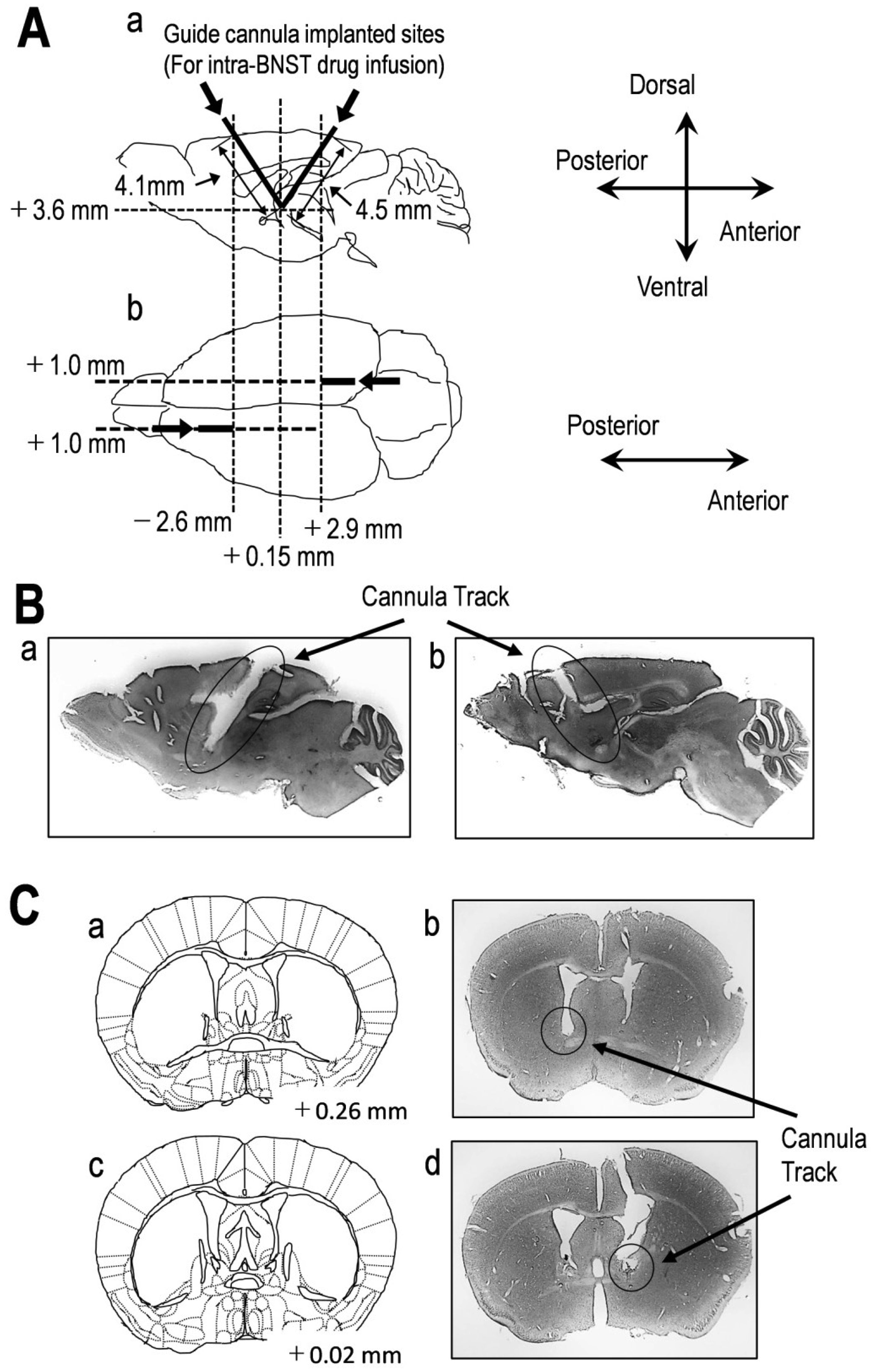
    [34] Franklin K, Paxinos G (2008) The Coronal Plates and Diagrams. The Mouse Brain in Stereotaxic Coordinates, Compact Academic Press, 1-256.
    [35] Williams RW (2000) Mapping genes that modulate mouse brain development: a quantitative genetic approach. Results Probl Cell Differ 30: 21-49. doi: 10.1007/978-3-540-48002-0_2
    [36] James TA, Starr MS (1978) Effects of the rate and volume of injection on the pharmacological response elicited by intranigral microapplication of drugs in the rat. J Pharmacol Methods 1: 197-202. doi: 10.1016/0160-5402(78)90049-9
    [37] Yang L, Shi LJ, Yu J, et al. (2016) Activation of protein kinase A in the amygdala modulates anxiety-like behaviors in social defeat exposed mice. Mol Brain 9: 3. doi: 10.1186/s13041-015-0181-3
    [38] Mul JD, Zheng J, Goodyear LJ (2016) Validity Assessment of 5 Day Repeated Forced-Swim Stress to Model Human Depression in Young-Adult C57BL/6J and BALB/cJ Mice. eNeuro 3: 1-9.
    [39] Kanda Y (2013) Investigation of the freely available easy-to-use software ‘EZR’ for medical statistics. Bone Marrow Transplant 48: 452-458. doi: 10.1038/bmt.2012.244
    [40] Pelloux Y, Hagues G, Costentin J, et al. (2005) Helplessness in the tail suspension test is associated with an increase in ethanol intake and its rewarding effect in female mice. Alcohol Clin Exp Res 29: 378-388. doi: 10.1097/01.ALC.0000156123.10298.FA
    [41] Perona MT, Waters S, Hall FS, et al. (2008) Animal models of depression in dopamine, serotonin, and norepinephrine transporter knockout mice: prominent effects of dopamine transporter deletions. Behav Pharmacol 19: 566-574. doi: 10.1097/FBP.0b013e32830cd80f
    [42] Galeotti N, Ghelardini C (2011) Antidepressant phenotype by inhibiting the phospholipase Cβ(1)—protein kinase Cγ pathway in the forced swim test. Neuropharmacology 60: 937-943. doi: 10.1016/j.neuropharm.2011.01.037
    [43] Chauhan VP (1990) Phosphatidylinositol 4,5-bisphosphate stimulates protein kinase C-mediated phosphorylation of soluble brain proteins. Inhibition by neomycin. FEBS Lett 272: 99-102. doi: 10.1016/0014-5793(90)80457-T
    [44] Descalzi G, Li XY, Chen T, et al. (2012) Rapid synaptic potentiation within the anterior cingulate cortex mediates trace fear learning. Mol Brain 5: 6. doi: 10.1186/1756-6606-5-6
    [45] Kodani S, Soya S, Sakurai T (2017) Excitation of GABAergic Neurons in the Bed Nucleus of the Stria Terminalis Triggers Immediate Transition from Non-Rapid Eye Movement Sleep to Wakefulness in Mice. J Neurosci 37: 7164-7176. doi: 10.1523/JNEUROSCI.0245-17.2017
    [46] Woulfe JM, Hrycyshyn AW, Flumerfelt BA (1988) Collateral axonal projections from the A1 noradrenergic cell group to the paraventricular nucleus and bed nucleus of the stria terminalis in the rat. Exp Neurol 102: 121-124. doi: 10.1016/0014-4886(88)90084-2
    [47] Mejías-Aponte CA, Drouin C, Aston-Jones G (2009) Adrenergic and noradrenergic innervation of the midbrain ventral tegmental area and retrorubral field: prominent inputs from medullary homeostatic centers. J Neurosci 29: 3613-3626. doi: 10.1523/JNEUROSCI.4632-08.2009
    [48] Dumont EC, Williams JT (2004) Noradrenaline triggers GABAA inhibition of bed nucleus of the stria terminalis neurons projecting to the ventral tegmental area. J Neurosci 24: 8198-8204. doi: 10.1523/JNEUROSCI.0425-04.2004
    [49] Velásquez-Martínez MC, Vázquez-Torres R, Rojas LV, et al. (2015) Alpha-1 adrenoreceptors modulate GABA release onto ventral tegmental area dopamine neurons. Neuropharmacology 88: 110-121. doi: 10.1016/j.neuropharm.2014.09.002
    [50] Paladini CA, Williams JT (2004) Noradrenergic inhibition of midbrain dopamine neurons. J Neurosci 24: 4568-4575. doi: 10.1523/JNEUROSCI.5735-03.2004
    [51] Mejias-Aponte CA (2016) Specificity and impact of adrenergic projections to the midbrain dopamine system. Brain Res 1641: 258-273. doi: 10.1016/j.brainres.2016.01.036
    [52] Dyer-Reaves K, Goodman AM, Nelson AR, et al. (2019) Alpha1-Adrenergic Receptor Mediated Long-Term Depression at CA3-CA1 Synapses Can Be Induced via Accumulation of Endogenous Norepinephrine and Is Preserved Following Noradrenergic Denervation. Front Synaptic Neurosci 11: 27. doi: 10.3389/fnsyn.2019.00027
    [53] Scheiderer CL, Dobrunz LE, McMahon LL (2004) Novel form of long-term synaptic depression in rat hippocampus induced by activation of alpha 1 adrenergic receptors. J Neurophysiol 91: 1071-1077. doi: 10.1152/jn.00420.2003
    [54] Choi SY, Chang J, Jiang B, et al. (2005) Multiple receptors coupled to phospholipase C gate long-term depression in visual cortex. J Neurosci 25: 11433-11443. doi: 10.1523/JNEUROSCI.4084-05.2005
    [55] Kirkwood A, Rozas C, Kirkwood J, et al. (1999) Modulation of long-term synaptic depression in visual cortex by acetylcholine and norepinephrine. J Neurosci 19: 1599-1609. doi: 10.1523/JNEUROSCI.19-05-01599.1999
    [56] Jo JH, Park EJ, Lee JK, et al. (2001) Lipopolysaccharide inhibits induction of long-term potentiation and depression in the rat hippocampal CA1 area. Eur J Pharmacol 422: 69-76. doi: 10.1016/S0014-2999(01)01075-5
    [57] Sagar SM, Price KJ, Kasting NW, et al. (1995) Anatomic patterns of Fos immunostaining in rat brain following systemic endotoxin administration. Brain Res Bull 36: 381-392. doi: 10.1016/0361-9230(94)00217-O
    [58] Gaykema RP, Daniels TE, Shapiro NJ, et al. (2009) Immune challenge and satiety-related activation of both distinct and overlapping neuronal populations in the brainstem indicate parallel pathways for viscerosensory signaling. Brain Res 1294: 61-79. doi: 10.1016/j.brainres.2009.07.076
    [59] Tanaka M, Yoshida M, Emoto H, et al. (2000) Noradrenaline systems in the hypothalamus, amygdala and locus coeruleus are involved in the provocation of anxiety: basic studies. Eur J Pharmacol 405: 397-406. doi: 10.1016/S0014-2999(00)00569-0
    [60] Southwick SM, Bremner JD, Rasmusson A, et al. (1999) Role of norepinephrine in the pathophysiology and treatment of posttraumatic stress disorder. Biol Psychiatry 46: 1192-1204. doi: 10.1016/S0006-3223(99)00219-X
    [61] Perahia DG, Quail D, Desaiah D, et al. (2008) Switching to duloxetine from selective serotonin reuptake inhibitor antidepressants: a multicenter trial comparing 2 switching techniques. J Clin Psychiatry 69: 95-105. doi: 10.4088/JCP.v69n0113
    [62] Nutt DJ (2002) The neuropharmacology of serotonin and noradrenaline in depression. Int Clin Psychopharmacol 17 Suppl 1: S1-S12. doi: 10.1097/00004850-200206001-00002
    [63] Sethy VH, Day JS, Cooper MM (1988) Dose-dependent down-regulation of beta-adrenergic receptors after chronic intravenous infusion of antidepressants. Prog Neuropsychopharmacol Biol Psychiatry 12: 673-682. doi: 10.1016/0278-5846(88)90011-5
    [64] Kreiner G, Zelek-Molik A, Kowalska M, et al. (2011) Effects of the noradrenergic neurotoxin DSP-4 on the expression of α1-adrenoceptor subtypes after antidepressant treatment. Pharmacol Rep 63: 1349-1358. doi: 10.1016/S1734-1140(11)70699-5
    [65] Heisler LK, Zhou L, Bajwa P, et al. (2007) Serotonin 5-HT(2C) receptors regulate anxiety-like behavior. Genes Brain Behav 6: 491-496. doi: 10.1111/j.1601-183X.2007.00316.x
    [66] Lebow MA, Chen A (2016) Overshadowed by the amygdala: the bed nucleus of the stria terminalis emerges as key to psychiatric disorders. Mol Psychiatry 21: 450-463. doi: 10.1038/mp.2016.1
    [67] Kim SY, Adhikari A, Lee SY, et al. (2013) Diverging neural pathways assemble a behavioural state from separable features in anxiety. Nature 496: 219-223. doi: 10.1038/nature12018
    [68] Dong HW, Petrovich GD, Watts AG, et al. (2001) Basic organization of projections from the oval and fusiform nuclei of the bed nuclei of the stria terminalis in adult rat brain. J Comp Neurol 436: 430-455. doi: 10.1002/cne.1079
    [69] Choi DC, Furay AR, Evanson NK, et al. (2007) Bed nucleus of the stria terminalis subregions differentially regulate hypothalamic-pituitary-adrenal axis activity: implications for the integration of limbic inputs. J Neurosci 27: 2025-2034. doi: 10.1523/JNEUROSCI.4301-06.2007
  • Reader Comments
  • © 2020 the Author(s), licensee AIMS Press. This is an open access article distributed under the terms of the Creative Commons Attribution License (http://creativecommons.org/licenses/by/4.0)
通讯作者: 陈斌, bchen63@163.com
  • 1. 

    沈阳化工大学材料科学与工程学院 沈阳 110142

  1. 本站搜索
  2. 百度学术搜索
  3. 万方数据库搜索
  4. CNKI搜索

Metrics

Article views(4674) PDF downloads(82) Cited by(3)

Figures and Tables

Figures(4)

Other Articles By Authors

/

DownLoad:  Full-Size Img  PowerPoint
Return
Return

Catalog