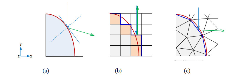
Understanding light propagation in skin tissues with complex blood vessels can help improve clinical efficacy in the laser treatment of cutaneous vascular lesions. The voxel-based Monte Carlo (VMC) algorithm with simple blood vessel geometry is commonly used in studying the law of light propagation in tissues. However, unavoidable errors are expected in VMC because of the zigzag polygonal interface. A tetrahedron-based Monte Carlo with extended boundary condition (TMCE) solver is developed to discretize complex tissue boundaries accurately. Tetrahedra are generated along the interface, resulting in a polyhedron approximation to match the real interface. A comparison between TMCE and VMC shows neglected differences in the overall distribution of energy deposition of different models, but poor adaptability of the curved tissue interface in VMC leads to a higher energy deposition error than TMCE in a mostly deposited region in blood vessels. Replacing the real blood vessel with a cylinder-shaped vessel shows an error lower than that caused by VMC. Statistical significance analysis of energy deposition by TMCE shows that mean curvature has stronger relationship with energy deposition than the Gaussian curvature, which indicates the importance of this geometric parameter in predicting photon behavior in vascular lesions.
Citation: Hao Jia, Bin Chen, Dong Li, Yuzhen Jin. Strategy of boundary discretization in numerical simulation of laser propagation in skin tissue with vascular lesions[J]. Mathematical Biosciences and Engineering, 2021, 18(3): 2455-2472. doi: 10.3934/mbe.2021125
Understanding light propagation in skin tissues with complex blood vessels can help improve clinical efficacy in the laser treatment of cutaneous vascular lesions. The voxel-based Monte Carlo (VMC) algorithm with simple blood vessel geometry is commonly used in studying the law of light propagation in tissues. However, unavoidable errors are expected in VMC because of the zigzag polygonal interface. A tetrahedron-based Monte Carlo with extended boundary condition (TMCE) solver is developed to discretize complex tissue boundaries accurately. Tetrahedra are generated along the interface, resulting in a polyhedron approximation to match the real interface. A comparison between TMCE and VMC shows neglected differences in the overall distribution of energy deposition of different models, but poor adaptability of the curved tissue interface in VMC leads to a higher energy deposition error than TMCE in a mostly deposited region in blood vessels. Replacing the real blood vessel with a cylinder-shaped vessel shows an error lower than that caused by VMC. Statistical significance analysis of energy deposition by TMCE shows that mean curvature has stronger relationship with energy deposition than the Gaussian curvature, which indicates the importance of this geometric parameter in predicting photon behavior in vascular lesions.

| [1] | G. Aguilar, B. Choi, M. Broekgaarden, O. Yang, B. Yang, P. Ghasri, et al., An overview of three promising mechanical, optical, and biochemical engineering approaches to improve selective photothermolysis of refractory port wine stains, Ann. Biomed. Eng., 40 (2012), 486-506. |
| [2] | D. Li, G. X. Wang, Y. L. He, K. M. Kelly, W. J. Wu, Y. X. Wang, et al., A two-temperature model for selective photothermolysis laser treatment of port wine stains, Appl. Therm. Eng., 59 (2013), 41-51. |
| [3] |
D. Yudovsky, A. J. Durkin, Hybrid diffusion and two-flux approximation for multilayered tissue light propagation modeling, Appl. Optics, 50 (2011), 4237-4245. doi: 10.1364/AO.50.004237

|
| [4] |
D. J. Smithies, P. H. Butler, Modelling the distribution of laser light in port-wine stains with the Monte Carlo method, Phys. Med. Biol., 40 (1995), 701-731. doi: 10.1088/0031-9155/40/5/001
|
| [5] |
L. H. Wang, S. L. Jacques, L. Q. Zheng, MCML—Monte Carlo modeling of light transport in multi-layered tissues, Comput. Meth. Prog. Biol., 47 (1995), 131-146. doi: 10.1016/0169-2607(95)01640-F
|
| [6] |
B. Chen, S. L. Thomsen, R. J. Thomas, J. Oliver, A. J. Welch, Histological and modeling study of skin thermal injury to 2.0 microm laser irradiation, Lasers Surg. Med., 40 (2008), 358-370. doi: 10.1002/lsm.20630
|
| [7] |
J. Zhou, J. Liu, Numerical study on 3-D light and heat transport in biological tissues embedded with large blood vessels during laser-induced thermotherapy, Numer. Heat Tr. Appl., 45 (2004), 415-449. doi: 10.1080/10407780490269030
|
| [8] | T. J. Pfefer, J. K. Barton, E. K. Chan, M. G. Ducros, B. S. Sorg, T. E. Milner, et al., A three-dimensional modular adaptable grid numerical model for light propagation during laser irradiation of skin tissue, IEEE J. Sel. Top. Quant., 2 (1996), 934-942. |
| [9] |
T. Binzoni, T. S. Leung, R. Giust, D. Rufenacht, A. H. Gandjbakhche, Light transport in tissue by 3D Monte Carlo: Influence of boundary voxelization, Comput. Meth. Prog. Biol., 89 (2008), 14-23. doi: 10.1016/j.cmpb.2007.10.008
|
| [10] | J. Premru, M. Milanič, B. Majaron, Monte Carlo simulation of radiation transfer in human skin with geometrically correct treatment of boundaries between different tissues, Proceedings of SPIE —The International Society for Optical Engineering, 8579 (2013), 85790Z. |
| [11] |
B. Majaron, M. Milanič, J. Premru, Monte Carlo simulation of radiation transport in human skin with rigorous treatment of curved tissue boundaries, J. Biomed. Opt., 20 (2015), 015002. doi: 10.1117/1.JBO.20.1.015002
|
| [12] |
Q. Q. Fang, Mesh-based Monte Carlo method using fast ray-tracing in Plücker coordinates, Biomed. Opt. Express, 1 (2010), 165-175. doi: 10.1364/BOE.1.000165
|
| [13] |
Q. Q. Fang, D. R. Kaeli, Accelerating mesh-based Monte Carlo method on modern CPU architectures, Biomed. Opt. Express, 3 (2012), 3223-3230. doi: 10.1364/BOE.3.003223
|
| [14] |
H. Shen, G. Wang, A tetrahedron-based inhomogeneous Monte Carlo optical simulator, Phys. Med. Biol., 55 (2010), 947-962. doi: 10.1088/0031-9155/55/4/003
|
| [15] | R. Y. Yao, X. Intes, Q. Q. Fang, Generalized mesh-based Monte Carlo for wide-field illumination and detection via mesh retessellation, Biomed. Opt. Express, 7 (2015), 171-184. |
| [16] |
H. Jia, B. Chen, D. Li, Y. Zhang, Boundary discretization in the numerical simulation of light propagation in skin tissue: problem and strategy, J. Biomed. Opt., 20 (2015), 25007. doi: 10.1117/1.JBO.20.2.025007
|
| [17] |
T. Li, C. Xue, P. Wang, Y. Li, L. H. Wu, Photon penetration depth in human brain for light stimulation and treatment: A realistic Monte Carlo simulation study, J. Innov. Opt. Heal. Sci., 10 (2017), 1743002. doi: 10.1142/S1793545817430027
|
| [18] |
M. Milanic, B. Majaron, Three-dimensional Monte Carlo model of pulsed-laser treatment of cutaneous vascular lesions, J. Biomed. Opt, 16 (2011), 128002. doi: 10.1117/1.3659205
|
| [19] |
H. C. Han, J. K. W. Chesnutt, J. R. Garcia, Q. Liu, Q. Wen, Artery buckling: New phenotypes, models, and applications, Ann. biomed. eng., 41 (2013), 1399-1410. doi: 10.1007/s10439-012-0707-0
|
| [20] |
D. Li, B. Chen, W. J. Wu, G. X. Wang, Y. L. He, Multi-scale modeling of tissue freezing during cryogen spray cooling with R134a, R407c and R404a, Appl. Therm. Eng., 73 (2014), 1489-1500. doi: 10.1016/j.applthermaleng.2014.03.034
|
| [21] |
H. Jia, B. Chen, D. Li, Theoretical study on pressure damage based on clinical purpura during the laser irradiation of port wine stains with real complex vessels, Appl. Sci-Basel., 9 (2019), 5478. doi: 10.3390/app9245478
|
| [22] | V. Periyasamy, M. Pramanik, Monte Carlo simulation of light transport in turbid medium with embedded object—spherical, cylindrical, ellipsoidal, or cuboidal objects embedded within multilayered tissues, J. Biomed. Opt., 19 (2014), 045003. |
| [23] | L. Z. Mu, H. W. Shao, H. E. Ying, O. Toshiaki, X. M. Jia, Construction of anatomically accurate finite element models of the human hand and a rat kidney, J. Mech. Med. Biol., 11 (2012), 1141-1164. |
| [24] | M. Meyer, M. Desbrun, P. Schrö der, A. H. Barr, Discrete differential-geometry operators for triangulated 2-manifolds, Visualization and mathematics Ⅲ, Springer, (2003), 35-57. |
| [25] |
Y. Zhang, B. Chen, D. Li, G. X. Wang, Efficient and accurate simulation of light propagation in bio-tissues using the three-dimensional geometric Monte Carlo method, Numer. Heat Tr. A-Appl., 68 (2015), 827-846. doi: 10.1080/10407782.2015.1023140
|
| [26] | R. A. Groeneveld, G. Meeden, Measuring skewness and kurtosis, J. Roy. Stat. Soc. D-sta, 33 (1984), 391-399. |


Figures(17) / Tables(3)

Hao Jia, Bin Chen, Dong Li, Yuzhen Jin. Strategy of boundary discretization in numerical simulation of laser propagation in skin tissue with vascular lesions[J]. Mathematical Biosciences and Engineering, 2021, 18(3): 2455-2472. doi: 10.3934/mbe.2021125


 DownLoad: