
Citation: Nosheen Aslam, Muhammad Sarfaraz Iqbal, Syed Makhdoom Hussain, Muhammad Rizwan, Qurat-Ul-Ain Naseer, Muhammad Afzal, Rizwan Muneer, Farzana Batool. Effects of chelating agents on heavy metals in Hepatitis C Virus (HCV) patients[J]. Mathematical Biosciences and Engineering, 2019, 16(3): 1138-1149. doi: 10.3934/mbe.2019054
[1] | Amar Nath Chatterjee, Fahad Al Basir, Yasuhiro Takeuchi . Effect of DAA therapy in hepatitis C treatment — an impulsive control approach. Mathematical Biosciences and Engineering, 2021, 18(2): 1450-1464. doi: 10.3934/mbe.2021075 |
[2] | Elamin H. Elbasha . Model for hepatitis C virus transmissions. Mathematical Biosciences and Engineering, 2013, 10(4): 1045-1065. doi: 10.3934/mbe.2013.10.1045 |
[3] | Dwi Lestari, Noorma Yulia Megawati, Nanang Susyanto, Fajar Adi-Kusumo . Qualitative behaviour of a stochastic hepatitis C epidemic model in cellular level. Mathematical Biosciences and Engineering, 2022, 19(2): 1515-1535. doi: 10.3934/mbe.2022070 |
[4] | Xiaowen Xiong, Yanqiu Li, Bingliang Li . Global stability of age-of-infection multiscale HCV model with therapy. Mathematical Biosciences and Engineering, 2021, 18(3): 2182-2205. doi: 10.3934/mbe.2021110 |
[5] | Dong-Me Li, Bing Chai, Qi Wang . A model of hepatitis B virus with random interference infection rate. Mathematical Biosciences and Engineering, 2021, 18(6): 8257-8297. doi: 10.3934/mbe.2021410 |
[6] | Sarah Kadelka, Stanca M Ciupe . Mathematical investigation of HBeAg seroclearance. Mathematical Biosciences and Engineering, 2019, 16(6): 7616-7658. doi: 10.3934/mbe.2019382 |
[7] | Tailei Zhang, Hui Li, Na Xie, Wenhui Fu, Kai Wang, Xiongjie Ding . Mathematical analysis and simulation of a Hepatitis B model with time delay: A case study for Xinjiang, China. Mathematical Biosciences and Engineering, 2020, 17(2): 1757-1775. doi: 10.3934/mbe.2020092 |
[8] | Maysaa Al Qurashi, Saima Rashid, Fahd Jarad . A computational study of a stochastic fractal-fractional hepatitis B virus infection incorporating delayed immune reactions via the exponential decay. Mathematical Biosciences and Engineering, 2022, 19(12): 12950-12980. doi: 10.3934/mbe.2022605 |
[9] | Tingting Xue, Long Zhang, Xiaolin Fan . Dynamic modeling and analysis of Hepatitis B epidemic with general incidence. Mathematical Biosciences and Engineering, 2023, 20(6): 10883-10908. doi: 10.3934/mbe.2023483 |
[10] | Suqi Ma . Low viral persistence of an immunological model. Mathematical Biosciences and Engineering, 2012, 9(4): 809-817. doi: 10.3934/mbe.2012.9.809 |
Hepatitis is the inflammation of liver and it is characterized by the injurious to liver cells. Viral hepatitis is a big health problem around the globe. It is more prevalent in developing countries. In Faisalabad district of Pakistan 10–12% population is suffering from HCV [1,2,3,4]. HCV infection leads to chronic hepatitis which increases the risk of hepatic steatosis, fibrosis, cirrhosis, and hepatocellular carcinoma [5,6,7]. Hepatic diseases modify the regulation of heavy metals metabolism and any deregulation in homeostasis can cause inflammatory changes and oxidative stress, which leads to enhanced HCV replication reduced the efficacy of anti-viral therapy in chronic hepatitis C (CHC) patients [1,8]. The emergence of new chelating agents may have a major impact on the treatment of many diseases to prevent serious complications.
It was generally seen that concentration of heavy metals are enhanced in HCV patients. As these heavy metals are very toxic [9] they cause pathological change of organs which ultimately leads to cancer [10]. Despite their hazards some metals have essential function in humans life, it was investigated that low amount of trace elements and infectious diseases often coexist and had very complex interactions. These are involved in most of the immunological, physiological and biochemical activities. They are carrying out many metabolic functions in the liver e.g. enzymatic functions, oxidative damage, protein synthesis, anti-oxidant defense, interferon therapy response, immunological competence and changes in virus genomes [11]. A large numbers of trace elements such as zinc (Zn), manganese (Mn), selenium (Sn), copper (Cu), have immunomodulatory functions and thus influenced variety of viral infections. It was also correlated that Selenium level decreases in HCV patients due to defense strategies of organisms induced by the various carotenoids. Similarly zinc levels in the serum of HCV patients lowers with severity of disease, Copper accumulation in fibrotic livers caused by chronic HCV infection may be contributed to hepatic injury and thus its presence increases the chances of HCV infection [12]. Cadmium placed a unique level among the metals due to its toxification. Other metals like Cu, Co, Pd, Nickle (Ni), iron (Fe) and Mn have a toxic role as well as act as immunomodulatory in living beings. For the detoxification of these metals chelation therapy are used in which the toxic metal complex are ligated with biological ligands which can further be excreted from body through urinary system [13]. In this therapy chelates (detoxificant) are injected or engulfed orally [14].
Cytotoxicity mechanism through virus core proteins, recurring immune-mediated progressions, prolonged inflammation, and triggering oxidative pressure through Fe overload appear to play an significant role in HCV infection-induced liver impairment [15]. Oxidative damage that is caused by due to pro-oxidants and anti-oxidant. Recent study has shown that pointedly advanced oxidative stress status was present in prolonged hepatitis c patients [16]. Prolonged inflammation is responsible for increased oxidative stress because polymorphonuclear cells and Kupffer cells stimulate nicotinamide adenine dinucleotide phosphate (NADPH) oxidase to constantly produce reactive oxygen radicals. Oxidative damage not only disturbs the effectiveness of immune response mechanism [17], but also enhance the progress of liver impairment. It is supposed that aspects causative to oxidative damage are dynamic to the incidence and pathogenesis of prolonged hepatitis c virus. Multiple physiologic progressions in the liver is influenced by numerous vital trace metals like copper (Cu), Fe and zinc (Z). These progressions consists of enzymatic activities, immune responses, oxidative-antioxidant status, erection and function of proteins [18]. Consociate of nonessential metals can also produce oxidative damage that could enter the body via contaminated food, dust, water and air. Lead (Pb), aluminum (Al), arsenic and cadmium (Cd) in precise may persuade toxic possessions escorted by liver injury and inflammation [19]. The contaminated indices might be triggered principally by an inequality of immunodisruptive effects, antioxidant defenses and interactions among toxic and vital trace metals [20]. This recommends that an amendment in the prestige of trace metals subsidizes pointedly to augmented oxidative pressure which can in turn lead to an upsurge in pathological infection, reliable with the liver swelling and grievance frequently seen in patients with chronic hepatitis C.
The present study explore the heavy metal concentrations in HCV patients and established correlation of heavy metals between healthy persons and HCV patients. monoisoamyl 2, 3-dimercaptosuccinic acid (lipophilic chelator) was used in chelation therapy to prevent health problems in HCV patients which are due to poisonous concentration of heavy metals. This chelation therapy will be helpful to reverse the HCV related health problems.
Study population was divided into healthy control subjects (n = 25) and hepatitis c patient (n = 25); All patients and control subjects were age and sex matched disparate individuals from Faisalabad region. Enrolment of patients was carried out after detailed demonstration of study subjects and submission of signed consent letter and questionnaire about the demographic conditions, health status, disease history, medication record and smoking status of patients. The diagnosis of hepatitis c is based on history of chronic significant, clinical signs of liver disease and supporting laboratory test which are AST, ALT and bilirubin level and gamma glutamyltransferase and ultrasonographic features. The height, weight, BP and BMI were recorded in both the groups.
A 5 ml blood sample was drawn from every patient and control individual. The blood was transferred into plain tube and allowed to clot at 37 ℃ then centrifuged at 4600 rpm for 10 minutes. Serum was separated and stored at −40 ℃until analyses. The nails & hairs samples from control and patients were collected after 7 days from last cut, and then these samples were washed and stored for further processing. Blood, nails and hair samples were digested by using nitric acid and Perchloric acid.
Concentrations of the heavy metals were determined in serum samples of hepatitis c patients. Analytical analysis was carried out in Hi-tech Laboratory of University of Agriculture Faisalabad (UAF) using Hitachi polarized Zeeman AAS, Z-8200, Japan model with standard burner as atomizer.
Dimercaptosuccinic acid (DMSA) was purchased from Sigma Aldrich. Monoisoamyl 2, 3-dimercaptosuccinic acid (MiADMSA) was synthesized by the controlled esterification of DMSA with the corresponding alcohol (isoamyl alcohol) in acidic medium (11).
The product was purified (purity 99.9%) and characterized using spectral and analytical methods before experimentation. The samples were stored, refrigerated in a dessicator to avoid oxidation and thermal decomposition. MiADMSA was dissolved in 5% sodium bicarbonate solution. The antidote solution was prepared immediately before use. MiADMSA was given 500 mg per day orally in two and three separated doses for every supplementary day for 5 weeks. It was given between meals. After chelation the urine samples were collected and analyzed. Heavy metals were analyzed from urine samples. Analytical analysis was performed with atomic absorption. Encet test was applied for the statistical analysis. The probability level of 95% was taken as significant in all cases.
The patients group was 25 patients with chronic HCV infections that included 12 males and 13 females (aged 35 ± 15 years), ranging between 20 and 65 years. The control group comprised 25 healthy volunteers that included 12 males and 13 females aged between 19 and 55 (mean of 40 ± 14) years (Table 1).
Group | Age in years | Gender | |||
N | Mean ± SD | Range of years | Male/Female | ||
Control | 25 | 40 ± 14 | 19–55 | 12/13 | |
Patients (HCV) | 25 | 35 ± 15 | 20–65 | 16/9 |
The findings of investigation indicated that Fe, Pb, Co, Cd, Mn and Cu concentration were significantly higher and their concentration in blood, nails and hair samples varied significantly (p < 0.05) in normal individuals and HCV patients. Ni was found in nearly normal concentration (1.34 ± 0.18) in blood samples but it varied significantly (p < 0.05) in nails and hairs samples (1.14 ± 0.39) in normal individuals and HCV patients. The serum level of Zn was low (42.9 ± 17.09), (129.8 ± 23.42) in blood and nails and hair samples of patients with chronic HCV infection. It significantly differs from that of control blood, nails and hair samples (81.8 ± 13) (Table 2).
Blood | Nails and Hairs | |||||
Normal | Hepatitis c patients | p-value | Normal | Hepatitis c patients | p-value | |
Iron (µg/dl) | 120.2 ± 36.11 | 150.22 ± 19.73 | 0.000** | 23.4 ± 5.18 | 25.55 ± 1.93 | 0.012** |
zinc (µg/dl) | 81.8 ± 13.52 | 42.9 ± 17.09 | 0.000** | 199 ± 19.11 | 129.8 ± 23.42 | 0.000** |
lead (µg/dl) | 24.5 ± 7.33 | 46.5 ± 6.73 | 0.000** | 3.04 ± 0.63 | 4.372 ± 0.93 | 0.002** |
nickel (ng/ml) | 1.34 ± 0.18 | 1.34 ± 0.58 | 0.784** | 0.96 ± 0.311 | 1.14 ± 0.39 | 0.013** |
Manganese (ng/ml) | 0.59 ± 0.179 | 1.5 ± 0.50 | 0.000** | 1.06 ± 0.673 | 4.4 ± 1.59 | 0.000** |
Copper (µg/L) | 110 ± 24.55 | 176 ± 17.44 | 0.000** | 70.68 ± 20.85 | 136.8 ± 22.72 | 0.000** |
Cobalt (ng/ml) | 0.4 ± 0.243 | 3.496 ± 1.105 | 0.000** | 0.092 ± 0.0571 | 0.20 ± 0.072 | 0.000** |
Cadmium (ng/ml) | 1.256 ± 0.515 | 4.828 ± 0.9825 | 0.000** | 0.23 ± 0.0426 | 0.403 ± 0.053 | 0.000** |
MiADMSA (orally) was used for the chelation of heavy metals Fe, Pb, Co, Cd, Mn and Cu. After the urine samples analysis, a significant amount of heavy metals were excreted in the urine against two different doses of MiADMSA. MiADMSA, which was given 500 mg in three separate doses, significantly lower the concentration of heavy metals as compared to given in two separate doses.
Heavy Metals | Before Chelation | After Chelation (Twice a day) BD | After Chelation (Thrice a day) TDS | p-value |
Iron (µg/dl) | 150.22 ± 19.73 | 133.6 ± 6.4 | 129.4 ± 9.1 | 0.000** |
lead (µg/dl) | 46.5 ± 6.73 | 30.2 ± 9.02 | 26.1 ± 3.1 | 0.000** |
Manganese (ng/ml) | 1.5 ± 0.50 | 0.9 ± 1.2 | 0.7 ± 5.8 | 0.002** |
Copper (µg/L) | 176 ± 17.44 | 135 ± 12.3 | 127 ± 2.9 | 0.001** |
Cobalt (ng/ml) | 3.496 ± 1.105 | 1.8 ± 1.4 | 1 ± 2.5 | 0.012** |
Cadmium (ng/ml) | 4.828 ± 0.9825 | 2.9 ± 1.3 | 2.1 ± 4.3 | 0.023** |
Chronic liver diseases are devastating health problems. Their prevalence depends upon many factors i.e., infectious, toxic and parasitic. Synergism of more than one factor may enhance the process of liver damage [21]. These damaged cells causes functional impairment of liver and alter the metabolism of trace elements [22]. In healthy persons and HCV patients zinc (Zn) concentration varied significantly with higher levels in healthy persons. This showed that Zn concentration was related with hepatitis, decreased with the progress of the disease and low liver function. Zinc is as an essential mineral and it occurs in almost every cell of the human body [23]. The deficiency of Zn occurs when its requirements increases or when it is poorly absorbed after its removal from the body [24]. Its shortage depresses immune function. Low Zn levels leads to reduction of inflammatory reaction in hepatitis patients [25]. In viral hepatitis metabolism of Fe is intermittent which results in the buildup of iron (Fe) in liver. Deposition of Fe in hepatocytes depends on the degree of liver inflammation and infected liver tissues damage in HCV infection. Accumulation of Fe also increase viral activity due to rise in mutation rates and have specific roles in their pathogenesis and progress [26].
Chronic hepatitis c virus infection is associated with the increased level of hepatic iron deposition and advance hepatic fibrosis in the patients. The prediction of the hepatic iron deposition and its severity of fibrosis is by the serum ferritin value and the predictor of severity of hepatic fibrosis in the patients of HCV infection. Copper (Cu) and lead (Pb) accumulation in fibrotic liver caused by chronic HCV may contribute to hepatic injury because they are linked to immune dysfunction and inflammatory responses in many diseases [27]. Cu in high concentration can cause Anemia, liver and kidney damage, stomach and intestinal irritation. Increased cu levels result from inflammatory responses and oxidative stress [28]. Pb competes in the body with calcium (Ca) causing numerous malfunctions in Ca facilitated cellular metabolism and Ca uptake and usage, including inhibition of neurotransmitter. Pb and Cu can interfere with Zn homeostasis [29] thereby contributing further Zn deficiency. Cadmium (Cd) is very toxic and its long-term exposure leads to build up in the kidney [30], lung damage and fragile bones.
Chelates complexes the metal and allow removal of excess or toxic metal from the system. It involves the use of chelates injected into the blood, muscles or taken orally to bind metals that are present in toxic concentration so they can be excreted from the body, most frequently in urine. Many studies suggested monoesters as one of potential and effective antidote [31]. Our results suggested that MiADMSA is highly effective in tumbling heavy metal load from hepatitis c virus patients. It contains sulfhydryl groups that bind or chelate heavy metals, and the resulting complex is excreted either renally or hepatically. In spite of reducing body stores of heavy metals in blood it also assemble skeletal stores of heavy metals [32]. The synthesis of MT in liver and kidneys was potentiate by the MIADMSA and in the liver and brain it reduce the oxidized glutathione (GSSG) level in tissues. MIADMSA is also has capacity to initiate the intracellular cadmium and it also perform a function to give antioxidant by releasing the cadmium where the deleterious oxidation reactions. In this compound the reduce sulfhydryl group may take part in the antioxidant reactions and the induced cadmium retard propagation [33]. Prolonged liver ailment is caused by chronic hepatitis c virus and in United States it's a common sign for liver relocation [34]. Almost 168 million people are suffering in this disease worldwide, including approximately 3% of universal population. Choo was the first person who isolated the hepatitis c virus from the person's serum with non-B, non-A in 1989 [10]. The virus of hepatitis c is ribonucleic acid virus that goes to Flaviviridae family. Hepatocytes cytoplasm is a place where the virus of hepatitis c is duplicates, however it does not show the effect of cytopathic. Tenacious infection acts to depend on fast production of virus and constant cell-to-cell spread, alongside by a deficiency of energetic T-cell immune reaction to hepatitis c virus antigens [35]. HCV ribonucleic acid genome transmutes often due to deficiency of inaccurate proofreading through the viral ribonucleic acid polymerase and fast viral duplication. It consists of 6 identified genotypes and additional 50 subtypes, for example 1a, 2a, 1b. Numerous hepatitis c virus transmutations and frequent subtypes obligate the examine for an HCV vaccine perplexing. The primary cause of HCV is revelation to septic blood. Following risk factor are responsible for the transmission of this virus, consist of sexual activity, usage of intravenous drug, compact organ relocation through diseased donor, hemodialysis, professional revelation, birth to an infected mother, domestic exposure and excessive use of intranasal cocaine. About 60% cause of this virus is drug and 20% is caused by sexual activity. And remaining 10% is caused by other modes of conduction (hemodialysis, professional, perinatal and domestic exposure) [36]. Severe onset of HCV septicity has been preeminent predictable, in the transfusion situation and 65%–80% of statuses were asymptotic. Nearby 21%–30% of adult through severe hepatitis c virus taint may progress medical sign and symptoms. After exposure of infection the symptom appear after 3–12 weeks. Sign and symptoms consists of anorexia, weakness, jaundice and malaise. After exposure of infection in 4 serum HCV ribonucleic acid can be identified [37]. The degree of development of liver fibrosis differs broadly in the background of stubborn hepatitis c viremia. For the categorization and performance of prolonged hepatitis c virus liver biopsy is used as gold standard. The physical liver injury, also recognized as fibrosis or phase, is mutable in prolonged HCV taint. Fibrosis suggests probable advancement to cirrhosis. Fibrosis is restricted to portal and periportal zones in minor situations. Further progressive variations are distinct by fibrosis that spreads from one gateway zone to another, correspondingly recognized as "bridging fibrosis". Substantial risk elements for hepatic fibrosis in hepatitis c virus coinfection through HBV OR HIV. Hepatitis c virus and hepatitis immunodeficiency virus coinfection is principally related amongst injection drug users and hemophiliacs [38]. The evolution to cirrhosis is frequently clinically inaudible, and several patients are not identified to have hepatitis c until them existent through the impediments of end-stage liver ailment or HCC.
Undiagnosed anti-HCV antibody progressive characters had disorders of trace metals, and disparity of antioxidant resistance, and augmented inflammatory retorts when related to controls. Present study indicate that HCV-RNA levels determination trials were not available; though, the occurrence of anti-HCV antibodies in plasma delivered an suggestion of acquaintance to HCV, which may characterize either an vigorous or a preceding HCV contamination. While severe infection with HCV is generally asymptomatic, nearby 80% of patients recurrently are at threat of rising cirrhosis, liver failure and hepatocellular carcinoma [39]. It was stated that conflicts in certain trace metals may subsidize to the extremely anti-HCV commonness [40]. Comparative to controls, these anti-HCV antibody-positive characters had meaningfully lower deliberations of plasma Zn, while Fe and plasma Cu meditations were higher. Here is no statistically momentous variance in Zn meditation amid prolonged hepatitis c patients and vigorous subjects, though the patients had higher Cu deliberations [41]. In chronic hepatitis c patients the concentration of Fe was increased and ominously decreased Zn concentration whereas the Cu concentrations lingered unaffected. In addition, the connotations of an upsurge in Cu with a decline in Zn with together raised alanine aminotransferase and -glutamyl transferase have been perceived in long-lasting hepatitis c patients [42]. Serum Zn deliberations were reduced in patients with hepatic fibrosis as linked with healthy controls [43]. Through this study we concluded that in liver cirrhosis and hepatic fibrosis concentration of zinc was decreased and Fe was increased. An accretion of Cu in fibrotic livers in enduring hepatitis c patients can subsidize to hepatic damage. This results indicate that instabilities in essential metals create eclectic array of liver harm. These vital metals play a vital role in natural processes. Zn persuades anti-inflammatory and antioxidant possessions that consequence in compact hepatocyte damage [44].
MiADMSA was perceived to be faintly more harmful in terms of copper impairment and some biochemical mutable in the hepatic matter in females as compared to male mice [45]. Through the study it was concluded that MiADMSA administration in female mice is perplexed through side effects and may oblige attention during its usage. No pragmatic contrary possessions points for parental and developing MiADMSA toxicity remain 47.5 mg/kg and 94 mg/kg/day, correspondingly, representing that MiADMSA would not create progressive toxicity in rats in the deficiency of maternal toxicity [45,46]. Mehta et al. [46] have recommended that MiADMSA had no consequence on gestational length, lactation, litter size, sexual relationship and feasibility. Mehta et al.have recommended that MiADMSA had no consequence on the length of gestation, litter size, sex ratio, feasibility and lactation [46].
Chelation therapy recombines metals and allows the removal of excess or toxic substances. In the world of increased metal exposure, chelation therapy is an important tool in fighting metal storage disorders, but still the lack of large-scale clinical trials still raises controversy regarding its clinical therapeutic benefit. The accessibility of new chelators' offerings new opportunities to the clinician treating HCV patients with metals overload. The significant amount of heavy metals was excreted in the urine after a challenged dose of chelating agent, so it can thus be concluded from the present study that MiADMSA TDS is a better treatment option than BD for chelation of heavy metals in hepatitis c virus patients. The combination of therapy of different chelating agents carries both the beneficial effects on the liver and a more rapid removal of toxic metals from the liver. Future studies should support to determine the long-term success of chelating agents, the role of several agents in patients with less severe iron overload, and the effectiveness and safety of new combinations of these chelating agents. Further, newer therapeutic strategies should be explored that may have a better therapeutic effect. Combination therapies with multiple chelating agents and/or prescription antioxidants or nutraceuticals may be considered more critical as a key recommendation for chelation therapy.
The authors declare that there is no conflict of interests in this paper.
[1] | M. S. Iqbal, U. A. Ashfaq and S. Khaliq, et al., Toll-like receptor 4 polymorphism as pretreatment predictor of response to HCV genotype 3a interferon-based treatment, Future. Med., 12 (2017). |
[2] | A. Arshad and U. A. Ashfaq, Epidemiology of hepatitis c infection in pakistan: Current estimate and major risk factors, 27 (2017), 63–77. |
[3] | M. S. Iqbal, U. A. Ashfaq and S. Aslam, et al., Analysis of polymorphism rs1990760 of IFIH1 gene and treatment outcomes in HCV infection, Future. Virol., 13 (2018). |
[4] | N. Jiwani, N. J. Mscn and R. Gul, A silent storm : Hepatitis C in Pakistan, 1 (2011), 89–91. |
[5] | M. U. Ghani, A. Haque and M. Qasim, et al., Involvement of vascular endothelial growth factor (VEGF) gene polymorphism in hepatocellular carcinoma of HCV patients from local population, Pure. Appl. Biol., 6 (2017), 725–732. |
[6] | R. Aslam, S. M. Raza and H. Naeemi, et al., SOCS3 mRNA expression and polymorphisms as pretreatment predictor of response to HCV genotype 3a IFN-based treatment, SpringerPlus, 5 (2016), 1826. |
[7] | K. Tarao, S. Ohkawa and Y. Miyagi, et al., Inflammation in background cirrhosis evokes malignant progression in HCC development from HCV-associated liver cirrhosis, Scand. J Gastroentero., 48 (2013), 729–735. |
[8] | N. Aslam, F. Batool and M. S. Iqbal, et al., Analysis of Toll-like receptors-9 (TLR9) gene polymorphism (rs5743836) in Pakistani patients with HCV, Pak. J. Pharm. Sci., 31 (2018), 2709–2714. |
[9] | L. Piekuse, M. Kreile and A. Zarina, et al., Association between inherited monogenic liver disorders and chronic hepatitis C, World. J. Hepatol., 6 (2014), 92–97. |
[10] | T. G. Kazi and N. F. Kolachi, Effects of mineral supplementation on liver cirrhotic/cancer male patients, Biol. Trace. Elem. Res., 150 (2012), 81–90. |
[11] | M. Afify, M. Diaa and E. A. Elmaksoud, et al., Serum levels of trace elements in Egyptian patients with chronic hepatitis C under interferon therapy. J. Genet. Eng. Biotechnol., 10 (2012), 81–86. |
[12] | J. E. Gall, R. S. Boyd and N. Rajakaruna, Transfer of heavy metals through terrestrial food webs : A review, Environ. Monit. Assess., 187 (2015), 201. |
[13] | V. M. Varnai, M. Piasek and K. Kostial, Chelators as antidotes of metal toxicity : Therapeutic and experimental aspects, Curr. Med. Chem., 12 (2005), 2771–2794. |
[14] | H. V. Aposhian, R. M. Maiorino and R. C. Dart, et al., Urinary excretion of 1989, 520–526. |
[15] | M. P. Simula and V. De Re, Hepatitis c virus-induced oxidative stress and mitochondrial dysfunction: A focus on recent advances in proteomics, Clin. Appl., 4 (2010), 782–793. |
[16] | M. Vidali, M. F. Tripodi and A. Ivaldi, et al., Interplay between oxidative stress and hepatic steatosis in the progression of chronic hepatitis C, J. Hepatol., 48 (2008), 399–406. |
[17] | S. J. Polyak, C. Morishima and M. C. Shuhart, et al., Inhibition of T-Cell Inflammatory Cytokines, Hepatocyte NF-κB Signaling, and HCV Infection by Standardized Silymarin, Gastroenterology, 132 (2007), 1925–1936. |
[18] | K. H. Ibs and L. Rink, Zinc-Altered Immune function, J. Nutr., 133 (2003), 1452S–1456S. |
[19] | T. Liu, W. He and C. Yan, et al., Roles of reactive oxygen species and mitochondria in cadmium-induced injury of liver cells, Toxicol. Ind. Health, 27 (2010), 249–256. |
[20] | S. Turgut, A. Polat and M. Inan, et al., Interaction between anemia and blood levels of iron, zinc, copper, cadmium and lead in children, Ind. J. Pediatr., 74 (2007), 827–830. |
[21] | B. Halliwell and J. M. C. Gutteridge, Role of free radicals and catalytic metal ions in human disease: An overview, Method. Enzymol., 186 (1990), 1–85. |
[22] | C. Pramoolsinsap, N. Promvanit and S. Komindr, et al., Gastroenterology serum trace metals in chronic viral hepatitis and hepatocellular carcinoma in Thailand, J. Gastroenterol., 29 (1994), 610–615. |
[23] | A. Ipek, E. Barut and H. Gulen, et al., Assessment of inter- and intra-cultivar variations in olive using SSR markers, Sci. Agri., 69 (2012), 327–335. |
[24] | B. M. Science and K. Medical, Selenium, iron, copper, and zinc levels and copper-to-zinc ratios in serum of patients at different stages of viral hepatic diseases, Biol. Trace. Elem. Res., 109 (2006), 15–23. |
[25] | B. Lo, Phytic acid-trace element (Zn, Cu, Mn) interactions, Int. J. Food Sci. Technol., 37 (2002), 749–758. |
[26] | C. M. Lange, Z. Kutalik and K. Morikawa, et al., Serum ferritin levels are associated with a distinct phenotype of chronic hepatitis C poorly responding to pegylated interferon-alpha and ribavirin therapy, Hepatology, 55 (2012), 1038–1047. |
[27] | N. Ganne-Carrié, C. Christidis and C. Chastang, et al., Liver iron is predictive of death in alcoholic cirrhosis: A multivariate study of 229 consecutive patients with alcoholic and/or hepatitis c virus cirrhosis: A prospective follow up study, Gut, 46 (2000), 277–282. |
[28] | H. Ataee-Esfahani, L. Wang and Y. Nemoto, et al., Synthesis of bimetallic Au@Pt nanoparticles with Au core and nanostructured Pt shell toward highly active electrocatalysts, Chem. Mater., 22 (2010), 6310–6318. |
[29] | D. Ozcelik, R. Ozaras and Z. Gurel, et al., Copper-mediated oxidative stress in rat liver, Biol. Trace. Elem. Res., 96 (2003), 209–215. |
[30] | O. Tschritter, A. Fritsche and C. Thamer, et al., Plasma adiponectin concentrations predict insulin sensitivity of both glucose and lipid metabolism, Diabetes, 52 (2003), 239–243. |
[31] | H. Kinoshita, Y. Hori and T. Fukumoto, et al., Novel assessment of hepatic iron distribution by synchrotron radiation X-ray fluorescence microscopy, Med. Mol. Morphol., 43 (2010), 19–25. |
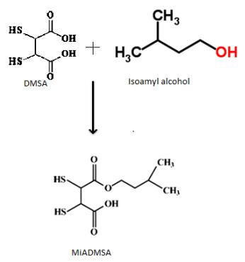
[32] | V. Pachauri and S. Flora, Combined efficacy of gallic acid and MiADMSA with limited beneficial effects over MiADMSA against arsenic-induced oxidative stress in mouse, Biochem. Insights, 8 (2015), 1–10. |
[33] | C. Loguercio, V. De Girolamo and A. Federico, et al., Relationship of blood trace elements to liver damage, nutritional status, and oxidative stress in chronic nonalcoholic liver disease, Biol. Trace. Elem. Res., 81 (2001), 245–254. |
[34] | C. Guo, P. Chen and K. Lin, et al., Trace metal imbalance associated with oxidative stress and inflammatory status in anti-hepatitis c virus antibody, Environ. Toxicol. Phar., 33 (2011), 288–296. |
[35] | A. U. Neumann, N. P. Lam and H. Dahari, et al., Hepatitis c viral dynamics in vivo and the antiviral efficacy of interferon-α therapy, Science, 282 (1998), 103–107. |
[36] | R. Y. Dodd and S. L. Stramer, Transfusion complications, 42 (2002), 975–979. |
[37] | R. Thimme, D, Oldach and K. M. Chang, et al., Determinants of viral clearance and Persistence during Acute hepatitis c virus Infection, J. Exp. Med., 194 (2001), 1395–1406. |
[38] | O. Lesens, M. Desche and M. Steben, Hepatitis c virus is related to progressive liver disease in human immunodeficiency virus-positive hemophiliacs and should be treated as an opportunistic infection, J. Infect. Dis., 179 (1999), 1254–1258. |
[39] | G. D. Kelen, G. B. Green and R. H. Purcell, et al., Hepatitis B and Hepatitis C in emergency department patients, New Engl. J. Med., 326 (1992), 1399–1404. |
[40] | M. El Sayed Zaki and W. Othman, Role of hepatitis E infection in acute on chronic liver failure in Egyptian patients, Liver Int., 31 (2011), 1001–1005. |
[41] | K. Grüngreiff, T. Hebell and K. Gutensohn, et al., Plasma concentrations of zinc, copper, interleukin-6 and interferon-γ, and plasma dipeptidyl peptidase IV activity in chronic hepatitis C, Mol. Med. Rep., 2 (2009), 63–68. |
[42] | K. C. Sorensen, S. Venn-Watson and S. H. Ridgway, Trace and non-trace elements in blood cells of bottlenose dolphins (Tursiops truncatus): Variations with values from liver function indicators, J. Wildlife Dis., 44 (2008), 304–317. |
[43] | M. Takahashi, H. Saito and M. Higashimoto, et al., Possible inhibitory effect of oral zinc supplementation on hepatic fibrosis through downregulation of TIMP-1: A pilot study, Hepatol. Res., 37 (2007), 405–409. |
[44] | T. Himoto, N. Hosomi and S. Nakai, et al., Efficacy of zinc administration in patients with hepatitis c virus-related chronic liver disease, Scand. J. Gastroentero., 42 (2007), 1078–1087. |
[45] | M. Blanuša, L. Prester and M. Piasek, et al., Monoisoamyl ester of DMSA reduces 203Hg(NO3)2 retention in rats: 1. Chelation therapy during pregnancy, J. Trace. Elem. Exp. Med., 10 (1997), 173–181. |
[46] | A. Mehta, S. C. Pant and S. J. S. Flora, Monoisoamyl dimercaptosuccinic acid-induced changes in pregnant female rats during late gestation and lactation, Reprod. Toxicol., 21 (2006), 94–103. |
1. | Miguel Melendez-Zamudio, Kevina Chavda, Michael A. Brook, Chelating Silicone Dendrons: Trying to Impact Organisms by Disrupting Ions at Interfaces, 2022, 27, 1420-3049, 1869, 10.3390/molecules27061869 | |
2. | Swaran J. S. Flora, Keerti Jain, Archna Panghal, Jayant Patwa, Chemistry, Pharmacology, and Toxicology of Monoisoamyl Dimercaptosuccinic Acid: A Chelating Agent for Chronic Metal Poisoning, 2022, 35, 0893-228X, 1701, 10.1021/acs.chemrestox.2c00129 | |
3. | Benjamin Rosen, Alberim Kurtishi, Gonzalo R. Vazquez-Jimenez, Simon Geir Møller, The Intersection of Parkinson’s Disease, Viral Infections, and COVID-19, 2021, 58, 0893-7648, 4477, 10.1007/s12035-021-02408-8 | |
4. | Suryakant Niture, Minghui Lin, Qi Qi, John T. Moore, Keith E. Levine, Reshan A. Fernando, Deepak Kumar, Orish Ebere Orisakwe, Role of Autophagy in Cadmium-Induced Hepatotoxicity and Liver Diseases, 2021, 2021, 1687-8205, 1, 10.1155/2021/9564297 | |
5. | Hadi Darvishi-Khezri, Hossein Karami, Mohammad Naderisorki, Mobin Ghazaiean, Mehrnoush Kosaryan, Amir Mosanejad-Galchali, Aily Aliasgharian, Hasan Karami, Alok Raghav, Two risk factors for hypozincemia in diabetic β-thalassemia patients: Hepatitis C and deferasirox, 2024, 19, 1932-6203, e0284267, 10.1371/journal.pone.0284267 | |
6. | Maryam Shamseddini Lori, Azade Kalantari Khandani, Gholamreza Dehghannoudeh, Mandana Ohadi, Mehdi Ansari, Therapeutic Potential of Iron Chelators in Viral Diseases: A Systematic Review, 2024, 31, 09298673, 4383, 10.2174/0109298673259596231211113211 |
Group | Age in years | Gender | |||
N | Mean ± SD | Range of years | Male/Female | ||
Control | 25 | 40 ± 14 | 19–55 | 12/13 | |
Patients (HCV) | 25 | 35 ± 15 | 20–65 | 16/9 |
Blood | Nails and Hairs | |||||
Normal | Hepatitis c patients | p-value | Normal | Hepatitis c patients | p-value | |
Iron (µg/dl) | 120.2 ± 36.11 | 150.22 ± 19.73 | 0.000** | 23.4 ± 5.18 | 25.55 ± 1.93 | 0.012** |
zinc (µg/dl) | 81.8 ± 13.52 | 42.9 ± 17.09 | 0.000** | 199 ± 19.11 | 129.8 ± 23.42 | 0.000** |
lead (µg/dl) | 24.5 ± 7.33 | 46.5 ± 6.73 | 0.000** | 3.04 ± 0.63 | 4.372 ± 0.93 | 0.002** |
nickel (ng/ml) | 1.34 ± 0.18 | 1.34 ± 0.58 | 0.784** | 0.96 ± 0.311 | 1.14 ± 0.39 | 0.013** |
Manganese (ng/ml) | 0.59 ± 0.179 | 1.5 ± 0.50 | 0.000** | 1.06 ± 0.673 | 4.4 ± 1.59 | 0.000** |
Copper (µg/L) | 110 ± 24.55 | 176 ± 17.44 | 0.000** | 70.68 ± 20.85 | 136.8 ± 22.72 | 0.000** |
Cobalt (ng/ml) | 0.4 ± 0.243 | 3.496 ± 1.105 | 0.000** | 0.092 ± 0.0571 | 0.20 ± 0.072 | 0.000** |
Cadmium (ng/ml) | 1.256 ± 0.515 | 4.828 ± 0.9825 | 0.000** | 0.23 ± 0.0426 | 0.403 ± 0.053 | 0.000** |
Heavy Metals | Before Chelation | After Chelation (Twice a day) BD | After Chelation (Thrice a day) TDS | p-value |
Iron (µg/dl) | 150.22 ± 19.73 | 133.6 ± 6.4 | 129.4 ± 9.1 | 0.000** |
lead (µg/dl) | 46.5 ± 6.73 | 30.2 ± 9.02 | 26.1 ± 3.1 | 0.000** |
Manganese (ng/ml) | 1.5 ± 0.50 | 0.9 ± 1.2 | 0.7 ± 5.8 | 0.002** |
Copper (µg/L) | 176 ± 17.44 | 135 ± 12.3 | 127 ± 2.9 | 0.001** |
Cobalt (ng/ml) | 3.496 ± 1.105 | 1.8 ± 1.4 | 1 ± 2.5 | 0.012** |
Cadmium (ng/ml) | 4.828 ± 0.9825 | 2.9 ± 1.3 | 2.1 ± 4.3 | 0.023** |