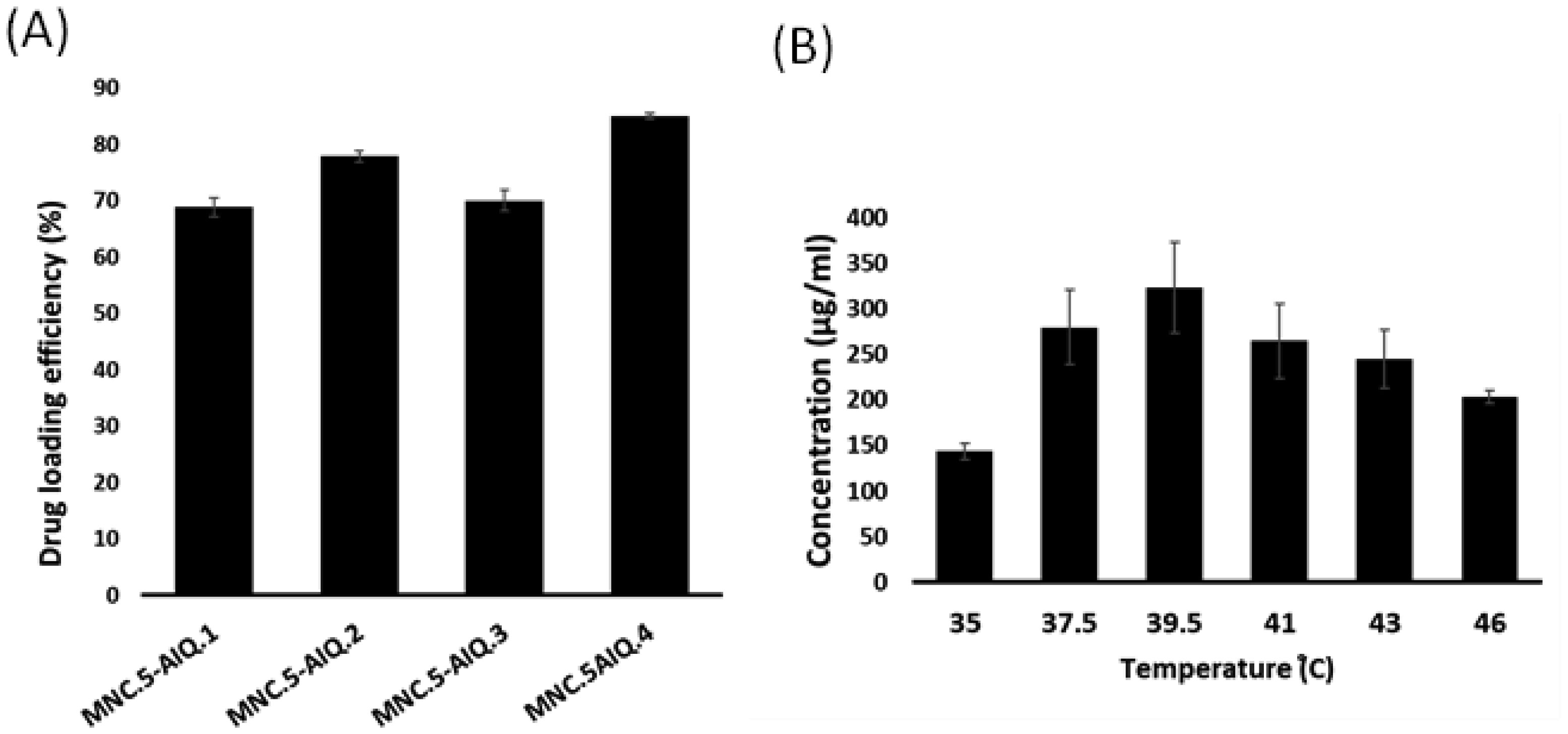
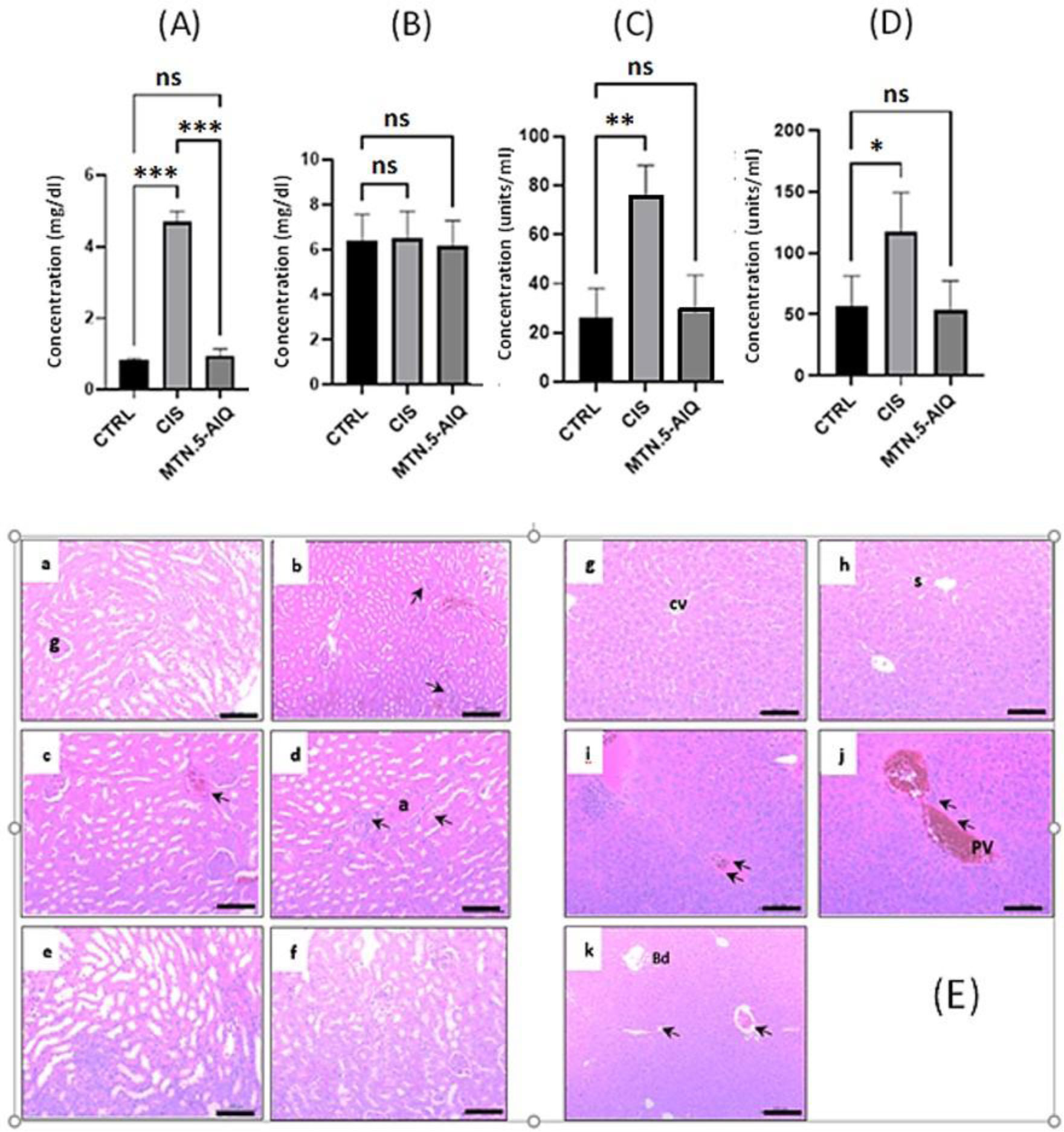
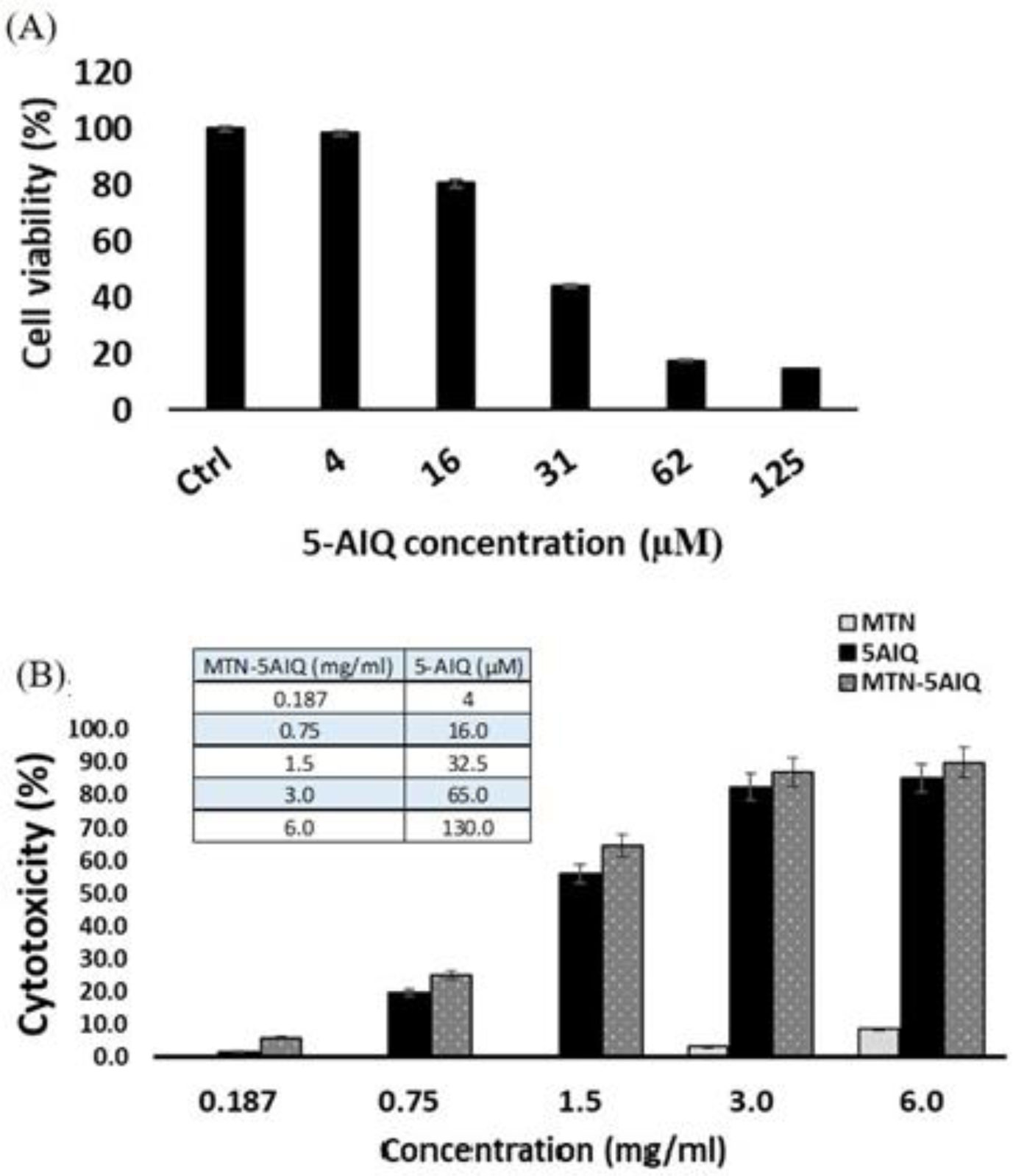
The current study presents a bimodal therapeutic platform for cancer treatment. Bimodal implies that the presented drug loaded core-shell structure is capable of elevating the tumor tissue temperature (hyperthermia) through the superparamagnetic iron oxide core and simultaneously release a Poly (ADP-ribose) polymerase-1(PARP-1)-modifying agent from the thermoresponsive shell. Magnetic thermoresponsive nanocomposite MTN was synthesized via an in situ free radical polymerization of thermo-responsive (N-isopropylacrylamide) (NIPAAm) monomer in the presence of 11-nm monodisperse SPIONs. The composite was allowed to swell in various concentrations of the PARP inhibitor: 5-aminoisoquinoline (5-AIQ) forming drug-loaded magnetic thermoresponsive nanocomposite (MTN-5.AIQ). Structural characterization of the formed composite is studied via various experimental tools. To assess the coil to globule transition temperature, the lower critical solution temperature (LCST) is determined by differential scanning calorimetry (DSC) method and the cloud point (Tp) is determined by turbidometry. Magnetic thermoresponsive nanocomposite (MTN) is formed with excellent potential for hyperthermia. A high drug loading efficiency (85.72%) is obtained with convenient temperature dependent drug release kinetics. Biocompatibility and cytotoxic efficacy are tested on an in vivo and in vitro colorectal-adenocarcinoma model, respectively. MTN.5-AIQ administration exhibits normal hepatic and renal functions as well as lower toxic effect on normal tissue. In addition, the composite effectively inhibits Caco-2 cells viability upon incubation. Based on the obtained results, the proposed therapeutic platform can be considered as a novel, promising candidate for dual therapy of colorectal adenocarcinoma exhibiting a PARP-1 overexpression. as well as increased the inhabiting efficacy of 5-AIQ.
Citation: Alaa AL-Rahman Gamal, El-Sayed Mahmoud El-Sayed, Tarek El-Hamoly, Heba Kahil. Development and bioevaluation of controlled release 5-aminoisoquinoline nanocomposite: a synergistic anticancer activity against human colon cancer[J]. AIMS Biophysics, 2022, 9(1): 21-41. doi: 10.3934/biophy.2022003
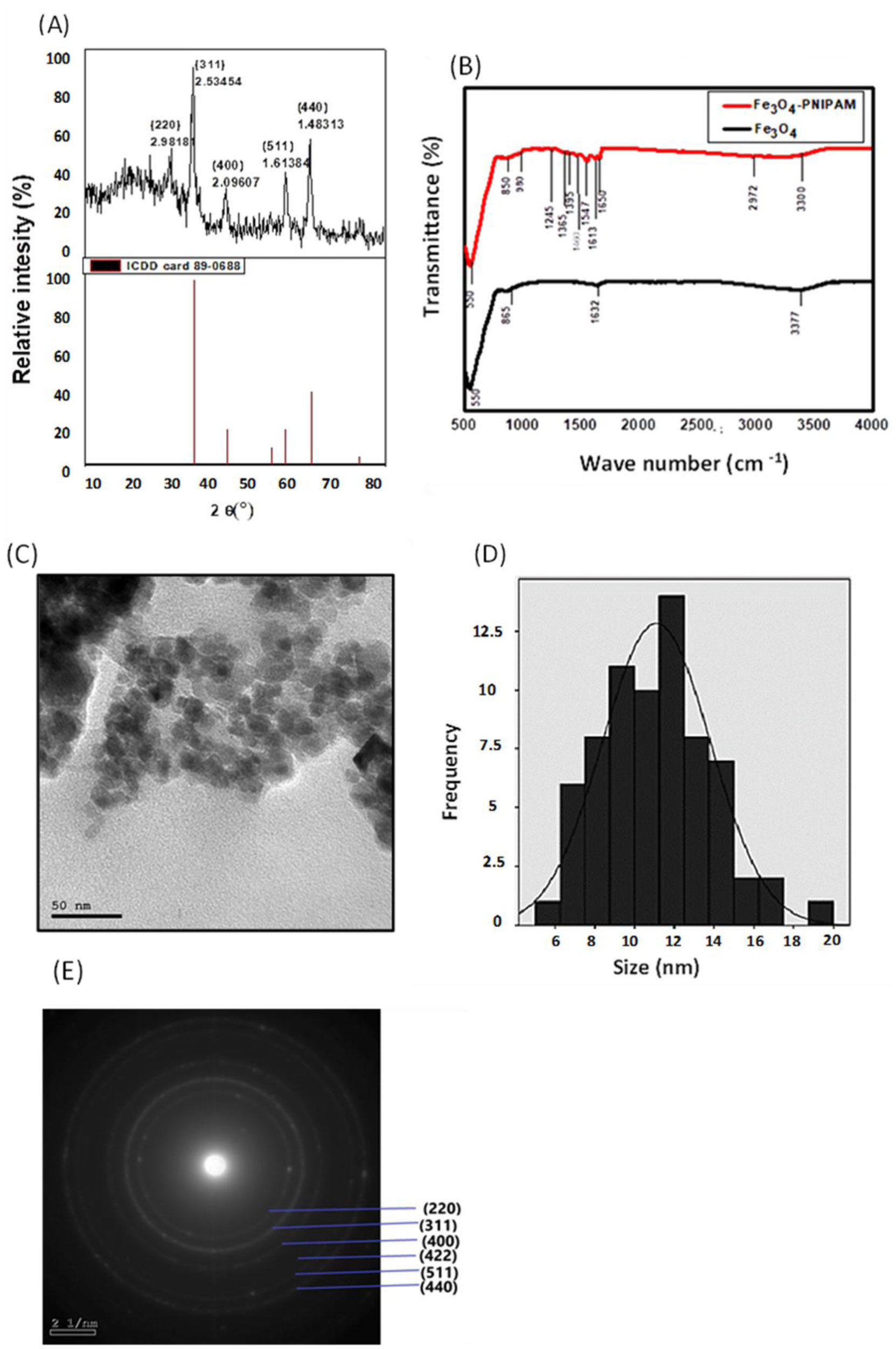
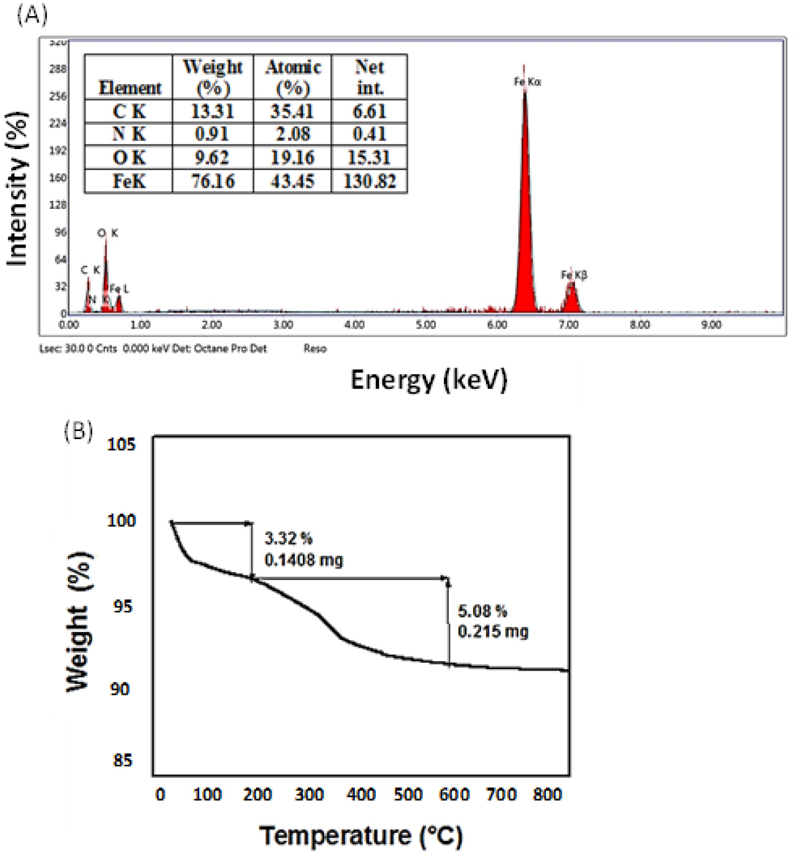
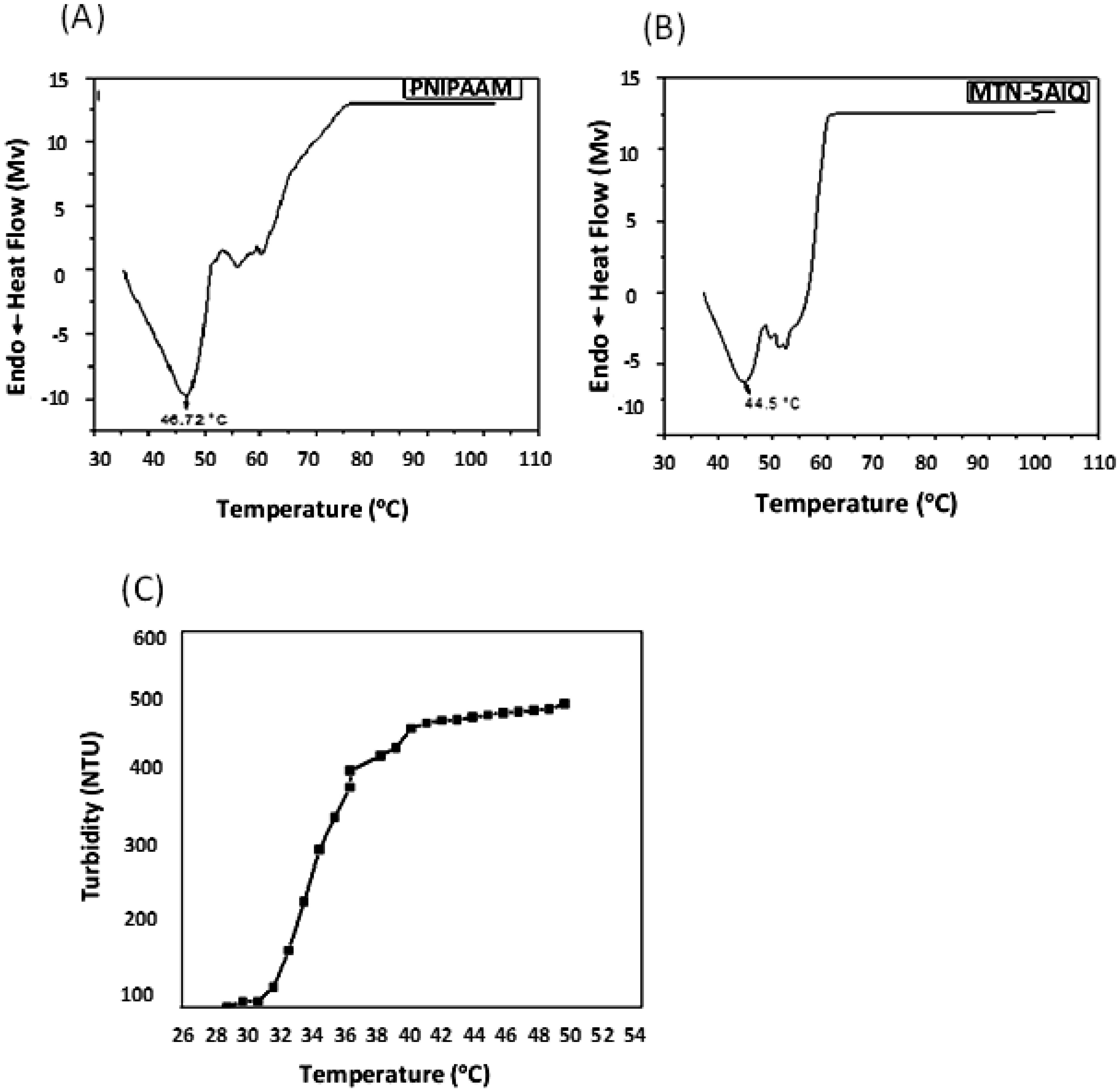
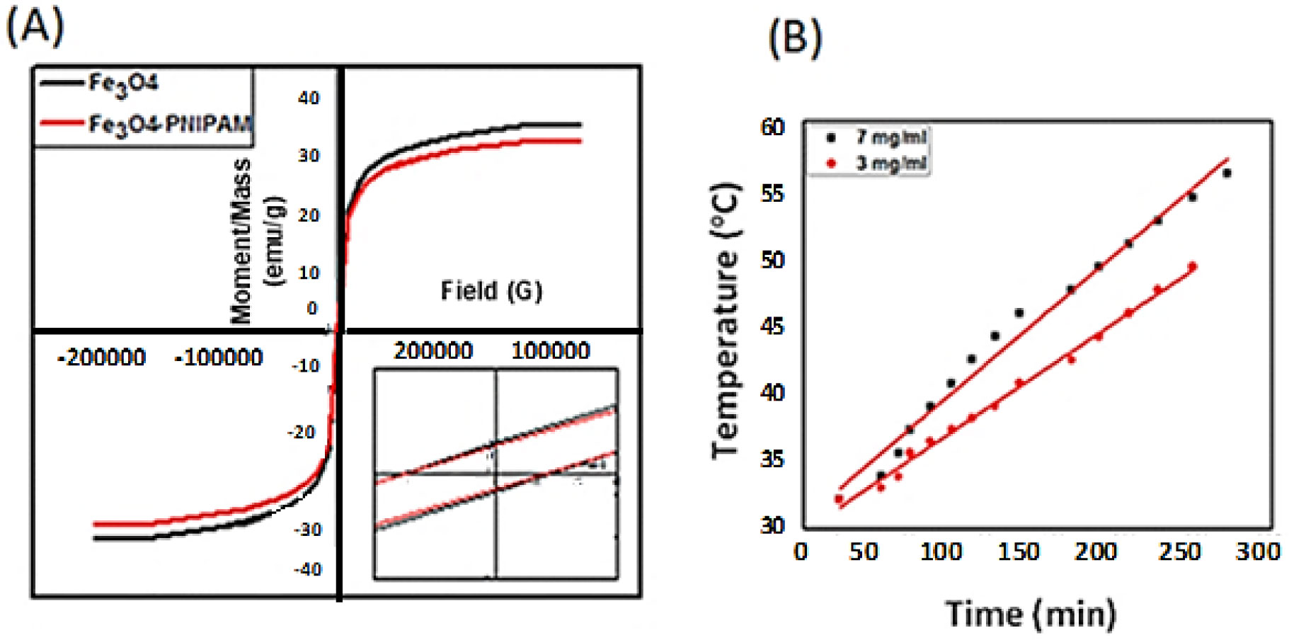
The current study presents a bimodal therapeutic platform for cancer treatment. Bimodal implies that the presented drug loaded core-shell structure is capable of elevating the tumor tissue temperature (hyperthermia) through the superparamagnetic iron oxide core and simultaneously release a Poly (ADP-ribose) polymerase-1(PARP-1)-modifying agent from the thermoresponsive shell. Magnetic thermoresponsive nanocomposite MTN was synthesized via an in situ free radical polymerization of thermo-responsive (N-isopropylacrylamide) (NIPAAm) monomer in the presence of 11-nm monodisperse SPIONs. The composite was allowed to swell in various concentrations of the PARP inhibitor: 5-aminoisoquinoline (5-AIQ) forming drug-loaded magnetic thermoresponsive nanocomposite (MTN-5.AIQ). Structural characterization of the formed composite is studied via various experimental tools. To assess the coil to globule transition temperature, the lower critical solution temperature (LCST) is determined by differential scanning calorimetry (DSC) method and the cloud point (Tp) is determined by turbidometry. Magnetic thermoresponsive nanocomposite (MTN) is formed with excellent potential for hyperthermia. A high drug loading efficiency (85.72%) is obtained with convenient temperature dependent drug release kinetics. Biocompatibility and cytotoxic efficacy are tested on an in vivo and in vitro colorectal-adenocarcinoma model, respectively. MTN.5-AIQ administration exhibits normal hepatic and renal functions as well as lower toxic effect on normal tissue. In addition, the composite effectively inhibits Caco-2 cells viability upon incubation. Based on the obtained results, the proposed therapeutic platform can be considered as a novel, promising candidate for dual therapy of colorectal adenocarcinoma exhibiting a PARP-1 overexpression. as well as increased the inhabiting efficacy of 5-AIQ.
5-aminoisoquinoline
Alternating magnetic field
Energy dispersive X-ray
Face centered cubic
Fourier transform infrared spectroscopy
Glutamic oxaloacetic transaminase
Glutamic pyruvic transaminase
High performance liquid chromatography
High resolution transmission electron microscope
Lower critical solution temperature
Magnetic thermoresponsive nanocomposite
Poly (ADP-ribose) polymerase-1
Poly(N-isopropylacrylamide)
Specific absorption rate
Superparamagnetic iron oxide nanoparticles
Cloud point temperature
X-ray diffraction

| [1] |
James HP, John R, Alex A, et al. (2014) Smart polymers for the controlled delivery of drugs–a concise overview. Acta Pharm Sin B 4: 120-127. https://doi.org/10.1016/j.apsb.2014.02.005 
|
| [2] |
Ward MA, Georgiou TK (2011) Thermoresponsive polymers for biomedical applications. Polymers (Basel) 3: 1215-1242. https://doi.org/10.3390/polym3031215
|
| [3] | Gould P (2006) Nanomagnetism shows in vivo potential. Nano Today 1: 34-39. https://doi.org/10.1016/S1748-0132(06)70115-3 |
| [4] |
Abenojar EC, Wickramasinghe S, Bas-Concepcion J, et al. (2016) Structural effects on the magnetic hyperthermia properties of iron oxide nanoparticles. Prog Nat Sci Mater Int 26: 440-448. https://doi.org/10.1016/j.pnsc.2016.09.004
|
| [5] |
Sharifi I, Shokrollahi H, Amiri S (2012) Ferrite-based magnetic nanofluids used in hyperthermia applications. J Magn Magn Mater 324: 903-915. https://doi.org/10.1016/j.jmmm.2011.10.017
|
| [6] |
Ba XQ, Garg NJ (2011) Signaling mechanism of poly(ADP-ribose) polymerase-1 (PARP-1) in inflammatory diseases. Am J Pathol 178: 946-955. https://doi.org/10.1016/j.ajpath.2010.12.004
|
| [7] |
Virág L, Szabó C (2002) The therapeutic potential of poly (ADP-ribose) polymerase inhibitors. Pharmacol Rev 54: 375-429. https://doi.org/10.1124/pr.54.3.375
|
| [8] |
D Threadgill M (2015) 5-Aminoisoquinolin-1-one (5-AIQ), a water-soluble inhibitor of the poly(ADP-Ribose)polymerases (PARPs). Curr Med Chem 22: 3807-3829. https://doi.org/10.2174/0929867322666151002110602
|
| [9] |
Vinod KR, Chandra S, Sharma SK (2010) Evaluation of 5-aminoisoquinoline (5-AIQ), a novel PARP-1 inhibitor for genotoxicity potential in vitro and in vivo. Toxicol Mech Methods 20: 90-95. https://doi.org/10.3109/15376510903572870
|
| [10] | Romano B, Pagano E, Iannotti FA, et al. (2021) N-Acylethanolamine acid amidase (NAAA) is dysregulated in colorectal cancer patients and its inhibition reduces experimental cancer growth. Brit J Pharmacol 1–16. https://doi.org/10.1111/bph.15737 |
| [11] |
Pagano E, Venneri T, Lucariello G, et al. (2021) Palmitoylethanolamide reduces colon cancer cell proliferation and migration, influences tumor cell cycle and exerts in vivo chemopreventive effects. Cancers (Basel) 13: 1923. https://doi.org/10.3390/cancers13081923
|
| [12] |
Pagano E, Borrelli F, Orlando P, et al. (2017) Pharmacological inhibition of MAGL attenuates experimental colon carcinogenesis. Pharmacol Res 119: 227-236. https://doi.org/10.1016/j.phrs.2017.02.002
|
| [13] |
Toğaçar M (2021) Disease type detection in lung and colon cancer images using the complement approach of inefficient sets. Comput Biol Med 137: 104827. https://doi.org/10.1016/j.compbiomed.2021.104827
|
| [14] |
Raftery L, Goldberg RM (2010) Optimal delivery of cytotoxic chemotherapy for colon cancer. Cancer J 16: 214-219. https://doi.org/10.1097/PPO.0b013e3181ddc5ac
|
| [15] |
Fernández J, Silván B, Entrialgo-Cadierno R, et al. (2021) Antiproliferative and palliative activity of flavonoids in colorectal cancer. Biomed Pharmacother 143: 112241. https://doi.org/10.1016/j.biopha.2021.112241
|
| [16] |
Küpeli Akkol E, Genç Y, Karpuz B, et al. (2020) Coumarins and coumarin-related compounds in pharmacotherapy of cancer. Cancers (Basel) 12: 1959. https://doi.org/10.3390/cancers12071959
|
| [17] |
Ahmed S, Khan H, Aschner M, et al. (2020) Anticancer potential of furanocoumarins: mechanistic and therapeutic aspects. Int J Mol Sci 21: 5622. https://doi.org/10.3390/ijms21165622
|
| [18] |
Nosho K, Yamamoto H, Mikami M, et al. (2006) Overexpression of poly(ADP-ribose) polymerase-1 (PARP-1) in the early stage of colorectal carcinogenesis. Eur J Cancer 42: 2374-2381. https://doi.org/10.1016/j.ejca.2006.01.061
|
| [19] |
Augustine T, Maitra R, Zhang J, et al. (2019) Sensitization of colorectal cancer to irinotecan therapy by PARP inhibitor rucaparib. Invest New Drug 37: 948-960. https://doi.org/10.1007/s10637-018-00717-9
|
| [20] |
Zhang J, Misra RDK (2007) Magnetic drug-targeting carrier encapsulated with thermosensitive smart polymer: core-shell nanoparticle carrier and drug release response. Acta Biomater 3: 838-850. https://doi.org/10.1016/j.actbio.2007.05.011
|
| [21] |
Hegazy M, Zhou P, Wu G, et al. (2017) Construction of polymer coated core-shell magnetic mesoporous silica nanoparticles with triple responsive drug delivery. Polym Chem 8: 5852-5864. https://doi.org/10.1039/C7PY01179B
|
| [22] |
Purushotham S, Ramanujan RV (2010) Thermoresponsive magnetic composite nanomaterials for multimodal cancer therapy. Acta Biomater 6: 502-510. https://doi.org/10.1016/j.actbio.2009.07.004
|
| [23] |
Meerod S, Rutnakornpituk B, Wichai U, et al. (2015) Hydrophilic magnetic nanoclusters with thermo-responsive properties and their drug controlled release. J Magn Magn Mater 392: 83-90. https://doi.org/10.1016/j.jmmm.2015.05.022
|
| [24] | Sharma R, Bisen DP, Shukla U, et al. (2012) X-ray diffraction: a powerful method of characterizing nanomaterials. Recent Res Sci Technol 4: 77-79. |
| [25] | Suryanarayana C, Norton MG (2013) X-ray diffraction: A practical approach. Springer Science & Business Media. |
| [26] |
Spirou SV, Basini M, Lascialfari A, et al. (2018) Magnetic hyperthermia and radiation therapy: Radiobiological principles and current practice. Nanomaterials 8: 401. https://doi.org/10.3390/nano8060401
|
| [27] | Page DL Theory and Practice of Histological Techniques (1983). https://doi.org/10.1016/S0046-8177(83)80171-3 |
| [28] | Wang B, Wei Q, Qu S (2013) Synthesis and characterization of uniform and crystalline magnetite nanoparticles via oxidation-precipitation and modified co-precipitation methods. Int J Electrochem Sci 8: 786-3793. |
| [29] |
Idris MI, Zaloga J, Detsch R, et al. (2018) Surface modification of SPIONs in PHBV microspheres for biomedical applications. Sci Rep 8: 7286. https://doi.org/10.1038/s41598-018-25243-9
|
| [30] | Lopez JA, González F, Bonilla FA, et al. (2010) Synthesis and characterization of Fe3O4 magnetic nanofluid. Rev Latinoam Metal y Mater 30: 60-66. |
| [31] | Coates J (2000) Interpretation of infrared spectra, a practical approach. Encycl Anal Chem 10815–10837. https://doi.org/10.1002/9780470027318.a5606 |
| [32] |
Omer M, Haider S, Park SY (2011) A novel route for the preparation of thermally sensitive core-shell magnetic nanoparticles. Polymer 52: 91-97. https://doi.org/10.1016/j.polymer.2010.11.011
|
| [33] | Narain R Engineered carbohydrate-based materials for biomedical applications: polymers, surfaces, dendrimers, nanoparticles, and hydrogels (2010). https://doi.org/10.1002/9780470944349 |
| [34] |
Mohapatra S, Rout SR, Panda AB (2011) One-pot synthesis of uniform and spherically assembled functionalized MFe2O4 (M = Co, Mn, Ni) nanoparticles. Colloids Surfaces A Physicochem Eng Asp 384: 453-460. https://doi.org/10.1016/j.colsurfa.2011.05.001
|
| [35] | Shindo D, Oikawa T (2013) Analytical Electron Microscopy for Materials Science. Springer Science & Business Media. |
| [36] |
Mutharani B, Ranganathan P, Chen SM (2019) Highly sensitive and selective electrochemical detection of antipsychotic drug chlorpromazine in biological samples based on poly-N-isopropylacrylamide microgel. J Taiwan Inst Chem Eng 96: 599-609. https://doi.org/10.1016/j.jtice.2018.10.029
|
| [37] |
Osváth Z, Iván B (2017) The dependence of the cloud point, clearing point, and hysteresis of poly (N-isopropylacrylamide) on experimental conditions: The need for standardization of thermoresponsive transition determinations. Macromol Chem Phys 218: 1600470. https://doi.org/10.1002/macp.201600470
|
| [38] |
Xia Y, Burke NAD, Stöver HDH (2006) End group effect on the thermal response of narrow-disperse poly(N-isopropylacrylamide) prepared by atom transfer radical polymerization. Macromolecules 39: 2275-2283. https://doi.org/10.1021/ma0519617
|
| [39] |
Shokrollahi H (2017) A review of the magnetic properties, synthesis methods and applications of maghemite. J Magn Magn Mater 426: 74-81. https://doi.org/10.1016/j.jmmm.2016.11.033
|
| [40] | Khairy M (2013) Synthesis, characterization and magnetic properties of gamma irradiated and unirradiated magnetic nanopowders. Int J Mater Chem 3: 106-111. https://doi.org/10.5923/j.ijmc.20130305.04 |
| [41] |
Li Q, Kartikowati CW, Horie S, et al. (2017) Correlation between particle size/domain structure and magnetic properties of highly crystalline Fe3O4 nanoparticles. Sci Rep 7: 9894. https://doi.org/10.1038/s41598-017-09897-5
|
| [42] |
Piñeiro-Redondo Y, Bañobre-López M, Pardiñas-Blanco I, et al. (2011) The influence of colloidal parameters on the specific power absorption of PAA-coated magnetite nanoparticles. Nanoscale Res Lett 6: 383. https://doi.org/10.1186/1556-276X-6-383
|
| [43] |
Wu WF, Wang JN, Li Z, et al. (2020) 7-Hydroxycoumarin protects against cisplatin-induced acute kidney injury by inhibiting necroptosis and promoting Sox9-mediated tubular epithelial cell proliferation. Phytomedicine 69: 153202. https://doi.org/10.1016/j.phymed.2020.153202
|
| [44] | Le Chang XLL, Dai Di Fan YQM, Huan Zhang HPM, et al. (2016) The efficiency of magnetic hyperthermia and in vivo histocompatibility for human-like collagen protein-coated magnetic nanoparticles. Int J Nanomed 11: 1175-1185. https://doi.org/10.2147/IJN.S101741 |
| [45] |
Vihola H, Laukkanen A, Valtola L, et al. (2005) Cytotoxicity of thermosensitive polymers poly(N-isopropylacrylamide), poly(N-vinylcaprolactam) and amphiphilically modified poly(N-vinylcaprolactam). Biomaterials 26: 3055-3064. https://doi.org/10.1016/j.biomaterials.2004.09.008
|
| [46] | Gamal A, El-sayed ES, El-Hamoly T, et al. Magnetic thermoresponsive nanocomposite for targeted PARP-1 in colorectal adenocarcinoma, an approach for tumor dual therapy research square (2021). https://doi.org/10.21203/rs.3.rs-295938/v1 |
 biophy-09-01-003-s001.pdf biophy-09-01-003-s001.pdf |
 |
Figures(7) / Tables(1)

Alaa AL-Rahman Gamal, El-Sayed Mahmoud El-Sayed, Tarek El-Hamoly, Heba Kahil. Development and bioevaluation of controlled release 5-aminoisoquinoline nanocomposite: a synergistic anticancer activity against human colon cancer[J]. AIMS Biophysics, 2022, 9(1): 21-41. doi: 10.3934/biophy.2022003


 DownLoad: