This study focuses on a simple, non-toxic, and environmentally friendly method for the green synthesis of silver nanoparticles using Dicranum scoparium moss extract. It includes the characterization of the biosynthesized nanoparticles and an evaluation of their antibacterial, antifungal, and anticancer activities. Transmission electron microscopy (TEM) and dynamic light scattering (DLS) analyses confirmed that the biosynthesized silver nanoparticles were within the nanoscale range (50–100 nm) and exhibited an irregular morphology. The biogenic nanoparticles demonstrate antibacterial activity against bacterial strains Staphylococcus aureus, Bacillus mesentericus, Escherichia coli, and Pseudomonas aeruginosa. The results indicate a pronounced antibacterial activity against E. coli and P. aeruginosa compared to the tested Gram-positive bacteria, which is attributed to differences in the bacterial cell wall structure. Additionally, the green synthesized silver nanoparticles inhibited the growth of Mucor plumber, Geotrichum candidum, Cladosporium herbarum, and Aspergillus flavus mold fungi. Additionally, they expressed considerable cytotoxic properties against cancer cells.
Citation: Gayane Semerjyan, Inesa Semerjyan, Mikayel Ginovyan, Nikolay Avtandilyan. Characterization and antibacterial/cytotoxic activity of silver nanoparticles synthesized from Dicranum scoparium moss extracts growing in Armenia[J]. AIMS Biophysics, 2025, 12(1): 29-42. doi: 10.3934/biophy.2025003
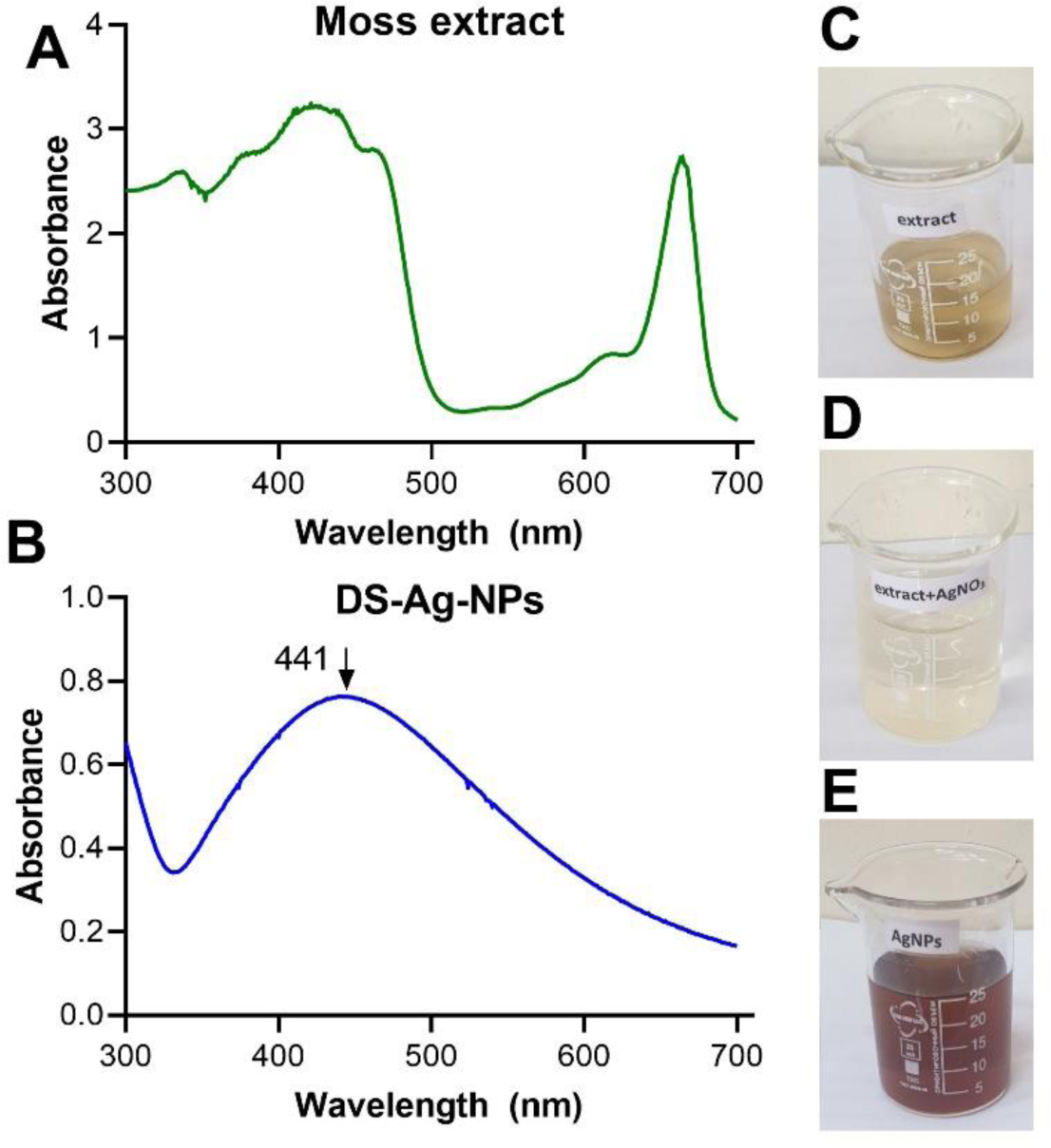
This study focuses on a simple, non-toxic, and environmentally friendly method for the green synthesis of silver nanoparticles using Dicranum scoparium moss extract. It includes the characterization of the biosynthesized nanoparticles and an evaluation of their antibacterial, antifungal, and anticancer activities. Transmission electron microscopy (TEM) and dynamic light scattering (DLS) analyses confirmed that the biosynthesized silver nanoparticles were within the nanoscale range (50–100 nm) and exhibited an irregular morphology. The biogenic nanoparticles demonstrate antibacterial activity against bacterial strains Staphylococcus aureus, Bacillus mesentericus, Escherichia coli, and Pseudomonas aeruginosa. The results indicate a pronounced antibacterial activity against E. coli and P. aeruginosa compared to the tested Gram-positive bacteria, which is attributed to differences in the bacterial cell wall structure. Additionally, the green synthesized silver nanoparticles inhibited the growth of Mucor plumber, Geotrichum candidum, Cladosporium herbarum, and Aspergillus flavus mold fungi. Additionally, they expressed considerable cytotoxic properties against cancer cells.

| [1] |
Yaqoob AA, Ahmad H, Parveen T, et al. (2020) Recent advances in metal decorated nanomaterials and their various biological applications: a review. Front Chem 8: 341. https://doi.org/10.3389/fchem.2020.00341 
|
| [2] |
Kocharyan M, Marutyan S, Nadiryan E, et al. (2024) Royal Jelly–mediated silver nanoparticles show promising anticancer effect on heLa and A549 cells through modulation of the VEGFa/PI3K/Akt/MMP-2 pathway. Appl Organomet Chem 38: e7726. https://doi.org/10.1002/aoc.7726
|
| [3] |
Selvaraj S, Krishnaswamy S, Devashya V, et al. (2014) Flavonoid-metal ion complexes: a novel class of therapeutic agents. Med Res Rev 34: 677-702. https://doi.org/10.1002/med.21301
|
| [4] |
Samuel MS, Ravikumar M, John JA, et al. (2022) A review on green synthesis of nanoparticles and their diverse biomedical and environmental applications. Catalysts 12: 459. https://doi.org/10.3390/catal12050459
|
| [5] |
Dahoumane SA, Wijesekera K, Filipe CDM, et al. (2014) Stoichiometrically controlled production of bimetallic gold-silver alloy colloids using micro-alga cultures. J Colloid Interf Sci 416: 67-72. https://doi.org/10.1016/j.jcis.2013.10.048
|
| [6] | Lavate RA, Sathe SS, Kumbhar D (2016) Bryophytes as Source of Silver Nanoparticles: A Review. Proceeding of International conference on Advances in Materials Science : 431-434. |
| [7] |
Gabrielyan L, Trchounian A (2019) Antibacterial activities of transient metals nanoparticles and membranous mechanisms of action. World J Microb Biot 35: 162. https://doi.org/10.1007/s11274-019-2742-6
|
| [8] |
Mba IE, Nweze EI (2021) Nanoparticles as therapeutic options for treating multidrug-resistant bacteria: research progress, challenges, and prospects. World J Microb Biot 37: 108. https://doi.org/10.1007/s11274-021-03070-x
|
| [9] |
Bonilla-Gameros L, Chevallier P, Sarkissian A, et al. (2020) Silver-based antibacterial strategies for healthcare-associated infections: processes, challenges, and regulations. An integrated review. Nanomedicine 24: 102142. https://doi.org/10.1016/j.nano.2019.102142
|
| [10] |
El-Gendy AO, Samir A, Ahmed E, et al. (2021) The antimicrobial effect of 400 nm femtosecond laser and silver nanoparticles on gram-positive and gram-negative bacteria. J Photoch Photobio B 223: 112300. https://doi.org/10.1016/j.jphotobiol.2021.112300
|
| [11] |
Muraro PCL, Pinheiro LDSM, Chuy G, et al. (2022) Silver nanoparticles from residual biomass: biosynthesis, characterization and antimicrobial activity. J Biotechnol 343: 47-51. https://doi.org/10.1016/j.jbiotec.2021.11.003
|
| [12] | Alam A, Shrama V, Rawat KK, et al. (2015) Bryophytes-the ignored medicinal plants. Biomed J 2: 299-316. |
| [13] | Alam A, Baliyan P, Sharma V, et al. (2021) Potential of bryophytes in nanotechnolohy: an overview. J Phytonanotechnology Pharm Sci 1: 1-3. http://dx.doi.org/10.21276/jpps.2021.1.1.1 |
| [14] | Sabovljevic A, Sabovljevic M, Jockovic N (2009) In vitro culture and secondary metabolite isolation in bryophytes. Protocols for In Vitro Cultures and Secondary Metabolite Analysis of Aromatic and Medicinal Plants : 117-128. https://doi.org/10.1007/978-1-60327-287-2_10 |
| [15] |
Novakovic M, Bukvicki D, Andjelkovic B, et al. (2019) Cytotoxic activity of riccardin and perrottetin derivatives from the liverwort Lunularia cruciata. J Nat Prod 82: 694-701. https://doi.org/10.1021/acs.jnatprod.8b00390
|
| [16] | Semerjyan I, Semerjyan G, Semerjyan H, et al. (2020) Antibacterial properties and flavonoids content of some mosses common in Armenia. Iran J Pharm Sci 2020: 31-42. https://doi.org/10.22037/ijps.v16.40308 |
| [17] |
Timotina M, Aghajanyan A, Schubert R, et al. (2022) Biosynthesis of silver nanoparticles using extracts of Stevia rebaudiana and evaluation of antibacterial activity. World J Microb Biot 38: 196. https://doi.org/10.1007/s11274-022-03393-3
|
| [18] |
Aghajanyan A, Gabrielyan L, Schubert R, et al. (2020) Silver ion bioreduction in nanoparticles using Artemisia annua L. extract: characterization and application as antibacterial agents. AMB Express 10: 66. https://doi.org/10.1186/s13568-020-01002-w
|
| [19] |
Gabrielyan L, Badalyan H, Gevorgyan V, et al. (2020) Comparable antibacterial effects and action mechanisms of silver and iron oxide nanoparticles on Escherichia coli and Salmonella typhimurium. Sci Rep 10: 13145. https://doi.org/10.1038/s41598-020-70211-x
|
| [20] |
Ginovyan M, Hovhannisyan S, Javrushyan H, et al. (2022) Screening revealed the strong cytotoxic activity of Alchemilla smirnovii and Hypericum alpestre ethanol extracts on different cancer cell lines. AIMS Biophys 10: 12-22. https://doi.org/10.3934/biophy.2023002
|
| [21] |
Almatroudi A (2024) Unlocking the potential of silver nanoparticles: from synthesis to versatile bio-applications. Pharmaceutics 16: 1232. https://doi.org/10.3390/pharmaceutics16091232
|
| [22] |
Hambardzumyan S, Sahakyan N, Petrosyan M, et al. (2020) Origanum vulgare L. extract-mediated synthesis of silver nanoparticles, their characterization and antibacterial activities. AMB Express 10: 162. https://doi.org/10.1186/s13568-020-01100-9
|


Figures(5)

Gayane Semerjyan, Inesa Semerjyan, Mikayel Ginovyan, Nikolay Avtandilyan. Characterization and antibacterial/cytotoxic activity of silver nanoparticles synthesized from Dicranum scoparium moss extracts growing in Armenia[J]. AIMS Biophysics, 2025, 12(1): 29-42. doi: 10.3934/biophy.2025003


 DownLoad: