Citation: William A. Toscano, Jr., Hitakshi Sehgal, Emily Yang, Lindsey Spaude, A. Frank Bettmann. Environment-Gene interaction in common complex diseases: New approaches[J]. AIMS Molecular Science, 2014, 1(4): 126-140. doi: 10.3934/molsci.2014.4.126
[1] | Md. Mamun Al-Amin, Robert K. P. Sullivan, Suzy Alexander, David A. Carter, DanaKai Bradford, Thomas H. J. Burne . Impaired spatial memory in adult vitamin D deficient BALB/c mice is associated with reductions in spine density, nitric oxide, and neural nitric oxide synthase in the hippocampus. AIMS Neuroscience, 2022, 9(1): 31-56. doi: 10.3934/Neuroscience.2022004 |
[2] | Sara Arciniegas Ruiz, Eliav Tikochinsky, Vardit Rubovitch, Chaim G Pick, Bernard Attali . Contextual fear response is modulated by M-type K+ channels and is associated with subtle structural changes of the axon initial segment in hippocampal GABAergic neurons. AIMS Neuroscience, 2023, 10(1): 33-51. doi: 10.3934/Neuroscience.2023003 |
[3] | Roya. Askari, Mohadeseh. NasrAbadi, Amir Hossein. Haghighi, Mohammad Jahan Mahin, Rajabi Somayeh, Matteo. Pusceddu . Effect of combined training in water on hippocampal neuronal Plasticity and memory function in healthy elderly rats. AIMS Neuroscience, 2024, 11(3): 260-274. doi: 10.3934/Neuroscience.2024017 |
[4] | Laaziz Abderrahim, El Mostafi Hicham, Elhessni Aboubaker, Azeroil Fatima, Touil Tarik, Boumlah Soufiane, Mesfioui Abdelhalim . Sex differences in behavioral, cognitive and voluntary ethanol-intake effects in Dexamethasone-induced depression-like state in Wistar rat. AIMS Neuroscience, 2022, 9(2): 228-249. doi: 10.3934/Neuroscience.2022012 |
[5] | Nouhaila Chaoui, Hammou Anarghou, Meriem Laaroussi, Oumaima Essaidi, Mohamed Najimi, Fatiha Chigr . Long lasting effect of acute restraint stress on behavior and brain anti-oxidative status. AIMS Neuroscience, 2022, 9(1): 57-75. doi: 10.3934/Neuroscience.2022005 |
[6] | I. Prieto, A.B. Segarra, M. de Gasparo, M. Ramírez-Sánchez . Neuropeptidases, Stress, and Memory—A Promising Perspective. AIMS Neuroscience, 2016, 3(4): 487-501. doi: 10.3934/Neuroscience.2016.4.487 |
[7] | Laura Serra, Sara Raimondi, Carlotta di Domenico, Silvia Maffei, Anna Lardone, Marianna Liparoti, Pierpaolo Sorrentino, Carlo Caltagirone, Laura Petrosini, Laura Mandolesi . The beneficial effects of physical exercise on visuospatial working memory in preadolescent children. AIMS Neuroscience, 2021, 8(4): 496-509. doi: 10.3934/Neuroscience.2021026 |
[8] | Robert A. Moss . A Theory on the Singular Function of the Hippocampus: Facilitating the Binding of New Circuits of Cortical Columns. AIMS Neuroscience, 2016, 3(3): 264-305. doi: 10.3934/Neuroscience.2016.3.264 |
[9] | Mirela Dubravac, Beat Meier . Stimulating the parietal cortex by transcranial direct current stimulation (tDCS): no effects on attention and memory. AIMS Neuroscience, 2021, 8(1): 33-46. doi: 10.3934/Neuroscience.2021002 |
[10] | UCHEWA O. Obinna, EMECHETA S. Shallom, EGWU A. Ogugua, EDE C. Joy, IBEGBU O. Augustine . Neuromodulatory roles of PIPER GUINEENSE and honey against Lead-Induced neurotoxicity in social interactive behaviors and motor activities in rat models. AIMS Neuroscience, 2022, 9(4): 460-478. doi: 10.3934/Neuroscience.2022026 |
The mammalian central nervous system produces and modulates behaviour through cycles of complex interactions involving neurotransmitters and their receptors. Its primary excitatory neurotransmitter, glutamate, is important in synaptic plasticity, learning and development [1], and has been implicated in the pathogenesis of learning, memory and anxiety disorders [2,3]. Interaction of glutamate at the N-Methyl-D-Aspartate (NMDA) receptors forms the basis for long-term potentiation/depression in memory processes. Glutamate acts as a neurotransmitter at most of the excitatory synapses in the central nervous system, and glutamatergic neurons are widely distributed in the forebrain [4,5] and hippocampus [5,6].
Glutamate is derivable from exogenous sources, such as dietary monosodium glutamate (MSG), a culinary flavour-enhancer. MSG is metabolised to yield free glutamate, which is not biochemically different from the endogenous ligand, and studies have shown that MSG administration results in reproducible behavioural changes in rodents [7,8]. Studies have shown that daily MSG consumption in human averages about 0.3-1.0 g [9], although there are variations amongst different nationalities all over the world. MSG consumption ranges from 0.5g/day in America [10] to 0.3-0.5 mg/day amongst the Europeans. In Asia, average consumption ranges from between 1.2-1.7 g/day [11)] in Korea and Japan, to 3-3.8 g/day in Taiwan and China [12]. Amongst the Thai population, MSG consumption ranges between 0.4-14 g/day, with an average daily intake of 6 g [13]. Advances in food-production practices worldwide and the need to improve the taste of food, in recent times, could mean that the permissible levels of consumption may be significantly exceeded within short periods of time. Hence, there is the need to study not only the morphological changes associated with MSG, but also its effects on hippocampal-dependent behaviours.
A number of studies have sought to explore the relationship between MSG ingestion (at increasing doses) and memory/anxiety, with diverse results. Available data show that ingestion of MSG at extremely high doses (levels significantly higher than average daily human consumption) led to a decrease in spatial-memory and learning tasks performance in adult/neonatal rats or mice [1,7,14,15,16,17]. The deficits in spatial memory and learning in MSG-treated mice have several possible explanations. A deficiency of NMDA glutamate receptors in the hippocampus, degenerative retinal function/impaired vision, cholinergic deficiency that leads to reduced acetylcholine synthesis (especially in the hippocampus), or a combination of some or all of these performance inhibitors are possible explanations [1,17]. Studies have also demonstrated that MSG, at very high doses was anxiogenic [18]; this is notable, as imbalances in glutamate/gamma-amino-butyric acid ratio have been implicated in anxiety disorders.
Interactions of glutamate with its ionotropic (mainly NMDA) receptors have sometimes been found to lead to neurotoxic changes, due to glutamate’s effect on calcium availability at the neurons [19]. Excessive activation of glutamate receptors is believed to contribute to neuronal injury, as persistent or overwhelming activation of glutamate-gated ion channels can result in degeneration of neurons, either through necrosis or apoptosis [20,21]. This phenomenon called‘‘excitotoxicity, ’’has been linked to the final common pathway of neuronal death in a number of disorders including Huntington disease, Alzheimer disease, amyotrophic lateral sclerosis (ALS), and stroke [19].
Early studies had described anatomical changes in specific brain regions secondary to MSG administration [22]. Some of these studies also showed that the hippocampus is one of the most susceptible regions of the brain, and that of several animal species; mice were the most susceptible to MSG neurotoxicity [23]. Hence, it is obvious from earlier studies using MSG that ingestion of high doses led to obvious anatomical changes in the hippocampi of susceptible species, in addition to behavioural changes. In a more recent study by us, repeated administration of MSG at the same doses used in the present study (equivalent to 0.7 g-5.6 g/day in a 70 kg man; doses within and below average daily consumption levels) was observed to cause hippocampal injury in mice, at the upper limit of dosing [8]. The justification for this study therefore was the need to evaluate the effects of repeated administration of MSG (at doses chosen with reference to previous studies [8,24,25,26] on hippocampal-dependent (recognition memory in the Y-maze, spatial memory in the Y and radial-arm mazes, and anxiety test in the elevated-plus maze) behaviours; and to compare effects seen, with standard reference drugs (scopolamine and diazepam respectively). Our hypothesis is that repeated administration of MSG at concentrations currently associated with typical dietary consumption may significantly alter recognition-memory, working-memory and anxiety related behaviours.
MSG (99.9% purity, Ajinomoto®), Diazepam (Valium® Roche) and Scopolamine hydrobromide (Guangzhou Pharmaceuticals, China).
Adult Swiss albino mice from Empire Breeders, Osogbo, Osun State, Nigeria, weighing 20-22 g each were used for this study. Mice were housed in groups of six in plastic cages located in a temperature-controlled quarters (22-25 degree Celsius) with 12 hour light/dark cycle (lights off at 7.00 a.m). All animals were fed commercial standard chow (Calories: 29% Protein, 13% Fat, 58% Carbohydrate) from weaning. Mice had free access to food and drinking water except during the behavioural tests. All procedures were conducted in accordance with the approved institutional protocols and within the provisions outlined in the“Guidelines for the Use of Animals in Neuroscience and Behavioural Research”prepared by the Committee on Guidelines for the Use of Animals in Neuroscience and Behavioural Research; National Research council of the National Academies (2003).
One hundred and eighty mice each were used for this study. Animals were divided into three main groups (Table 1): Y-maze recognition/working-memory (60), radial-arm maze working-memory (60) and EPM anxiety tests (60). They were further divided into 6 groups of 10 animals each. Animals in each group received vehicle (distilled water) at 10 ml/kg, a standard anxiolytic (diazepam at 0.5 mg/kg i.p. for the anxiety test) and a standard amnesic (scopolamine at 1 mg/kg i.p. for cognition test) or one of four selected doses of MSG (10, 20, 40 and 80 mg/kg) daily for three weeks with an oral cannula. Doses of MSG were calculated by dissolving measured quantities of the salt (Ajinomoto®) in distilled water. MSG was administered via gavage to: accurately determine how much was consumed, closely simulate means of administration in humans, and finally because studies have shown that administration of MSG by gavage achieves higher plasma levels than when MSG is mixed with food [27]. Mice were weighed weekly, while the behavioural tests were carried out after the first and last dose of vehicle or drug.
Groups | Test | ||
Y-maze | Radial-arm maze | EPM | |
VEH(10 ml/kg) | n = 10 | n = 10 | n = 10 |
10 mg/kg | n = 10 | n = 10 | n = 10 |
20 mg/kg | n = 10 | n = 10 | n = 10 |
40 mg/kg | n = 10 | n = 10 | n = 10 |
80 mg/kg | n = 10 | n = 10 | n = 10 |
SCOP(1 mg/kg) | n = 10 | n = 10 | |
DIZ (0.5 mg/kg) | n = 10 | ||
TOTAL | 60 | 60 | 60 |
Neurobehavioural tests were carried out in a quiet room between the hours of 8 a.m. and 2 p.m. On each of the test days, animals were transported to the testing room in their home cages, and allowed to acclimatize for 30 minutes before behavioural tests, after which drug or vehicle was administered. Behavioural tests were conducted after 30 minutes of administration of MSG, based on the pharmacokinetics of oral MSG. At the onset of the tests, each animal was placed in the apparatus and its behaviour videotaped for subsequent analysis. After testing, each mouse was removed from the maze, returned to its home cage, the maze cleaned thoroughly with 70 % ethanol, and then wiped dry to remove any trace of odour.
Hippocampus-dependent Y-maze task characterizes spatial recognition-memory, which is based on the propensity of rodents to gravitate toward novel spatial environments, which is not contingent upon either reward, or water-escape associated stress. The two-trial Y-maze test consisted of two trials separated by an inter-trial interval of 30 minutes to assess spatial recognition-memory. At the start of each session, each animal was gently placed at the end of the start arm, facing away from the central platform. The number of entries and time spent in each arm of the maze by each mouse were recorded over the trial session. Arm entry is defined as the entry of all four paws into one arm. For the first trial, mice were placed inside the start arm, while the novel arm is closed with a wooden block (the same arm was closed for all animals in all test groups). Therefore, mice were able to explore the start and other arm, but not the novel arm [28,29]. Memory-retrieval (second trial) was evaluated in a test session carried out 30 minutes after the first trial [30]. For this trial, trained animals were placed back in the maze in the same starting arm, with free access to all three arms. Choice of novel arm and time spent in the novel arm were then scored. The entire task is based on the innate explorative behaviour of rodents. If the animals have good recognition-memory, they will spend more time in the novel arm relative to the previously explored arms. Number of entries, and time spent in each arm was scored. Percentage time spent in the novel arm was scored as: time spent in the novel arm relative to the average time×by 100. Novel arm choice on retrial was scored as the number of times animals in the group entered the novel arm first/number of animals in the group×100.
The Y-maze was also used as a measure of spatial working-memory, measured using the spontaneous alternation. Spontaneous alternation was measured using a Y-maze made up of three equally spaced arms (120°, 41 cm long and 15 cm high). Each mouse was placed in one of the arm compartments and allowed to move freely until its tail completely entered another arm. An alternation was defined as entry into all three arms consecutively [31]. The number of actual alternations is number of sequential arm entries into three arms, designated A, B and C. The percentage alternation is calculated as {(Actual alternations/Total arm entry minus two)×100} in a 5 minute interval.
Working-memory in the radial-arm maze was measured as alternation index, which is the ratio of sequential arm entries before error and total arm entry. The apparatus is made up of eight equidistantly spaced arms, each about 33 cm long, all radiating from a small circular central platform. Working-memory was assessed when the rat enters each arm a single time over a 5 minute period. Re-entry into the arms would result in a working-memory error [31].
Anxiety-related behaviours was measured in the elevated plus maze {EPM}. The EPM is plus-shaped, with two open arms measuring 25×5×5 cm lying across from each other and perpendicular to two closed arms measuring 25×5×16 cm with a centre platform (5×5×0.5 cm). The closed arms are enclosed by 2 high walls (16 cm) while the open arms have no side wall. Animals are placed in the central platform facing the open arm and behaviours recorded for 5 minutes. The criterion for arm visit was considered only when the animal decisively moved all its four limbs into an arm. The following measures were measured: Percent open arm entries (open arm entries/total arm entry multiplied by 100) and percent time spent in the open arms (time in open arms/ total time spent in the maze multiplied by 100) [32]. Administration of an anxiolytic drug e.g. (diazepam) in rodents significantly increased the percentage of entries into, and time spent in the open arms.
At the end of the experimental period, animals were euthanized using diethyl-ether anesthesia and perfused transcardially with ice-cold saline. Whole brains were dissected out and the hippocampus sectioned and blotted dry. A 10 % hippocampal homogenate was prepared with ice-cold phosphate buffered saline using Teflon-glass homogenizer. The homogenate was centrifuged at 5, 000 rpm (at 4 degrees Celsius), for 15 min. and the pellet discarded. The supernatant was used to assay brain glutamate and glutamine levels [8].
Plasma and hippocampal glutamate and glutamine levels were assayed using glutamate and glutamine assay kit following the manufacturer’s instructions. The glutamate assay kit provides a sensitive detection method of the glutamate in a variety of samples. The glutamate enzyme mix recognizes glutamate as a specific substrate leading to proportional colour development. The glutamine assay is based on the hydrolysis of glutamine to glutamate which produces a stable signal, which is directly proportional to the quantity of glutamine in the sample [8].
Data was analysed using Chris Rorden’s ezANOVA for windows, version 0.98. Hypothesis testing was performed using analysis of variance (ANOVA).We tested the hypothesis, that acute and repeated administration of low doses of MSG causes significant changes in hippocampal-dependent behaviours, using a two-way analysis of variance (ANOVA). One way ANOVA was used for analysis of body weight, plasma and hippocampal glutamate and glutamine level data. Tukey highly significant difference (HSD) test was used for pairwise comparisons. Results are expressed as mean±S.E.M, p values less than 0.05 were considered statistically significant.
Figure 1 represents the percentage body weight change, defined as the percentage difference between the final and initial body weights, divided by the initial body weight, multiplied by 100. There was no significant (F (4, 45)=1.56, p < 0.786) difference in % weight increase at any of the doses compared to either vehicle or MSG.
Figure 2 shows one way ANOVA analysis the effect of acute or repeated administration of MSG on novel arm choice on retrial in the 2-trial Y-maze test. Acute (F (5, 48)=23.27, p < 0.015) and repeated (F (5, 48)=12.10, p < 0.001) administration of MSG resulted in a significant increase in recognition-memory (measured as choice of the novel arm on retrial) at 10 mg/kg and a decrease at 20, 40 and 80 mg/kg, compared to vehicle respectively; compared to scopolamine however, there was an increase at 10, 20 and 40 mg/kg with acute administration, while with repeated administration significant increase was seen at 10, 20 and 40 mg/kg and a decrease at 80 mg/kg.
Figure 3 shows the effect of MSG on time spent in the novel arm on retrial. Two-factor ANOVA assessing the effects of two main factors (MSG dose and duration of administration) revealed a significant effect of MSG dose (F (5, 108)=456, p < 0.001) and duration of administration (F (1, 108)=5.88, p < 0.020), with significant interactions between MSG dose×duration of administration (F (5, 108)=2.64, p < 0.030). Pairwise comparisons of the effects of scopolamine (SCOP) or MSG dose and vehicle (control) following acute and repeated administration revealed a significant decrease in recognition-memory measured as % time spent in the novel arm with SCOP (0.001) and with MSG at 40 (0.010) and 80 mg/kg (p < 0.001), while at 10 mg/kg (p < 0.001), a significant increase was seen compared to vehicle. Compared to SCOP however, there was a significant increase in recognition-memory at 10 (p < 0.130), 20 (p < 0.001) and 40 mg/kg (p < 0.001) of MSG. Pairwise comparisons of the effect of acute vs. repeated dosing revealed no significant difference at any of the doses of MSG.
Figure 4 shows the effect of MSG on spatial working-memory in the Y-maze. Two-factor ANOVA assessing the effect of two factors (MSG dose and duration of administration) revealed a significant effect of MSG dose (F (5, 108)=23.1, p < 0.001) and duration of administration (F (1, 108)=11.20, p < 0.003), with a significant interaction between MSG dose×duration of administration (F (5, 108)=23.90, p < 0.001). Pairwise comparisons of the effect of SCOP or MSG doses and vehicle (control) following acute administration, revealed a significant decrease in spatial memory scores (measured as % alternation) with SCOP (p < 0.002), at 40 (p < 0.001) and 80 mg/kg (p < 0.001) of MSG, while at 10 (p < 0.001) and 20 mg/kg (p < 0.001) of MSG, a significant increase was seen. Repeated administration resulted in a significant decrease in spatial memory with SCOP and following MSG at 20 (p < 0.018), 40 (p < 0.020) and 80 mg/kg (p < 0.001), while at 10 mg/kg (p < 0.001) there was a significant increase compared to vehicle. Compared to scopolamine, there was a significant increase in memory at 10 (p < 0.001), 20 (p < 0.001) and 40 mg/kg (p < 0.001), following both acute and repeated administration. Pairwise comparisons of the effect of acute and repeated dosing revealed a significant decrease in spatial memory with repeated administration at 20 mg/kg (p < 0.001) compared to acute administration.
Figure 5 shows the effect of MSG on spatial working-memory in the radial arm-maze, measured as alternation index/5 minute, which is a fraction of number of correct alternation before first error and total number of alternations made in a five minute period. Two-factor ANOVA assessing the effect of two factors (MSG dose and duration of administration) revealed a significant effect of MSG dose (F (5, 108)=6.70, p < 0.010) and duration of administration (F (1, 108)=5.32, p < 0.020), with significant interactions between MSG dose x duration of administration (F (5, 108)=3.88, p < 0.002). Pairwise comparisons of the effect of SCOP or MSG dose and vehicle (control) revealed a significant decrease in spatial memory with SCOP (p < 0.012, p < 0.001)) and at 80 mg/kg (p < 0.021, p < 0.001) of MSG, while at 10 mg/kg (p < 0.001, p < 0.001), a significant increase was seen following both acute and repeated administration respectively. Compared to SCOP however, a significant increase in spatial memory was seen at 10 (p < 0.001, p < 0.001), 20 (p < 0.013, p < 0.001) and 40 mg/kg (p < 0.001, p < 0.001) following acute and repeated administration respectively. Pairwise comparisons of the effect of acute and repeated dosing revealed no significant difference at any of the doses of MSG.
Figure 6 shows the effect of MSG on % open-arm entries in the elevated plus maze. Two-factor ANOVA assessing the effect of two factors (MSG dose and duration of administration) revealed a significant effect of MSG dose (F (5, 108)=9.40, p < 0.001) and duration of administration (F (1, 108)=5.38, p < 0.002), with no significant interaction between MSG dose×duration of administration (F (5, 108)=0.85, p < 0.342). Pairwise comparisons of the effect of diazepam (DIZ) or MSG dose and vehicle (control) revealed a significant increase in open arms entry with DIZ (p < 0.001, p < 0.001), at 10 (p < 0.001, p < 0.001) and 20 mg/kg (p < 0.001, p < 0.001) of MSG, while at 80 mg/kg (p < 0.001, p < 0.001), a significant decrease was seen following acute and repeated administration respectively. In comparison to DIZ however, there was a significant decrease in number of open arm entries at all doses of MSG (p < 0.001) following acute and repeated administration respectively. Comparisons of the effect of acute and repeated dosing revealed no significant difference in number of open arm entry at any of the doses of MSG with repeated compared to acute administration.
Figure 7 shows the effect of MSG on % time spent in the open-arm of the EPM. Two-factor ANOVA assessing the effect of two factors (MSG dose and duration of administration) revealed a significant effect of MSG dose (F (5, 108)=12.20, p < 0.001) and duration of administration (F (1, 108)=7.44, p < 0.010), with significant interactions between MSG dose×duration of administration (F (5, 108)=4.55, p < 0.002). Pairwise comparisons of the effect of DIZ or MSG dose and vehicle (control) revealed a significant increase in time spent in the open arms with DIZ (p < 0.001, p < 0.001), at 10 (p < 0.001, p < 0.015) and 20 mg/kg (p < 0.001, p < 0.001) of MSG, while at 40 (p < 0.001, p < 0.001) and 80 mg/kg (p < 0.024, p < 0.013) a significant decrease was seen, following acute and repeated administration respectively. In comparison to DIZ, there was a significant decrease in time spent in the open arms at all doses of MSG (p < 0.050) following acute and repeated administration. Pairwise comparisons of the effect of acute and repeated dosing revealed no significant difference in time spent in the open arms at any of the doses of MSG with repeated compared to acute administration.
Table 2 shows the effects of MSG on locomotor activity in the Y-maze and radial arm maze respectively following 5 minutes of exploration. Following exploration in the Y-maze, two-factor ANOVA revealed a significant effect of MSG dose (F (5, 108)=33.5, p < 0.001) and duration of administration (F (1, 108)=74.44, p < 0.001) with no significant interaction between MSG dose×duration of administration (F (5, 108)=9.45, p < 0.212). Pairwise comparisons of the effect of SCOP or MSG dose and vehicle following acute and repeated administration revealed a significant increase in locomotor activity with SCOP (p < 0.001, 0.021) and no significant difference at any of the doses of MSG respectively. Compared to SCOP, there was a significant decrease in locomotor activity at 10 (p < 0.002, p < 0.001), 20 mg/kg (p < 0.012, p < 0.031), 40 (p < 0.001, p < 0.001) and 80 (p < 0.001, p < 0.001)] of MSG following acute and repeated administration respectively. Comparison of the effect of acute and repeated dosing revealed no significant difference in locomotor activity at any of the doses of MSG.
Locomotor activity | Groups | Acute Mean±S.E.M | Repeated Mean±S.E.M |
Y-maze | VEH | 7.00±1.52 | 6.58±1.47 |
SCOP | 19.5±1.29* | 19.83±1.61* | |
10 | 9.25±1.46µ | 5.83±1.20*µ | |
20 | 8.20±1.51 µ | 7.60±1.06µ | |
40 | 7.25±1.51 µ | 7.22±1.25µ | |
80 | 7.98±0.96 µ | 8.31±1.55µ | |
Radial-arm | VEH | 11.00±1.14 | 11.75±1.05 |
SCOP | 43.00±1.85* | 44.92±2.63* | |
10 | 10.00±1.92 µ | 18.50±0.75* µ | |
20 | 13.25±1.72 µ | 15.607±1.77 µ | |
40 | 11.55±1.11 µ | 14.22±2.12 µ | |
80 | 9.22±1.32 µ | 11.41±1.89 µ | |
Comparisons: Differences between experimental groups (MSG or SCOP) and control (VEH) was marked by asterisk (post hoc test: p < 0.05). Differences between dosages of MSG, VEH and SCOP are marked by hash (post hoc test; p < 0.05). number of mice per treatment group=12; VEH: Vehicle, SCOP: scopolamine. |
Following exploration in the radial-arm maze, two-factor ANOVA revealed a significant effect of MSG dose (F (5, 108)=321, p < 0.001) and duration of administration (F (1, 108)=334.8, p < 0.001), with no significant interactions between MSG dose×duration of administration (F (5, 108)=1.25, p < 0.402). Pairwise comparisons of the effect of SCOP or MSG dose and vehicle revealed a significant increase in locomotor activity with SCOP (p < 0.001, p < 0.001) but no significant difference at either dose of MSG following acute and repeated administration respectively. In comparison to SCOP, there was a significant decrease in locomotor activity at 10 (p < 0.001; p < 0.001), 20 mg/kg (p < 0.001; p < 0.001), 40 (p < 0.001; p < 0.001) and 80 mg/kg (p < 0.001; p < 0.001) of MSG following acute and repeated administration respectively. Comparisons of the effect of acute and repeated dosing revealed no significant difference in locomotor activity at any of the doses of MSG.
Table 3 shows the effects of MSG on plasma and hippocampal levels of glutamate and glutamine at day 21. There was a significant increase in plasma glutamate (F (5, 45)=16.52, p < 0.001) and glutamine (F (5, 45)=12.52, p < 0.011) levels at 40 and 80 mg/kg respectively compared to vehicle. Hippocampal glutamate (F (5, 45)=1.10, p < 0.335) and glutamine (F (5, 45)=2.53, p < 0.112) levels did not differ significantly from vehicle, at any of the doses of MSG respectively.
Groups | Plasma glutamate µmoles/L | Plasma glutamine µmoles/L | Hippocampal glutamate µmoles/g | Hippocampal glutamine µmoles/g |
VEH | 13.21±0.62 | 6.41±0.4 | 1.12±0.11 | 0.75±1.2 |
10 | 15.96±0.27 | 6.89±1.3 | 1.16±0.21 | 0.88±0.34 |
20 | 16.20±0.31 | 7.12±1.9 | 1.25±1.01 | 0.86±1.00 |
40 | 28.88±0.28*# | 10.15±1.98* | 1.50±2.03 | 0.91±1.63 |
80 | 30.00±60.68*# | 12.41±0.4* | 1.62±3.10 | 0.92±1.50 |
Comparisons: Differences between experimental groups (MSG or SCOP) and control (VEH) was marked by asterisk (post hoc test: p < 0.05), number of mice per treatment group=12; VEH: Vehicle. |
Our study in mice (using test groups outlined in Table 1) revealed that acute and repeated administration of MSG was associated with: (1) no significant effect on body weight (2) enhancement of spatial recognition and spatial working-memory at 10 mg/kg while impairing both at higher doses (3) mixed anxiety-related response, (4) an increase in plasma glutamate and glutamine levels at high doses, while only a non-significant trend towards an increase in hippocampal levels was seen.
In this study, administration of MSG did not significantly alter body weight, compared to vehicle (figure 1). This corroborates an earlier study in which similar results were reported [8], although reports on the effects of MSG consumption on body weight continue to be conflicting, with more studies supporting an age-related effect on body weight. MSG effects on body weight have been reported to be mediated by gut L-glutamate receptors which are connected to the afferent fibres of the vagus nerve or via L-glutamate taste receptors (Umami) [33,34,35,36]. MSG administered by gavage delivers a loading dose of MSG to the gastrointestinal tract lumen, activating vagal afferents [36,37], indirectly through production and release of either nitric oxide and/or serotonin [38]; although MSG-mediated gastric response is concentration-dependent [34], which may account for the variations in body weight effects reported in a number of studies. Studies in which MSG was administered to neonatal or prenatal rodents reported increased weight gain at times bordering on obesity [39], whilst studies in which adults rats or mice were used either reported no weight change or a reduction [8,38,40]. In this study, food and water intake (not reported) did not differ significantly from control animals, which suggests that the mechanisms responsible for lesser or no weight gain may be associated with higher energy expenditure rather than increased energy intake.
In this study, MSG increased spatial-recognition (figure 2 and figure 3) and spatial working-memory (figure 4 and figure 5) at 10 mg/kg and impaired it at higher doses, following both acute and repeated administration. Learning and memory in the Y-maze and radial-arm maze are measured by motor reactions; and the results of Y-maze and radial-arm maze locomotor activity (Table 2) showed a non-significant trend towards increased locomotion, which affirms that memory changes seen in this study were not simply a consequence of motor impairment. The effect seen at 10 mg/kg is suggestive of a possible nootropic effect; although, this contravenes the results of a number of studies in which MSG was administered at extremely high doses (doses that far exceed average daily human consumption), where impaired memory [15,41,42,43] was reported irrespective of route of administration. In a previous study [44], acute administration of very low doses of MSG (0.5-1.5 mg/kg) showed no significant effect on spatial working-memory in the Y-maze. This also suggests that the possible nootropic effect of MSG is only observable within a narrow margin of doses. The possible mechanisms for this effect are still being studied. Studies have shown that the memory impairment seen with MSG are either as a result of interference with glutamate synthesis in the hippocampus [41,42], or through inhibition of the cholinergic system [41]. A number of the studies in which MSG has been associated with memory loss or no improvement in memory have either used very low doses [44] or very high doses [41]. At the high doses, memory impairment has also been associated with neuronal injury, possibly due to glutamate excitotoxicity or oxidative stress [8,45]. In a recently published study with doses similar to those used in this study, neuronal injury was not evident at doses less than 40 mg/kg [8], suggesting that at these levels, intensity of glutamate receptor stimulation is not high enough to causes excitotoxicity, although it is sufficient for memory enhancement. The results of hippocampal glutamate and glutamine levels (Table 3) in this study showed non-significant increases compared to vehicle. Recent experimental evidence supports the notion that ‘umami’ taste in the gastrointestinal tract can stimulate cortical and sub-cortical brain areas that are linked to working-memory [46]. Using functional magnetic resonance imaging, Meyer-Gerspach et al. [46] found that MSG administration via nasogastric tube in human subjects was associated with stimulation of neuron clusters in the pre-central gyrus, post-central gyrus, hemispheric operculum, precuneus and the cingulate gyrus; however this did not result in significant changes in working-memory performance [46]. It must be noted that a major limitation of their study is that MSG delivery through nasogastric tube bypasses the physiological route of nutrient intake, as a result, information from the tongue and the gut are not integrated; this failure of integration is likely to have reduced the strength of stimulatory impulses delivered to the brain. In contrast, MSG was administered by oral gavage in our study.
The impairment in working-memory seen at the higher doses of MSG could be secondary to a combined effect of slight elevation of glutamate and glutamine, coupled with increased oxidative stress that has been reported at these two doses [8]. The results of locomotor activity in this study showed no significant difference from vehicle. This implies that the memory-impairment seen at high doses were not locomotor/motor-related.
In terms of practical usefulness, the application of glutamic acid as a nootropic agent is a topic that has generated lots of debates; presently, it is used as an adjunctive memory-enhancing agent, however, its usage is still neither popular nor backed by convincing experimental evidences.
In the EPM, (figure 6 and figure 7) acute MSG administration led to an anxiolytic response at the lower doses (10 and 20 mg/kg) and an anxiogenic response at the higher doses, when compared to vehicle. However, the anxiolytic response seen at lower doses of MSG is not comparable to that of diazepam. Repeated administration of MSG resulted in an anxiogenic response across all doses tested in comparison to both vehicle and diazepam controls. The results seen with repeated administration in this study corroborates a number of studies that have demonstrated anxiety behaviour with MSG administration [18,47]. Increased glutamatergic transmission has been associated with anxiety disorders [48,49], and in a few studies, a correlation between increase in glutamate concentration in the hippocampus and/or amygdala and anxiety disorders in humans have been documented [50,51]. Glutamate-GABA imbalances have also been implicated in anxiety disorders and the response elicited following oral MSG could mean that administration of MSG might have resulted in an alteration in the glutamate-GABA balance in the brain that is enough to cause the observed effects. The loss of an anxiolytic and assumption of anxiogenic response with continuous administration of MSG suggests that even at low doses, repeated exposure of the brain to MSG may alter anxiety-related behaviours.
In conclusion, MSG at doses studied is nootropic at low doses in mice, suggesting a need for further studies. Although, the association of MSG with memory loss and anxiogenesis at higher doses still suggest that continued caution in its use is necessary.
Authors declare no conflicts of interest in this paper.
[1] |
Nakamura J, Mutlu E, Sharma V, et al. (2014) The Endogenous Exposome. DNA Repair (Amst) 19: 3-13. doi: 10.1016/j.dnarep.2014.03.031
![]() |
[2] |
Wild CP (2012) The exposome: From concept to utility. Int J Epidemiol 41: 24-32. doi: 10.1093/ije/dyr236
![]() |
[3] | EPA, Persistent Organic Pollutants: A Global Issue, A Global Response. EPA, 2009. Available from: http://www2.epa.gov/international-cooperation/persistent-organic-pollutants-global-issue-global-response#pops (Accessed July 17, 2014). |
[4] |
Magliano DJ, Loh VH, Harding JL, et al. (2014) Persistent Organic Pollutants and Diabetes: A Review of the Epidemiological Evidence. Diabetes Meab 40: 1-14. doi: 10.1016/j.diabet.2013.09.006
![]() |
[5] |
Myre M, Imbeault P (2014) Persistent Organic Pllutants Meet Adipose Tissue Hypoxia: Does Cross-talk Contribute to Obesity. Obes Res 15: 19-28. doi: 10.1111/obr.12086
![]() |
[6] |
Taylor KW, Novak RF, Anderson HA, et al. (2013) Evaluation of the Association Between Persistent Organic pollutants (POPs) and diabetes in Epidemiological Studies: A National Toxicology Program Workshop Review. Environ Health Perspect 121: 774-783. doi: 10.1289/ehp.1205502
![]() |
[7] |
Gascon M, Morales E, Sunyer J, et al. (2013) Effects of Persistent Organic Pollutants on the Developing Respiratory and Immune Systems: A Systematic Review. Environ Int 52: 51-65. doi: 10.1016/j.envint.2012.11.005
![]() |
[8] |
Li QQ, Loganath A, Chong YS, et al. (2006) Persistent Organic Pollutants and Adverse Health Effects in Humans. J Toxicol Env Health Part A 69: 1987-2005. doi: 10.1080/15287390600751447
![]() |
[9] |
Zielinski I, Tsui I-C (1995) Cystic Fibrosis: Genotypic and Phenotypic Variations. Ann, Rev Genet 29: 777-807. doi: 10.1146/annurev.ge.29.120195.004021
![]() |
[10] | Braun A, Roth R, McGinnis MJ (2003) Technology challenges in screening single gene disorders. Eur J Pediatr 162: S 13-16. |
[11] | WHO (2014) World Health Statistics 2014. Geneva, Switzerland: World Health Organization, 180. |
[12] | Russell JY (2010) A Philosophical Framework for an Open and Critical Transdisciplinary Inquuiry. In: Brown VA, Harris JA, Russell JY, editors. Tackling Wicked Problems Through the Transdisciplinary Imagination. NY, NY: Earthscan, 31-60. |
[13] | Stokols D, Hall KL, Vogel AL (2013) Transdisciplinary Public Health: Definitions, Core Characteristics, and Strategies for Success. In: Haire-Joshu D, McBride TD, editors. Transdisciplinary Public Health: Research, Education, and Practice. San Francisco, CA: Jossey-Bass, 1-30. |
[14] |
Jacobs JA, Frickel S (2009) Interdisciplinarity: A Critical Assessment. Annu Rev Sociol 35: 43-65. doi: 10.1146/annurev-soc-070308-115954
![]() |
[15] | Klein JT (2008) Evaluation of Interdisciplinary and Transdisciplinary Research - A Literature Review. Amer J Prev Med Suppl 2 35: S-116-S-123. |
[16] | Hays TC (2008) The Science of Team Science - Commentary on Measurements of Scientific Readiness. Amer J Prev Med Suppl 2 35: S-193-S195. |
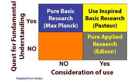
[17] | DE (1997) Pasteur's Quadrant: Basic Science and Technological Innovation: Brookings Institution, Washington, DC. |
[18] | NRC (2014) Convergence: Facilitating Transdisciplinary Integration of Life Sciences, Physical Sciences, Engineering and Beyond. Washington, DC: National Academies Press, 137. |
[19] | Roche M, Glick TF (1944) Thomas Edison and Basic Science, Edison: Myths and Reality. Arbor: 39-50. |
[20] | Duggan MB (2014) Prevention of childhood malnutrition: immensity of the challenge and variety of strategies. Paediatr Int Child Health 32: 190-203. |
[21] |
Blackburn GL (2001) Pasteur's Quadrant and Malnutrition. Nature 409: 397-401. doi: 10.1038/35053187
![]() |
[22] |
Verbeke W (2005) Consumer acceptance of functional foods: Socio-demographic, cognitive and attitudinal determinants. Food Quality and Preference 16: 45-57. doi: 10.1016/j.foodqual.2004.01.001
![]() |
[23] | Bhutta ZA, Darmstadt GL (2014) A Role for Science Investments in Advancing Newborn Health. Science Translational Medicine 6: 253-258. |
[24] | Childress DS (1999) Working in Pasteur's Quadrant. J Rehabilitation Res Dev 36: xi-xii. |
[25] | De Luigi AJ, Cooper RA (2014) Adaptive sports technology and biomechanics: prosthetics. PM R 8 Suppl.: S40-S57. |
[26] |
Hardbower DM, Peek RM, Jr., Wilson KT (2014) At the Bench: Helicobacter pylori, dysregulated host responses, DNA damage, and gastric cancer. J Leukoc Biol 96: 201-212. doi: 10.1189/jlb.4BT0214-099R
![]() |
[27] |
Dalal RS, Moss SF (2014) At the Bedside: Helicobacter pylori, dysregulated host responses, DNA damage, and gastric cancer. J Leukoc Biol 96: 213-224. doi: 10.1189/jlb.4BT0214-100R
![]() |
[28] | Nobel Committee, The Nobel Prize in Physiology or Medicine Prize Announcement. The Nobel Committee for Physiology or Medicine,2005. Available from:http://www.nobelprize.org/nobel_prizes/medicine/laureates/2005/announcement.html. |
[29] | Konturek SJ, Konturek PC, Brzozowski T, et al. (2005) From nerves and hormones to bacteria in the stomach; Nobel prize for achievements in gastrology during last century. J Physiol Pharmacol 56: 507-536. |
[30] |
Marshall B (2006) Helicobacter connections. Chem Med Chem 1: 783-802. doi: 10.1002/cmdc.200600153
![]() |
[31] | Woo KT, Lau YK, Yap HK, et al. (2006) 3rd College of Physicians' lecture--translational research: From bench to bedside and from bedside to bench; incorporating a clinical research journey in IgA nephritis (1976 to 2006). Ann Acad Med Singapore 35: 735-741. |
[32] |
Hamilton JG, Edwards HM, Khoury MJ, et al. (2014) Cancer screening and genetics: a tale of two paradigms. Cancer Epidemiol Biomarkers Prev 23: 909-916. doi: 10.1158/1055-9965.EPI-13-1016
![]() |
[33] | Preston R (2000) The Genome Warrier. The New Yorker 76: 66-83. |
[34] | Tchounwou PB, Toscano WA (2011) Environmental Epidemiology and Human Health-Biomarkers of Disease and Genetic Susceptibility In: Nriagu JO, editor. Encyclopedia of Environmentl Health Sciences. San Diego, CA: Elsevier, 357-366. |
[35] |
Teo S, Morgan M, Stirling D, et al. (2000) Assessment of the In Vitro and In Vivo Genotoxicity of Thalomid (Thalidomide). Teratog Carcinog Mutagen 20: 301-311. doi: 10.1002/1520-6866(2000)20:5<301::AID-TCM6>3.0.CO;2-2
![]() |
[36] |
Portin P (2002) Historical Development of the Concept of the Gene. J Med Philosophy 27: 257-286. doi: 10.1076/jmep.27.3.257.2980
![]() |
[37] | Crick FHC (1958) Central Dogma of Molecular Biology. Symp Soc Exp Biol XII 12: 139-173. |
[38] |
Crick FHC (1970) Central Dogma of Molecular Biology. Nature (London) 227: 561-563. doi: 10.1038/227561a0
![]() |
[39] |
Hahn WC, Weinberg RA (2002) Modeling the Molecular Circuitry of Cancer. Nat Rec Cancer 2: 331-341. doi: 10.1038/nrc795
![]() |
[40] | Hocquette JF (2005) Where are we in genomics? J Physiol Pharmacol Suppl 3: 37-70. |
[41] |
Sharma S, Kelly TK, Jones PA (2010) Epigenetics in cancer. Carcinogenesis 31: 27-36. doi: 10.1093/carcin/bgp220
![]() |
[42] |
Long CR, Westhusin ME, Golding MC (2014) Reshaping the transcriptional frontier: epigenetics and somatic cell nuclear transfer. Mol Reprod Dev 81: 183-193. doi: 10.1002/mrd.22271
![]() |
[43] | Nilsson E, Skinner MK (2014) Definitions and History of Generational Epigeneti Inheritance. In: Tollefsbol TO, editor. Translational Epigenetics: Evidence and Debate. San Diego, CA: Academic Press, 11-24. |
[44] | Hoile SP, Lillycrop KA, Grenfel LR, et al. (2014) Phenotypic and Epigenetic Inheritance Across Multiple Generations in Mammals through the Female Line. In: Tollefsbol TO, editor. Transgenerational Epigenetics: Evidence and Debate. San Diego, CA: Academic Press, 269-278. |
[45] | Mashoodh R, Champagne FA (2014) Paternal Epigenetic Inheritance. In: Tollefsbol TO, editor. Transgenerational Epigenetics: Evidence and Debate, 231-236. |
[46] | Toscano WA, Oehlke KP (2011) Nutrigenomics: A New Frontier in Environmental Health Sciences. In: Nriagu JO, editor. Encyclopedia of Environmental Health Burlington, VT: Elsevier, 202-205. |
[47] | Toscano WA, Oehlke KP, Kafouri R (2010) An Environmental Systems Biology Approach to the Study of Asthma. In: Pawnkar R, Holgate S, Rosenwasser LJ, editors. Allergy Frontiers: Future Perspectives. Springer: NY, NY, 239-252. |
[48] | Toscano WAJ, Lee P, Oehlke KP (2008) The Role of Environmental Health Research in Understanding Chronic Conditions. Minn Physician 21: 27-28. |
[49] |
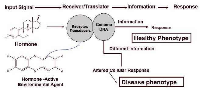
McLachlan JA (2001) Environmental Signaling: What Embryos and Evolution Teach us about Endocrine Disrupting Chemicals. Endocr Rev 22: 319-341. doi: 10.1210/edrv.22.3.0432
![]() |
[50] |
Vandenberg LN (2014) Non-monotonic Dose Responses in Studies of Endocrine Disrupting Chemicals: Bisphenol-A as a Case Study. Dose-Response 12: 259-276. doi: 10.2203/dose-response.13-020.Vandenberg
![]() |
[51] |
Vandenberg LN, Colborn T, Hayes TB, et al. (2012) Hormones and endocrine-disrupting chemicals: low-dose effects and nonmonotonic dose responses. Endocr Rev 33: 378-455. doi: 10.1210/er.2011-1050
![]() |
[52] | Vandenberg LN, Erlich S, Belcher SM, et al. (2013) Low-dose Effects of Bisphenol-A: An Integrated review of in vitro, Laboratory, Animal, and Epidemiology Studies. Endocrine Disruptors 1: 1-20. |
[53] | CDC (2014) National Diabetes Statistics Report, 2014. Center for Disease Control, Atlanta, GA. |
[54] |
Misra A, Bhardwai S (2014) Obesity and the Metbolic Syndrome in Developing Countries: Focus on South Asians. Nestle Nutr Inst Workshop Ser 78: 133-140. doi: 10.1159/000354952
![]() |
[55] |
Chase K, Sharma RP (2013) Epigenetic Developmental Programs and Adipogenesis - Implications for Psychotropic Induced Obesity. Epigenetics 8: 1133-1140. doi: 10.4161/epi.26027
![]() |
[56] | Janesick A, Blumberg B (2011) Minireview: PPARγ as the target of obesogens. J Ster Biochem & Mol Biol 127: 4-8. |
[57] | Janesick A, Blumberg B (2011) The role of environmental obesogens in the obesity epidemic. Obesity before birth: Springer. 383-399. |
[58] | Kishida K, Shimomura H, Nishizawa N, et al. (2001) Enhancement of the aquaporin adipose gene expression by a peroxisome proliferator-activated receptor gamma. J Biol Chem 276: 48572-48579. |
[59] |
Grun F, Blumberg B (2009) Minireview: the case for obesogens. Mol Endocrinol 23: 1127-1134. doi: 10.1210/me.2008-0485
![]() |
[60] |
Kemper MF, Stirone C, N. KD, et al. (2014) Genomic and non-genomic regulation of PGC1 isoforms by estrogen to increase cerebral vascular mitochondrial biogenesis and reactive oxygen species. Eur J Pharmacol 723: 322-329. doi: 10.1016/j.ejphar.2013.11.009
![]() |
[61] |
Riu A, Grimaldi M, le Maire A, et al. (2011) Peroxisome Proliferator-Activated Receptor γ Is a Target for Halogenated Analogs of Bisphenol A. Environ Health Perspect 119: 1227-1232. doi: 10.1289/ehp.1003328
![]() |
[62] |
Swedenborg E, Ruegg J, Mäkelä S, et al. (2009) Endocrine disruptive chemicals: mechanisms of action and involvement in metabolic disorders. J Mol Endovrinol 43: 1-10. doi: 10.1677/JME-08-0132
![]() |
[63] |
Willy PJ, Murray IR, Busch BB, et al. (2004) Regulation of PPARγ coactivator 1α (PGC-1α) signaling by an estrogen-related receptor α (ERRα) ligand. Proc Natl Acad Sci (USA) 101: 8912-8917. doi: 10.1073/pnas.0401420101
![]() |
[64] | Alaynick WA (2006) Nuclear Receptors, Mitochondria and Lipid Metabolism. Mitochondrion 8: 329-337. |
[65] |
Koo S-H, Satoh H, Herzig S, et al. (2004) PGC-1 Promotes Insulin Resistance in Liver through PPAR-alpha-dependent Induction of TRB-3. Nature Med 10: 530-534. doi: 10.1038/nm1044
![]() |
[66] |
Sargis RM, Johnson DN, Choudhury RA, et al. (2010) Environmental endocrine disruptors promote adipogenesis in the 3T3-L1 cell line through glucocorticoid receptor activation. Obesity 18: 1283-1288. doi: 10.1038/oby.2009.419
![]() |
[67] |
Unterberger C, Stapls KJ, Smallie T, et al. (2008) Role of STAT3 in Glucocorticoid-induced Expression of the Human IL-10 Gene. Mol Immunol 45: 3230-3237. doi: 10.1016/j.molimm.2008.02.020
![]() |
[68] |
Tanaka T, Nobuiaki Y, Kishimoto T, et al. (1997) Defective Adipocyte Differentiation in Mice Lacking the C/EBP or C/EBP Gene. EMBO J 16: 7432-7443. doi: 10.1093/emboj/16.24.7432
![]() |
[69] |
Berger J, Tanen M, Elbrecht A, et al. (2001) Peroxisome Proliferator-activated Receptor-γ Ligands Inhibit Adipocyte 11β-Hydroxysteroid Dehydrogenase Type 1 Expression and Activity. J Biol Chem 276: 12629-12635. doi: 10.1074/jbc.M003592200
![]() |
[70] |
Fukazawa H, Hoshino K, Shiozawa T, et al. (2001) Identification and Quantification of Chlorinated Bisphenol A in Wastewater from Wastepaper Recycling Plants. Chemosphere 44: 973-979. doi: 10.1016/S0045-6535(00)00507-5
![]() |
[71] |
Guerra P, Eljarrat E, Barcelo D (2010) Simultaneous Determination Hexabromocyclododecane, Tetrabromobisphenol A, and Related Compounds in Sewage Sludge and Sediment tsamples from the Ebro River Basin (Spain). Anal Bianal Chem 397: 2817-2824. doi: 10.1007/s00216-010-3670-3
![]() |
[72] | Kolpin DW, Furlong ET, Meyer MT, et al. (2002) Pharmaceuticals, Hormones, and Other Organic Wastewater Contaminants in U.S. Streams, 1999-2000: A National Reconnaissance. Environ Sci Technol 36: 1202-1211. |
[73] |
Korshin G, Kim J, Gan L (2006) Comparative Study of Reactions of Endocrine Eisruptors, Bisphenol-A and Diethylstilbestrol in Electrochemical Treatment and Chlorination. Water Res 40: 1070-1078. doi: 10.1016/j.watres.2006.01.003
![]() |
[74] |
Gallard H, Leclercq A, Croué J-P (2004) Chlorination of Bisphenol A: Kinetics and By-products Formation. Chemosphere 56: 465-473. doi: 10.1016/j.chemosphere.2004.03.001
![]() |
[75] | Levian C, Ruiz E, Yang X (2014) The Pathogenesis of Obesity from a Genomic and Systems Biology Perspective. Yale J Biol Med 87: 113-126. |
[76] |
Zhao S, Iyengar R (2012) Systems pharmacology: Network analysis to identify multiscale mechanisms of drug action. Ann Rev Pharmacol Toxicol 52: 505-521. doi: 10.1146/annurev-pharmtox-010611-134520
![]() |
[77] |
Toscano WA, Oehlke KP (2005) Systems Biology: New Approaches to Old Environmental Health Problems. Int J Environ Res Public Health 2: 4-9. doi: 10.3390/ijerph2005010004
![]() |
[78] | Leischow SJ, Best A, Trochim WM, et al. (2008) Systems Thinking to Improve the Public's Health. Amer J Prev Med Suppl 2 35: S-196 - S-203. |
[79] |
Cox J, Mann M (2011) Quantitative, High-Resolution Proteomics for Data-Driven Systems Biology. Annu Rev Biochem 80: 273-299. doi: 10.1146/annurev-biochem-061308-093216
![]() |
[80] |
Li H, Deng H (2010) Systems Genetics, Bioinformatics and eQTL Mapping. Genetica 138: 915-924. doi: 10.1007/s10709-010-9480-x
![]() |
[81] |
Majithia AR, Flannick J, Shahanian P, et al. (2014) Rare variants in PPARG with decreased activity in adipocyte differentiation are associated with increased risk of type 2 diabetes. Proc Natl Acad Sci (USA) 111: 13127-13132. doi: 10.1073/pnas.1410428111
![]() |
[82] |
Hoffman S (2011) Computational Analysis of High Throughput Sequencing Data. Methods Mol Biol 719: 199-217. doi: 10.1007/978-1-61779-027-0_9
![]() |
[83] | Dondorp WJ, de Wert GM (2013) The Thousand-Dollar Genome: An Ethical Exploration. Eur J Hum Genet Suppl 1: S6-S-26. |
[84] | Zhang L, Kim S (2014) Learning Gene Networks under SNP Perturbations using eQTL Datasets. PLoS Comput Biol 10: e1003420. |
[85] |
Meaburn E, Schulz R (2012) Next generation sequencing in epigenetics: insights and challenges. Semin Cell Dev Biol 23: 192-199. doi: 10.1016/j.semcdb.2011.10.010
![]() |
[86] |
Pellegrini M, Ferrri R (2012) Epigenetic analysis: ChIP-chip and ChIP-seq. Methods Mol Biol 802: 377-387. doi: 10.1007/978-1-61779-400-1_25
![]() |
[87] |
Ku CS, Naidoo N, Wu M, et al. (2011) Studying the epigenome using next generation sequencing. J Med Genet 48: 721-730. doi: 10.1136/jmedgenet-2011-100242
![]() |
[88] |
Patel CJ, Battacharya J, Butte AJ (2010) An Environment-Wide Association Study (EWAS) on Type 2 Diabetes Mellitus. PLoS One 5: e10746. doi: 10.1371/journal.pone.0010746
![]() |
[89] |
Manolio TA, Brooks LD, Collins F (2008) A HapMap harvest of insights into the genetics of common disease. J Clin Invest 118: 1590-1605. doi: 10.1172/JCI34772
![]() |
[90] | Zeggini E, Weeden MN, Lindgren CM, et al. Replication of Genome-Wide Association Signals in UK Samples Reveals Risk Loci for Type 2 Diabetes. Science (Washington) 316: 1336-1341. |
[91] | Ioannidis J, Loy EY, Poulton R, et al. (2009) Researching Nongenetic Determinants of Disease A Comparison and Proposed Unification. Sci Transl Med 1: 7ps8. |
[92] |
Davis AP, Rosenstein MC, Wiegers TC, et al. (2011) DiseaseComps: a metric that discovers similar diseases based upon common toxicogenomic profiles at CTD. Bioinformation 7: 154-156. doi: 10.6026/97320630007154
![]() |
1. | A.Y. Onaolapo, O.J. Onaolapo, Food additives, food and the concept of ‘food addiction’: Is stimulation of the brain reward circuit by food sufficient to trigger addiction?, 2018, 25, 09284680, 263, 10.1016/j.pathophys.2018.04.002 | |
2. | Adejoke Y. Onaolapo, Olakunle J. Onaolapo, Dietary glutamate and the brain: In the footprints of a Jekyll and Hyde molecule, 2020, 80, 0161813X, 93, 10.1016/j.neuro.2020.07.001 | |
3. | Olakunle James Onaolapo, Olayemi Quyyom Ademakinwa, Temitayo Opeyemi Olalekan, Adejoke Yetunde Onaolapo, Ketamine-induced behavioural and brain oxidative changes in mice: an assessment of possible beneficial effects of zinc as mono- or adjunct therapy, 2017, 234, 0033-3158, 2707, 10.1007/s00213-017-4666-x | |
4. | Olakunle James Onaolapo, Olaleye Samuel Aremu, Adejoke Yetunde Onaolapo, Monosodium glutamate-associated alterations in open field, anxiety-related and conditioned place preference behaviours in mice, 2017, 390, 0028-1298, 677, 10.1007/s00210-017-1371-6 | |
5. | O. J. Onaolapo, T. B. Paul, A. Y. Onaolapo, Comparative effects of sertraline, haloperidol or olanzapine treatments on ketamine-induced changes in mouse behaviours, 2017, 32, 0885-7490, 1475, 10.1007/s11011-017-0031-3 | |
6. | Onaolapo AY, Olawore OI, Yusuf FO, Adeyemo AM, Adewole IO, Onaolapo OJ, Oral Monosodium Glutamate Differentially Affects Open-Field Behaviours, Behavioural Despair and Place Preference in Male and Female Mice, 2019, 8, 22115560, 130, 10.2174/2211556008666181213160527 | |
7. | Adejoke Yetunde Onaolapo, Ebenezer Oladimeji Adebisi, Adegbayi Emmanuel Adeleye, Anthony Tope Olofinnade, Olakunle James Onaolapo, Dietary Melatonin Protects Against Behavioural, Metabolic, Oxidative, and Organ Morphological Changes in Mice that are Fed High-Fat, High- Sugar Diet, 2020, 20, 18715303, 570, 10.2174/1871530319666191009161228 | |
8. | Adejoke Y. Onaolapo, Olufemi A. Aina, Olakunle James Onaolapo, Melatonin attenuates behavioural deficits and reduces brain oxidative stress in a rodent model of schizophrenia, 2017, 92, 07533322, 373, 10.1016/j.biopha.2017.05.094 | |
9. | A.Y. Onaolapo, I. Odetunde, A.S. Akintola, M.O. Ogundeji, A. Ajao, A.Y. Obelawo, O.J. Onaolapo, Dietary composition modulates impact of food-added monosodium glutamate on behaviour, metabolic status and cerebral cortical morphology in mice, 2019, 109, 07533322, 417, 10.1016/j.biopha.2018.10.172 | |
10. | Olakunle J. Onaolapo, Oluwole I. Adeyemi, Omolola J. Amujoyegbe, Eunice A. Fasola, Anthony T. Olofinnade, Adejoke Y. Onaolapo, High Dietary Fat Modulates Neurobehavioural Effect of Lopinavir/ Ritonavir in Mice, 2020, 21, 13892010, 158, 10.2174/1389201020666191011144930 | |
11. | Adejoke Yetunde Onaolapo, Olakunle James Onaolapo, Glutamate and depression: Reflecting a deepening knowledge of the gut and brain effects of a ubiquitous molecule, 2021, 11, 2220-3206, 297, 10.5498/wjp.v11.i7.297 | |
12. | Anthony Tope Olofinnade, Adejoke Yetunde Onaolapo, Olakunle James Onaolapo, Olugbenga Adekunle Olowe, The potential toxicity of food-added sodium benzoate in mice is concentration-dependent, 2021, 10, 2045-4538, 561, 10.1093/toxres/tfab024 | |
13. | Karema Abu-Elfotuh, Somaia A. Abdel-Sattar, Ashwaq Najemaldeen Abbas, Yasmen F. Mahran, Aliah R. Alshanwani, Ahmed Mohsen Elsaid Hamdan, Ahmed M. Atwa, Enji Reda, Yasmin M. Ahmed, Sameh S. Zaghlool, Mahmoud Nour El-Din, The protective effect of thymoquinone or/and thymol against monosodium glutamate-induced attention-deficit/hyperactivity disorder (ADHD)-like behavior in rats: Modulation of Nrf2/HO-1, TLR4/NF-κB/NLRP3/caspase-1 and Wnt/β-Catenin signaling pathways in rat model, 2022, 155, 07533322, 113799, 10.1016/j.biopha.2022.113799 | |
14. | Robert Peter Biney, Francis Tanam Djankpa, Silas Acheampong Osei, Daniel Lawer Egbenya, Benjamin Aboagye, Akua Afriyie Karikari, Abdala Ussif, Gideon Akuamoah Wiafe, David Nuertey, Effects of in utero exposure to monosodium glutamate on locomotion, anxiety, depression, memory and KCC2 expression in offspring, 2022, 82, 0736-5748, 50, 10.1002/jdn.10158 | |
15. | Adina Lia Longodor, Aurelia Coroian, Igori Balta, Marian Taulescu, Corina Toma, Bogdan Sevastre, Zamfir Marchiș, Luisa Andronie, Ioana Pop, Florica Matei, Octavia Maria Tamas-Krumpe, Stefania Maris, Protective Effects of Dietary Supplement Spirulina (Spirulina platensis) against Toxically Impacts of Monosodium Glutamate in Blood and Behavior of Swiss mouse, 2021, 8, 2297-8739, 218, 10.3390/separations8110218 | |
16. | Sofia Nasini, Sara Tidei, Atea Shkodra, Danilo De Gregorio, Marco Cambiaghi, Stefano Comai, Age-Related Effects of Exogenous Melatonin on Anxiety-like Behavior in C57/B6J Mice, 2023, 11, 2227-9059, 1705, 10.3390/biomedicines11061705 | |
17. | Anthony Tope Olofinnade, Adejoke Yetunde Onaolapo, Olakunle James Onaolapo, Graded Effects of Dry-Feed Added Sodium Benzoate/Ascorbic Acid Combination on Neurobehaviour, Oxidative Stress, and Markers of Inflammation in Mice, 2024, 20, 15734072, 10.2174/1573407219666230706145617 | |
18. | Folake Olubukola Asejeje, Michael Abayomi Abiola, Oluwatobi Adewumi Adeyemo, Olalekan Bukunmi Ogunro, Abayomi Mayowa Ajayi, Exogenous monosodium glutamate exacerbates lipopolysaccharide-induced neurobehavioral deficits, oxidative damage, neuroinflammation, and cholinergic dysfunction in rat brain, 2024, 825, 03043940, 137710, 10.1016/j.neulet.2024.137710 | |
19. | Singh S. Ankul, Lakshmi Chandran, Singh Anuragh, Ilango Kaliappan, Rapuru Rushendran, Chitra Vellapandian, A systematic review of the neuropathology and memory decline induced by monosodium glutamate in the Alzheimer’s disease-like animal model, 2023, 14, 1663-9812, 10.3389/fphar.2023.1283440 | |
20. | Benjamin F. Gruenbaum, Alexander Zlotnik, Anna Oleshko, Frederic Matalon, Honore N. Shiyntum, Amit Frenkel, Matthew Boyko, The Relationship between Post-Traumatic Stress Disorder Due to Brain Injury and Glutamate Intake: A Systematic Review, 2024, 16, 2072-6643, 901, 10.3390/nu16060901 | |
21. | Matthew Boyko, Benjamin F. Gruenbaum, Anna Oleshko, Igor Merzlikin, Alexander Zlotnik, Diet’s Impact on Post-Traumatic Brain Injury Depression: Exploring Neurodegeneration, Chronic Blood–Brain Barrier Destruction, and Glutamate Neurotoxicity Mechanisms, 2023, 15, 2072-6643, 4681, 10.3390/nu15214681 | |
22. | Halil Şaban Erkartal, Seyma Toy, Seyda Secgin, Yusuf Secgin, Histopathological and biochemical evaluation of the effects of monosodium glutamate on cognitive memory and brain functions in rats, 2025, 1438-2377, 10.1007/s00217-025-04723-z | |
23. | Ogugua I. Nedozi, Temitope O. Sogbanmu, Muhammed M. Abdulrazaq, Ahmed K. Oloyo, Simulated occupational exposure to volatile organic compounds from industrial printing ink alter behavioral, neurological, oxidative, hepatic, and kidney indices in mice, 2025, 1933-8244, 1, 10.1080/19338244.2025.2503917 |
Groups | Test | ||
Y-maze | Radial-arm maze | EPM | |
VEH(10 ml/kg) | n = 10 | n = 10 | n = 10 |
10 mg/kg | n = 10 | n = 10 | n = 10 |
20 mg/kg | n = 10 | n = 10 | n = 10 |
40 mg/kg | n = 10 | n = 10 | n = 10 |
80 mg/kg | n = 10 | n = 10 | n = 10 |
SCOP(1 mg/kg) | n = 10 | n = 10 | |
DIZ (0.5 mg/kg) | n = 10 | ||
TOTAL | 60 | 60 | 60 |
Locomotor activity | Groups | Acute Mean±S.E.M | Repeated Mean±S.E.M |
Y-maze | VEH | 7.00±1.52 | 6.58±1.47 |
SCOP | 19.5±1.29* | 19.83±1.61* | |
10 | 9.25±1.46µ | 5.83±1.20*µ | |
20 | 8.20±1.51 µ | 7.60±1.06µ | |
40 | 7.25±1.51 µ | 7.22±1.25µ | |
80 | 7.98±0.96 µ | 8.31±1.55µ | |
Radial-arm | VEH | 11.00±1.14 | 11.75±1.05 |
SCOP | 43.00±1.85* | 44.92±2.63* | |
10 | 10.00±1.92 µ | 18.50±0.75* µ | |
20 | 13.25±1.72 µ | 15.607±1.77 µ | |
40 | 11.55±1.11 µ | 14.22±2.12 µ | |
80 | 9.22±1.32 µ | 11.41±1.89 µ | |
Comparisons: Differences between experimental groups (MSG or SCOP) and control (VEH) was marked by asterisk (post hoc test: p < 0.05). Differences between dosages of MSG, VEH and SCOP are marked by hash (post hoc test; p < 0.05). number of mice per treatment group=12; VEH: Vehicle, SCOP: scopolamine. |
Groups | Plasma glutamate µmoles/L | Plasma glutamine µmoles/L | Hippocampal glutamate µmoles/g | Hippocampal glutamine µmoles/g |
VEH | 13.21±0.62 | 6.41±0.4 | 1.12±0.11 | 0.75±1.2 |
10 | 15.96±0.27 | 6.89±1.3 | 1.16±0.21 | 0.88±0.34 |
20 | 16.20±0.31 | 7.12±1.9 | 1.25±1.01 | 0.86±1.00 |
40 | 28.88±0.28*# | 10.15±1.98* | 1.50±2.03 | 0.91±1.63 |
80 | 30.00±60.68*# | 12.41±0.4* | 1.62±3.10 | 0.92±1.50 |
Comparisons: Differences between experimental groups (MSG or SCOP) and control (VEH) was marked by asterisk (post hoc test: p < 0.05), number of mice per treatment group=12; VEH: Vehicle. |