
A strongly symmetric stress approximation is proposed for the Brinkman equations with mixed boundary conditions. The resulting formulation solves for the Cauchy stress using a symmetric interior penalty discontinuous Galerkin method. Pressure and velocity are readily post-processed from stress, and a second post-process is shown to produce exactly divergence-free discrete velocities. We demonstrate the stability of the method with respect to a DG-energy norm and obtain error estimates that are explicit with respect to the coefficients of the problem. We derive optimal rates of convergence for the stress and for the post-processed variables. Moreover, under appropriate assumptions on the mesh, we prove optimal
Citation: Salim Meddahi, Ricardo Ruiz-Baier. A new DG method for a pure–stress formulation of the Brinkman problem with strong symmetry[J]. Networks and Heterogeneous Media, 2022, 17(6): 893-916. doi: 10.3934/nhm.2022031
[1] | Abdul Saboor Karzad, Moussa Leblouba, Zaid A. Al-Sadoon, Mohamed Maalej, Salah Altoubat . Modeling the flexural strength of steel fibre reinforced concrete. AIMS Materials Science, 2023, 10(1): 86-111. doi: 10.3934/matersci.2023006 |
[2] | Albert Uchenna Ude, Che Husna Azhari . Lateral crashworthiness response of bombyx mori fibre/glass–fibre/epoxy hybrid composite cylindrical tubes-experimental. AIMS Materials Science, 2019, 6(6): 1227-1239. doi: 10.3934/matersci.2019.6.1227 |
[3] | Zulzamri Salleh, Md Mainul Islam, Jayantha Ananda Epaarachchi, Haibin Su . Mechanical properties of sandwich composite made of syntactic foam core and GFRP skins. AIMS Materials Science, 2016, 3(4): 1704-1727. doi: 10.3934/matersci.2016.4.1704 |
[4] | Sanna Virtanen, Lisa Wikström, Kirsi Immonen, Upi Anttila, Elias Retulainen . Cellulose kraft pulp reinforced polylactic acid (PLA) composites: effect of fibre moisture content. AIMS Materials Science, 2016, 3(3): 756-769. doi: 10.3934/matersci.2016.3.756 |
[5] | Md Shamsuddoha, Md Mainul Islam, Thiru Aravinthan, Allan Manalo, Luke P. Djukic . Effect of hygrothermal conditioning on the mechanical and thermal properties of epoxy grouts for offshore pipeline rehabilitation. AIMS Materials Science, 2016, 3(3): 832-850. doi: 10.3934/matersci.2016.3.832 |
[6] | Albert Uchenna Ude, Che Husna Azhari . Crashworthiness response of natural silk-fibre glass hybrid reinforced epoxy cylindrical composite tubes under quasi-static load. AIMS Materials Science, 2019, 6(5): 852-863. doi: 10.3934/matersci.2019.5.852 |
[7] | M. Rajanish, N. V. Nanjundaradhya, Ramesh S. Sharma, H. K. Shivananda, Alok Hegde . Directional Interlaminar Shear Strength (ILSS) of nano-modified epoxy/unidirectional glass fibre composite. AIMS Materials Science, 2018, 5(4): 603-613. doi: 10.3934/matersci.2018.4.603 |
[8] | Timothy K. Mulenga, Albert U. Ude, Chinnasamy Vivekanandhan . Concise review on the mechanical characteristics of hybrid natural fibres with filler content. AIMS Materials Science, 2020, 7(5): 650-664. doi: 10.3934/matersci.2020.5.650 |
[9] | M. A. Mendizabal, Maitane Garcia, Luis Palenzuela, Enrique Hernández . Obtaining preforms by additive fused deposition modelling (FDM) extrusion technology for the manufacture of high-performance composites. AIMS Materials Science, 2022, 9(3): 481-497. doi: 10.3934/matersci.2022028 |
[10] | Sohel Rana, Subramani P, Raul Fangueiro, Antonio Gomes Correia . A review on smart self-sensing composite materials for civil engineering applications. AIMS Materials Science, 2016, 3(2): 357-379. doi: 10.3934/matersci.2016.2.357 |
A strongly symmetric stress approximation is proposed for the Brinkman equations with mixed boundary conditions. The resulting formulation solves for the Cauchy stress using a symmetric interior penalty discontinuous Galerkin method. Pressure and velocity are readily post-processed from stress, and a second post-process is shown to produce exactly divergence-free discrete velocities. We demonstrate the stability of the method with respect to a DG-energy norm and obtain error estimates that are explicit with respect to the coefficients of the problem. We derive optimal rates of convergence for the stress and for the post-processed variables. Moreover, under appropriate assumptions on the mesh, we prove optimal
The shape transformation of erythrocytes is of great interest in the field of hematology since several clinical conditions are associated with erythrocytes shape alterations [1,2]. Microscopic observation of blood smear is still the gold standard for blood cell analysis. It is a time-consuming procedure and needs technical expertise to interpret what can be seen in the blood smear [3]. Recently hematological analyzers with automated image analysis systems have been introduced and become a new technology in pathology [4,5]. Once images are digitized, computer applications can be used to analyze the included information. Several tools, such as assisted image analysis and multispectral image analysis, have been developed that promise to improve accuracy, reliability, specificity, productivity—moreover, spatial and spectral image information to classify image contents [6].
Quantitative image analysis is becoming a vital technique for biological experiments. Recently many automated image analysis methods have been developed to quantify the contents of biological images [7]. Many parameters are needed to be extracted from the biological images, such as the number, size, and shape of the cells. A productive collaboration between biologists and imaging processing experts and algorithm developer has been established in order to develop imaging processing tools could be used effectively in the biological-image analysis [8]. Fractal analysis is commonly used in physics, image processing, and medical sciences [9]. Cancer research and medical images analysis are examples for which fractal analysis has proved its utility [10,11,12,13]. The fractal-based techniques have been applied in many areas of digital image processing, such as image segmentation, image analysis, image synthesis, computer graphics, and texture coding[14,15]. Based on the fractal theory, image context can be constructed by a set of model parameters that require fewer bits to describe than the original image [16,17]. Fractals are a rough, complex geometric shape that can be subdivided into parts. The fractals can be described using fractal measure (K) and fractal dimension (FD) [16,18,19,20,21,22,23,24,25,26]. There are different methods to estimated FD, such as walking divider method, box-counting, and fractional Brownian motion [27]. Box-counting is the most straightforward method algorithm for computing FD of 1D and 2D objects [28,29,30]. It works by covering fractal (its image) with boxes (squares) and then evaluating how many boxes are needed to cover fractal completely. Repeating this measurement with different sizes of boxes will result in the logarithmical function of box size (x-axis) and several boxes needed to cover fractal (y-axis). The Box dimension is taken as an appropriate approximation of the fractal dimension [16,31].
This study aims to evaluate the use of fractal dimension and shape descriptors in the analysis of erythrocytes images and examine their efficiency in the microscopic differentiation of erythrocytes. This is in addition to creating a discriminant model for testing that estimated parameters could be used as predictors for the morphology of erythrocytes.
Blood smear slides were collected from different medical laboratories that were already diagnosed, and It had confirmed to contain the desired erythrocytes. The slides were classified according to the erythrocyte's abnormalities into three groups: Group 1: control erythrocytes, Group 2: echinocyte, and Group 3: sickle cells. Images were captured from each blood smear slide with a digital microscopic system consists of a compound light microscope (Leica DM300, Leica-microsystem), eyepiece CCD camera, and computer. The images were captured under a 100X oil-immersed lens and with the magnification of 1000X. The native resolution of the images taken was 128 × 128 pixels (0.064 × 0.064 µm) in TIFF format. Automated imaging analysis of erythrocytes was done in three stages: preprocessing, segmentation, and fractal and shape descriptors analysis. MATLAB (MathWorks. (2011)) was used for image preprocessing, and segmentation while ImageJ was used in fractal analysis and shape descriptors determination [29]. Figure 1 showed the summary of the imaging preprocessing and processing procedures done in this study.
All images were processed and analyzed were generated in RGB color format, which is challenging to be segmented [32]. Thus, we converted these images into grayscale to facilitate image segmentation. The cell segmentation process was done based on the analysis of the image's histogram to identify the two major classes present in the image, foreground "the cells" (our region of interest (ROI)) and background. The background intensity values tend to be higher intensity, while the foreground was darker than the background, as shown in Figure 2. The optimal threshold for image segmentation was estimated based on the image histogram to minimize the intra-class intensity variance "homogeneity" and increase inter-classes variance between the background and the foreground. All images were converted to 8-bit grayscale. Then auto-correction of the brightness/contrast was done. The contrast enhancement helps in accurately specify an optimal threshold for segmentation. The blood smear images have Bi-modal histogram distribution. One of the peaks represents the cells, and the other represents the background, as shown in Figure 2. By contrast enhancement, the separation between the two peaks was increased.
Otsu's segmentation method is one of the popular segmentation methods that can efficiently segment the foreground from the background based on the image histogram [33]. Using the Otsu method, a robust threshold is estimated based on the enhanced histogram [34]. The optimal threshold is found using an exhaustive search based on all possible threshold values from 0 (minimum intensity value) to 255 (maximum intensity value). It shall minimize the intra-class class variability for both the background and foreground. For each threshold value "
(1) |
where
(2) |
(3) |
(4) |
Thus, the objective is to maximize
(5) |
(6) |
(7) |
The objective is to find "
The fractal dimension (FD) was calculated for the segmented cells using the Box counting method. The cell edge boundary was extracted by edge detection technique. A grid of 𝑁 squares was superimposed over the edges. The boxes occupied the cell edge were counted. The Hausdorff fractal dimension (FD) was calculated as the following:
(7) |
where, 𝑁 is the number of squares in the superimposed grid, and N(s) is boxes.
The segmented cells were represented with none zero pixels from the binary equivalent image. The shape features measured in this study were region and boundary-based. The features determined were roundness (RO) and aspect ratio (AR). RO and AR were estimated by the following formulas:
(8) |
(9) |
All the parameters were presented as mean ± SD. The statistical significance was considered as p ˂ 0.001. FD, RO, and AR were used as a predictor variable to build a discriminant model [35]. The proposed discriminant analysis formed from the composition of canonical discriminant functions of a linear combination of independent predictor variables. Predicator equations (PE) were built by the coefficients of predictor variables, which were the Fisher linear functions [35]. Casewise testing was performed to check the validity of PE. IBM SPSS 23 was used to perform statistical analysis.
Human erythrocytes were used in this study to examine that fractal dimension and shape descriptors parameters could reflect well the morphological characteristics of normal and abnormal erythrocytes. One hundred erythrocytes for each group were used in this study to be analyzed by the suggested image analysis parameters. As shown in Figure 5, FD for control was 1.825 ± 0.008, while it was 1.502 ± 0.019 and 1.620 ± 0.018 for echinocyte, and sickle cell, respectively. RO was 0.94 ± 0.05, 0.83 ± 0.04 and 0.56 ± 0.02 for control, echinocyte, and sickle cell, respectively. As shown in Figure 6, the difference between RO of control and echinocyte and the sickle cell was highly significant. The aspect ratio values were 1.005 ± 0.151, 1.046 ± 0.089 and 1.742 ± 0.162 for control, echinocyte, and sickle cell, respectively, and the differences were statistically significant, as shown in Figure 7.
The normality test of FD, RO, and AR showed that they were normally distributed. As normality is the prerequisite of discriminant analysis, FD, RO, and AR for the three examined groups were suitable to be used as discriminators parameters. Table 1 shows the two canonical discriminant functions. The discriminant analysis showed that the two discriminant functions cumulatively accounted for 100 % of the variance. Thus, these two functions could represent the suggested predictive model correctly. Table 2 represents the links between the canonical functions and the original variable by introducing the structure matrix of the model. A strong positive correlation was indicated between FD and first function, while the strong negative correlation between RO and second function was reported. A moderate correlation between AR and second function was reported. Fisher's function coefficients are listed in Table 3. These coefficients were used to build the predictor equation (PE) as the following [36]:
(10) |
Function | Eigenvalue | % of Variance | Cumulative % | Canonical Correlation |
1 | 53.173a | 78.3 | 78.3 | 0.991 |
2 | 14.705a | 21.7 | 100.0 | 0.968 |
a. First 2 canonical discriminant functions were used in the analysis. |
Variables | Function1 | Function2 |
FD | 0.857* | 0.507 |
RO | 0.365 | -0.744* |
AR | -0.163 | 0.561* |
Pooled within-groups correlations between discriminating variables and standardized canonical discriminant functions Variables ordered by absolute size of correlation within function. *. Largest absolute correlation between each variable and any discriminant function. |
Variables | Type | ||
Control | Echinocyte | Sickle cell | |
FD | 4524.468 | 3725.123 | 3957.578 |
RO | 806.539 | 694.857 | 555.051 |
AR | 12.752 | 22.147 | 55.269 |
(Constant) | -4517.802 | -3098.574 | -3410.204 |
Fisher's linear discriminant functions. |
where
PE was used in erythrocytes classification. The classification of the erythrocyte, according to the suggested model, was originated from the substitution for FD, RO, and AR and their coefficients given in Table 3 in equation 10. Classification of the erythrocytes was done according to the largest PE value. The summary of the classification results of casewise testing is reported in Table 4. The comparison between the observed and predicted erythrocytes was 96%, 94%, and 98 % for control, echinocyte, and sickle cell, respectively. The coefficient of the agreement was 0.75 (95% confidence interval, p < 0.001). In this study, evaluation of using imaging processing software in conjunction with the statistical tool was presented. Our results showed that the predictor function originated from the discriminant analysis effectively classified erythrocytes according to their morphological characteristics. Thus the discriminant analysis is proven to be a powerful statistical tool that overcomes the deficiency in the corresponding analysis tools [37,38]. The imaging analysis in this study was performed using two different applications MATLAB and ImageJ. This may be considered as a drawback of this study. Image segmentation is the most critical step in imaging processing, as well as choosing the ROI. We applied image segmentation by MATLAB to get reliable results with high efficiency due to the options offered in MATLAB. ImageJ was used in this study to estimate the fractal dimension and shape descriptors of erythrocytes. ImageJ offered considerable sensitivity and specificity in the morphological analysis of erythrocytes. The automated cell morphometric method based on discriminant analysis was suggested by Albertini et al. [36]. Their model was based on the morphometric parameters such as chromogenic index and density profile extracted from the image processing of erythrocytes [36]. The same analytical method was used in this study but with different predictor variables. By using FD with other shape descriptors from three different erythrocyte cell shape morphologies, three PE were constructed. PE sensitivity and specificity were higher than other classification functions used in the previous studies [36]. This can be explained as we entered the fractal dimension in the analysis with other shape descriptors. FD was used before to study living cells and tissues in different pathogenic cases [39,40,41]. Fractal analysis expresses well the details of the exterior features of the object [40,42]. The fractal dimension was one of the predictor variables used in the present study and improved remarkably the suggested classification PE results.
Predicted Group Membership | |||||
Type | Control | Echinocyte | Sickle cell | Total | |
Count % | Control | 96 % | 4 % | 0 | 100 % |
Echinocyte | 4 % | 94 % | 2 % | 100 % | |
Sickle cell | 0 % | 2 % | 98 % | 100 % |
The fractal analysis and shape descriptors in the present study reflected well the morphological characteristics of the erythrocytes. The discriminant analysis using fractal dimension and morphological parameters improved classifications results of erythrocytes. The method suggested in this study provides a suitable tool with high sensitivity and specificity to differentiate erythrocytes.
The authors would like to acknowledge the financial support of this work from the Deanship of Scientific Research (DSR), University of Tabuk (Tabuk, Saudi Arabia, under grant no. (S-1439-0133)). This study was done under the approval of the Research Ethics Committee, University of Tabuk (Approval No. UT-71-19-2018).
Authors declare no conflict of interest in this paper.
1. | D. Flore, K. Wegener, H. Mayer, U. Karr, C.C. Oetting, Investigation of the high and very high cycle fatigue behaviour of continuous fibre reinforced plastics by conventional and ultrasonic fatigue testing, 2017, 141, 02663538, 130, 10.1016/j.compscitech.2017.01.018 | |
2. | F. Delzendehrooy, M.R. Ayatollahi, A. Akhavan-Safar, L.F.M. da Silva, Strength improvement of adhesively bonded single lap joints with date palm fibers: Effect of type, size, treatment method and density of fibers, 2020, 188, 13598368, 107874, 10.1016/j.compositesb.2020.107874 | |
3. | Joginder Singh, M. R. Tyagi, 2020, Chapter 38, 978-981-15-2646-6, 427, 10.1007/978-981-15-2647-3_38 | |
4. | Wen Chen, Jianzhong Hao, Ming Tang, Improved estimate and accurate measurement of thermal stresses in FRP tendon, 2018, 164, 09500618, 620, 10.1016/j.conbuildmat.2017.12.151 | |
5. | Ranjan K. Behera, S.K. Parida, R.R. Das, Effect of using fibre reinforced epoxy adhesive on the strength of the adhesively bonded Single Lap Joints, 2023, 248, 13598368, 110358, 10.1016/j.compositesb.2022.110358 |
Function | Eigenvalue | % of Variance | Cumulative % | Canonical Correlation |
1 | 53.173a | 78.3 | 78.3 | 0.991 |
2 | 14.705a | 21.7 | 100.0 | 0.968 |
a. First 2 canonical discriminant functions were used in the analysis. |
Variables | Function1 | Function2 |
FD | 0.857* | 0.507 |
RO | 0.365 | -0.744* |
AR | -0.163 | 0.561* |
Pooled within-groups correlations between discriminating variables and standardized canonical discriminant functions Variables ordered by absolute size of correlation within function. *. Largest absolute correlation between each variable and any discriminant function. |
Variables | Type | ||
Control | Echinocyte | Sickle cell | |
FD | 4524.468 | 3725.123 | 3957.578 |
RO | 806.539 | 694.857 | 555.051 |
AR | 12.752 | 22.147 | 55.269 |
(Constant) | -4517.802 | -3098.574 | -3410.204 |
Fisher's linear discriminant functions. |
Predicted Group Membership | |||||
Type | Control | Echinocyte | Sickle cell | Total | |
Count % | Control | 96 % | 4 % | 0 | 100 % |
Echinocyte | 4 % | 94 % | 2 % | 100 % | |
Sickle cell | 0 % | 2 % | 98 % | 100 % |
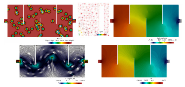
Flow on a maze-shaped domain. Heterogeneous permeability distribution, zoom to visualize the simplicial barycentric trisected mesh, Cauchy stress magnitude, first post-process of velocity, and post-processed pressure
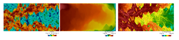
Channel flow with permeability from the SPE10–layer 45 benchmark data, and using a twice quadrisected crisscrossed mesh. Heterogeneous permeability distribution in log scale, Cauchy stress magnitude in log scale, and line integral convolution of second post-process of velocity in log scale
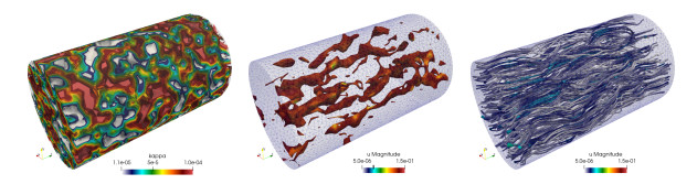
Channel flow with synthetic permeability. The mesh is of simplicial barycentric quadrisected type. Heterogeneous permeability distribution, contour iso-surfaces of velocity magnitude in log scale, and velocity streamlines