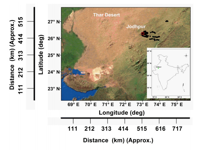
We evaluate temperature and humidity profiles retrieved from an INSAT-3DR sounder against radiosonde measurements for one year during 2017–2018. This evaluation is carried out in terms of slope, intercept, bias, Root Mean Square Error (RMSE), and correlation coefficient in temperature and relative humidity profiles. This comparison provided first-hand information about the performance of INSAT-3DR sounder retrievals. This validation exercise is unique in terms of comparing with the high resolution (1 Hz) radiosonde measurements performed in the afternoon time when there is maximum convection driven by solar radiation. 276 pairs of co-located temperatures have been compared. INSAT-3DR temperature retrievals are strongly linearly correlated to radiosonde measurements, with the slope as 1.001 and correlation coefficient of 0.99, while the mean temperature bias between INSAT-3DR and radiosonde is -0.23°C with RMSE of 1.9°C. Comparison of Relative humidity retrieved from INSAT-3DR to the radiosonde data results in RMSE of 9.8% with a slope of 0.99 with a correlation coefficient of 0.77. The collocation of data points for validation is done matching in space [Latitude, Longitude, Pressure level (height)] and time (±30 minutes). The effect of relaxing the space and time window on the validation statistics is also studied for the first time.
Citation: Hareef Baba Shaeb Kannemadugu, Manish Verma, Dibyendu Dutta, Lianne Rachel Johnson, Srinivasa Rao, Seshasai MVR. Comparison of temperature and humidity profiles retrieved from INSAT-3DR sounder with high resolution radiosonde measurements[J]. AIMS Geosciences, 2021, 7(2): 180-193. doi: 10.3934/geosci.2021011
We evaluate temperature and humidity profiles retrieved from an INSAT-3DR sounder against radiosonde measurements for one year during 2017–2018. This evaluation is carried out in terms of slope, intercept, bias, Root Mean Square Error (RMSE), and correlation coefficient in temperature and relative humidity profiles. This comparison provided first-hand information about the performance of INSAT-3DR sounder retrievals. This validation exercise is unique in terms of comparing with the high resolution (1 Hz) radiosonde measurements performed in the afternoon time when there is maximum convection driven by solar radiation. 276 pairs of co-located temperatures have been compared. INSAT-3DR temperature retrievals are strongly linearly correlated to radiosonde measurements, with the slope as 1.001 and correlation coefficient of 0.99, while the mean temperature bias between INSAT-3DR and radiosonde is -0.23°C with RMSE of 1.9°C. Comparison of Relative humidity retrieved from INSAT-3DR to the radiosonde data results in RMSE of 9.8% with a slope of 0.99 with a correlation coefficient of 0.77. The collocation of data points for validation is done matching in space [Latitude, Longitude, Pressure level (height)] and time (±30 minutes). The effect of relaxing the space and time window on the validation statistics is also studied for the first time.

| [1] | GCOS, The Global observing system for climate: Implementation needs, 2016. Available from: https://unfccc.int. |
| [2] |
Kaplan LD (1959) Inference of atmospheric structure from remote radiation measurements. J OPT Soc Am 49: 1004-1007. doi: 10.1364/JOSA.49.001004

|
| [3] | Houghton JT, Smith SD (1966) Infrared Physics, Oxford: Oxford University Press. |
| [4] | Peckham G, Rodgers CD, Houghton JT, et al. (1966) Remote Temperature Sensing of the Earth's Atmosphere Using a Selective Chopper Radiometer, Proceedings of the Symposium on Electromagnetic Sensing of the Earth from Satellites, Miami Beach, Florida. |
| [5] |
Ellis P, Holah G, Houghton JT, et al. (1973) Remote Sounding of Atmospheric Temperature from Satellites IV. The Selective Chopper Radiometer for Nimbus 5. Pro R Soc London Ser A 334: 149-170. doi: 10.1098/rspa.1973.0085
|
| [6] |
Cracknell AP, Varotsos CA (2021) Editorial Sir John Houghton. Remote Sensing Letters 12: 364-376. doi: 10.1080/2150704X.2021.1881649
|
| [7] |
Aumann HH, Chahine MT, Gautier C, et al. (2003) AIRS/AMSU/HSB on the AQUA Mission: Design, Science Objectives, Data Products, and Processing Systems. IEEE Trans Geosci Remote Sens 41: 253-264. doi: 10.1109/TGRS.2002.808356
|
| [8] | Blumstein D, Chalon G, Carlier T, et al. (2004) IASI Instrument: Technical Overview and Measured Performances. SPIE 5543: 196. |
| [9] |
Han Y, Rivercomb H, Cromp M, et al. (2013) Suomi NPP Crls measurements, sensor data record algorithm, calibration and validation activities, and record data quality. J Geophys Res Atmos 118: 12734-12748. doi: 10.1002/2013JD020344
|
| [10] |
Cracknell AP, Varotsos CA (2011) New Aspects of Global Climate-dynamics Research and Remote Sensing Preface. Int J Remote Sens 32: 579-600. doi: 10.1080/01431161.2010.517807
|
| [11] | Mitra A, Bhan S, Sharma A, et al. (2015) INSAT-3D vertical profile retrievals at IMDPS, New Delhi. Mausam 66: 687-694. |
| [12] |
Ratnam MV, Kumar AH, Jayaraman A (2016) Validation of INSAT-3D sounder data with in situ measurements and other similar satellite observations over India. Atmos Meas Tech 9: 5735-5745. doi: 10.5194/amt-9-5735-2016
|
| [13] |
Singh T, Mittal R, Shukla MV (2017) Validation of INSAT-3D temperature and moisture sounding retrievals using matched radiosonde measurements. Int J Remote Sens 3: 3333-3355. doi: 10.1080/01431161.2017.1294776
|
| [14] |
Durre I, Yin X (2008) Enhanced radiosonde data for studies of vertical structure. Bull Amer Meteor Soc 89: 1257-1262. doi: 10.1175/2008BAMS2603.1
|
| [15] |
Durre I, Yin, X, Vose RS, et al. (2018) Enhancing the Data Coverage in the Integrated Global Radiosonde Archive. J Atmos Oceanic Technol 35: 1753-1770. doi: 10.1175/JTECH-D-17-0223.1
|
| [16] | Satyanarayana S (2008) GPS radiosonde development. Technology Development for Atmospheric Research and Applications, ISRO, 142-188. |
| [17] |
Divya US, Girija J, Satyanarayana S, et al. (2014) An Advanced Radiosonde System for Aerospace Applications. J Atmos Oceanic Technol 31: 2067-2077. doi: 10.1175/JTECH-D-13-00050.1
|
| [18] |
Nair SK, Anurose TJ, Subrahamanyam DB, et al. (2011) Characterization of the vertical structure of coastal atmospheric boundary layer (cabl) over Thumba (8.5N, 76.9E) during different seasons. Adv Meteorol 2011: 1-9. doi: 10.1155/2011/390826
|
| [19] |
Kompalli SK, Suresh Babu S, Krishna Moorthy K, et al. (2014) Aerosol black carbon characteristics over Central India: Temporal variation and its dependence on mixed layer height. Atmos Res 147-148: 27-37. doi: 10.1016/j.atmosres.2014.04.015
|
| [20] | Kannemadugu HBS (2019) Seasonal Characteristics of Atmospheric Boundary Layer and its Associated Dynamics over Central India. Asia-Pacific J Atmos Sci. |
| [21] | Space Application Centre, Ahmedabad, India. INSAT-3D Algorithm Theoretical Basis Development Document (ATBD), 2015. Available from: http://mosdac.gov.in/data/doc/INSAT_3D_ATBD_MAY_2015.pdf. |
| [22] |
Li J, Wolf WW, Menzel WP, et al. (2000) Global soundings of the atmosphere from ATOVS measurements: The algorithm and validation. J Appl Meteorol 39: 1248-1268. doi: 10.1175/1520-0450(2000)039<1248:GSOTAF>2.0.CO;2
|
| [23] |
Ma XL, Schmit T, Smith W (1999) A non-linear physical retrieval algorithm-Its application to the GOES-8/9 sounder. J Appl Meteor 38: 501-503. doi: 10.1175/1520-0450(1999)038<0501:ANPRAI>2.0.CO;2
|
| [24] |
Hayden CM (1988) GOES-VAS simultaneous temperature-moisture retrieval algorithm. J Appl Meteor 27: 705-733. doi: 10.1175/1520-0450(1988)027<0705:GVSTMR>2.0.CO;2
|
| [25] |
Suneeth KV, Das SS, Das SK (2017) Diurnal variability of the global tropical tropopause: results inferred from COSMIC observations. Clim Dyn 49: 3277-3292. doi: 10.1007/s00382-016-3512-x
|
| [26] |
Meza A, Mendoza MP, Natali C, et al. (2020) Diurnal variation of precipitable water vapor over Central and South America. Geod Geodyn 11: 426-441. doi: 10.1016/j.geog.2020.04.005
|
| [27] |
Efstathiou MN, Varotsos CA (2010) On the altitude dependence of the temperature scaling behaviour at the global troposphere. Int J Remote Sens 31: 343-349. doi: 10.1080/01431160902882702
|
| [28] |
Vömel H, Selkirk H, Miloshecich, L, et al. (2007) Radiation dry bias of the Vaisala RS92 humidity sensor. J Atmos Oceanic Technol 24: 953-963. doi: 10.1175/JTECH2019.1
|


Figures(5) / Tables(2)

Hareef Baba Shaeb Kannemadugu, Manish Verma, Dibyendu Dutta, Lianne Rachel Johnson, Srinivasa Rao, Seshasai MVR. Comparison of temperature and humidity profiles retrieved from INSAT-3DR sounder with high resolution radiosonde measurements[J]. AIMS Geosciences, 2021, 7(2): 180-193. doi: 10.3934/geosci.2021011


 DownLoad: