Citation: Navid J. Ayon. Features, roles and chiral analyses of proteinogenic amino acids[J]. AIMS Molecular Science, 2020, 7(3): 229-268. doi: 10.3934/molsci.2020011

| [1] | Alberts B, Johnson A, Lewis J, et al. (2002) Molecular Biology of the Cell New York: Garland Science. |
| [2] |
Boyle J (2005) Lehninger principles of biochemistry (4th ed.): Nelson, D., and Cox, M. Biochem Mol Biol Educ 33: 74-75. doi: 10.1002/bmb.2005.494033010419

|
| [3] | Gutiérrez-Preciado A, Romero H, Peimbert M (2010) An Evolutionary Perspective on Amino Acids. Nature Education 3. |
| [4] |
Ambrogelly A, Palioura S, Söll D (2007) Natural expansion of the genetic code. Nat Chem Biol 3: 29-35. doi: 10.1038/nchembio847
|
| [5] |
Johansson L, Gafvelin G, Arnér ESJ (2005) Selenocysteine in proteins—properties and biotechnological use. Biochim Biophys Acta 1726: 1-13. doi: 10.1016/j.bbagen.2005.05.010
|
| [6] |
Rother M, Krzycki JA (2010) Selenocysteine, Pyrrolysine, and the Unique Energy Metabolism of Methanogenic Archaea. Archaea 2010: 453642. doi: 10.1155/2010/453642
|
| [7] |
Frenkel-Pinter M, Haynes JW, C M, et al. (2019) Selective incorporation of proteinaceous over nonproteinaceous cationic amino acids in model prebiotic oligomerization reactions. Proc Natl Acad Sci 116: 16338. doi: 10.1073/pnas.1904849116
|
| [8] |
Wu G (2009) Amino acids: metabolism, functions, and nutrition. Amino Acids 37: 1-17. doi: 10.1007/s00726-009-0269-0
|
| [9] | Ulbricht J (2018) Insights into Polymer Biodegradation - Investigations on oxidative, hydrolytic and enzymatic Pathways. |
| [10] |
Kaspar H, Dettmer K, Gronwald W, et al. (2008) Automated GC–MS analysis of free amino acids in biological fluids. J Chromatogr B 870: 222-232. doi: 10.1016/j.jchromb.2008.06.018
|
| [11] |
Armstrong DW (1984) Chiral Stationary Phases for High Performance Liquid Chromatographic Separation of Enantiomers: A Mini-Review. J Liq Chromatogr 7: 353-376. doi: 10.1080/01483918408073942
|
| [12] |
Prinsen H, Schiebergen-Bronkhorst BGM, Roeleveld MW, et al. (2016) Rapid quantification of underivatized amino acids in plasma by hydrophilic interaction liquid chromatography (HILIC) coupled with tandem mass-spectrometry. J Inherit Metab Dis 39: 651-660. doi: 10.1007/s10545-016-9935-z
|
| [13] |
Marcone GL, Rosini E, Crespi E, et al. (2020) D-amino acids in foods. Appl Microbiol Biotechnol 104: 555-574. doi: 10.1007/s00253-019-10264-9
|
| [14] |
Koga R, Yoshida H, Nohta H, et al. (2019) Multi-Dimensional HPLC Analysis of Metabolic Related Chiral Amino Acids -Method Development and Biological/Clinical Applications. Chromatogr 40: 1-8. doi: 10.15583/jpchrom.2019.002
|
| [15] |
Domínguez-Vega E, Crego AL, Lomsadze K, et al. (2011) Enantiomeric separation of FMOC-amino acids by nano-LC and CEC using a new chiral stationary phase, cellulose tris(3-chloro-4-methylphenylcarbamate). Electrophor 32: 2700-2707. doi: 10.1002/elps.201000701
|
| [16] |
Asbury GR, Hill HH (2000) Separation of amino acids by ion mobility spectrometry. J Chromatogr A 902: 433-437. doi: 10.1016/S0021-9673(00)00799-8
|
| [17] |
Nemkov T, D'Alessandro A, Hansen KC (2015) Three-minute method for amino acid analysis by UHPLC and high-resolution quadrupole orbitrap mass spectrometry. Amino acids 47: 2345-2357. doi: 10.1007/s00726-015-2019-9
|
| [18] |
Song Y, Xu C, Kuroki H, et al. (2018) Recent trends in analytical methods for the determination of amino acids in biological samples. J Pharm Biomed Anal 147: 35-49. doi: 10.1016/j.jpba.2017.08.050
|
| [19] |
Pisarewicz K, Mora D, Pflueger FC, et al. (2005) Polypeptide Chains Containing d-γ-Hydroxyvaline. J Am Chem Soc 127: 6207-6215. doi: 10.1021/ja050088m
|
| [20] |
Vollmer W, Blanot D, De Pedro MA (2008) Peptidoglycan structure and architecture. FEMS Microbiol Rev 32: 149-167. doi: 10.1111/j.1574-6976.2007.00094.x
|
| [21] |
Golovanov AP, Hautbergue GM, Wilson SA, et al. (2004) A Simple Method for Improving Protein Solubility and Long-Term Stability. J Am Chem Soc 126: 8933-8939. doi: 10.1021/ja049297h
|
| [22] |
Kato M, Yamazaki T, Kato H, et al. (2017) Effects of the pH and Concentration on the Stability of Standard Solutions of Proteinogenic Amino Acid Mixtures. Anal Sci 33: 1241-1245. doi: 10.2116/analsci.33.1241
|
| [23] |
Rowe L, Peller J, Mammoser C, et al. (2019) Stability of non-proteinogenic amino acids to UV and gamma irradiation. Int J Astrobiol 18: 426-435. doi: 10.1017/S1473550418000381
|
| [24] | Cataldo F, Iglesias-Groth S, Angelini G, et al. (2013) Stability toward High Energy Radiation of Non-Proteinogenic Amino Acids: Implications for the Origins of Life. Life (Basel) 3: 449-473. |
| [25] | Kendall C, McKenzie BF (1929) dl-Alanine. Organic Synthesis 9. |
| [26] | Litwack G (2018) Chapter 4 - Proteins. Human Biochemistry Boston: Academic Press, 63-94. |
| [27] | Meister A (1965) Biochemistry of the Amino Acids Academic Press. |
| [28] |
Wendisch VF (2007) Amino Acid Biosynthesis – Pathways, Regulation and Metabolic Engineering Springer Berlin Heidelberg. doi: 10.1007/978-3-540-48596-4
|
| [29] |
Shapiro BM, Stadtman ER (1970) The Regulation of Glutamine Synthesis in Microorganisms. Annu Rev Microbiol 24: 501-524. doi: 10.1146/annurev.mi.24.100170.002441
|
| [30] |
Ljungdahl PO, Daignan-Fornier B (2012) Regulation of Amino Acid, Nucleotide, and Phosphate Metabolism in Saccharomyces cerevisiae. Genetics 190: 885. doi: 10.1534/genetics.111.133306
|
| [31] |
Gutteridge A, Thornton JM (2005) Understanding nature's catalytic toolkit. Trends Biochem Sci 30: 622-629. doi: 10.1016/j.tibs.2005.09.006
|
| [32] |
Fichtner M, Voigt K, Schuster S (2017) The tip and hidden part of the iceberg: Proteinogenic and non-proteinogenic aliphatic amino acids. Biochim Biophys Acta 1861: 3258-3269. doi: 10.1016/j.bbagen.2016.08.008
|
| [33] |
Shelp BJ, Bown AW, McLean MD (1999) Metabolism and functions of gamma-aminobutyric acid. Trends Plant Sci 4: 446-452. doi: 10.1016/S1360-1385(99)01486-7
|
| [34] |
Tejero J, Biswas A, Wang Z-Q, et al. (2008) Stabilization and characterization of a heme-oxy reaction intermediate in inducible nitric-oxide synthase. J Biol Chem 283: 33498-33507. doi: 10.1074/jbc.M806122200
|
| [35] |
Bradley Levine A, Levine AB, Punihaole D, et al. (2012) Characterization of the Role of Nitric Oxide and Its Clinical Applications. Cardiology 122: 55-68. doi: 10.1159/000338150
|
| [36] |
Suárez-Marina I, Abul-Haija YM, Turk-MacLeod R, et al. (2019) Integrated synthesis of nucleotide and nucleosides influenced by amino acids. Commun Chem 2: 28. doi: 10.1038/s42004-019-0130-7
|
| [37] | Shemin D, Rittenberg D (1946) The biological utilization of glycine for the synthesis of the protoporphyrin of hemoglobin. J Biol Chem 166: 621-625. |
| [38] |
Fahn S, Jankovic J, Hallett M (2011) Chapter 20 - Myoclonus: Phenomenology, etiology, physiology, and treatment. Principles and Practice of Movement Disorders Edinburgh: W.B. Saunders, 447-464. doi: 10.1016/B978-1-4377-2369-4.00020-2
|
| [39] |
Whillier S, Garcia B, Chapman BE, et al. (2011) Glutamine and alpha-ketoglutarate as glutamate sources for glutathione synthesis in human erythrocytes. Febs J 278: 3152-3163. doi: 10.1111/j.1742-4658.2011.08241.x
|
| [40] | Brady S, Siegel G, Albers RW, et al. (2012) Basic Neurochemistry: Principles of Molecular, Cellular, and Medical Neurobiology Elsevier Science. |
| [41] |
Ducker GS, Rabinowitz JD (2017) One-Carbon Metabolism in Health and Disease. Cell Metab 25: 27-42. doi: 10.1016/j.cmet.2016.08.009
|
| [42] |
Bardal SK, Waechter JE, Martin DS (2011) Chapter 20 - Neoplasia. Applied Pharmacology Philadelphia: Content Repository Only, 305-324. doi: 10.1016/B978-1-4377-0310-8.00020-8
|
| [43] |
Barocelli E, Ballabeni V (2003) Histamine in the control of gastric acid secretion: a topic review. Pharmacol Res 47: 299-304. doi: 10.1016/S1043-6618(03)00009-4
|
| [44] |
Ashina K, Tsubosaka Y, Nakamura T, et al. (2015) Histamine Induces Vascular Hyperpermeability by Increasing Blood Flow and Endothelial Barrier Disruption In Vivo. PLoS One 10: e0132367-e0132367. doi: 10.1371/journal.pone.0132367
|
| [45] |
White MV (1990) The role of histamine in allergic diseases. J Allergy Clin Immunol 86: 599-605. doi: 10.1016/S0091-6749(05)80223-4
|
| [46] |
Zeisel SH, Blusztajn JK (1994) Choline and human nutrition. Annu Rev Nutr 14: 269-296. doi: 10.1146/annurev.nu.14.070194.001413
|
| [47] |
Wu G, Bazer FW, Burghardt RC, et al. (2011) Proline and hydroxyproline metabolism: implications for animal and human nutrition. Amino acids 40: 1053-1063. doi: 10.1007/s00726-010-0715-z
|
| [48] |
Puglisi-Allegra S, Ventura R (2012) Prefrontal/accumbal catecholamine system processes high motivational salience. Front Behav Neurosci 6: 31. doi: 10.3389/fnbeh.2012.00031
|
| [49] |
Axelsson J (1971) Catecholamine Functions. Annu Rev Physiol 33: 1-30. doi: 10.1146/annurev.ph.33.030171.000245
|
| [50] | Irsfeld M, Spadafore M, Prüß BM (2013) β-phenylethylamine, a small molecule with a large impact. Webmedcentral 4: 4409. |
| [51] |
Solecka D (1997) Role of phenylpropanoid compounds in plant responses to different stress factors. Acta Physiol Plant 19: 257-268. doi: 10.1007/s11738-997-0001-1
|
| [52] | Pawelek JM, Körner AM (1982) The Biosynthesis of Mammalian Melanin: The regulation of pigment formation, the key to disorders such as albinism and piebaldism, may also offer some clues for the treatment of melanoma. Am Sci 70: 136-145. |
| [53] |
Savelieva KV, Zhao S, Pogorelov VM, et al. (2008) Genetic Disruption of Both Tryptophan Hydroxylase Genes Dramatically Reduces Serotonin and Affects Behavior in Models Sensitive to Antidepressants. PLoS One 3: e3301. doi: 10.1371/journal.pone.0003301
|
| [54] |
Dhawan PS, Goodman BP (2014) Chapter 15 - Neurologic Manifestations of Nutritional Disorders. Aminoff's Neurology and General Medicine Boston: Academic Press, 273-290. doi: 10.1016/B978-0-12-407710-2.00015-1
|
| [55] |
Newsholme EA, Blomstrand E (1996) The plasma level of some amino acids and physical and mental fatigue. Experientia 52: 413-415. doi: 10.1007/BF01919308
|
| [56] | Voet D, Voet JG (1990) Biochemistry: Wiley. |
| [57] |
Maeda J, Higashiyama M, Imaizumi A, et al. (2010) Possibility of multivariate function composed of plasma amino acid profiles as a novel screening index for non-small cell lung cancer: a case control study. BMC Cancer 10: 690. doi: 10.1186/1471-2407-10-690
|
| [58] |
Kakazu E, Kanno N, Ueno Y, et al. (2007) Extracellular branched-chain amino acids, especially valine, regulate maturation and function of monocyte-derived dendritic cells. J Immunol 179: 7137-7146. doi: 10.4049/jimmunol.179.10.7137
|
| [59] |
Jagenburg OR (1965) Disturbances of amino acid metabolism in neurologic disorders. Acta Neurol Scand 41: 339-356. doi: 10.1111/j.1600-0404.1965.tb01896.x
|
| [60] | Griffin JWD, Bradshaw PC (2017) Amino Acid Catabolism in Alzheimer's Disease Brain: Friend or Foe? Oxid Med Cell Longevity 2017: 5472792. |
| [61] |
Sarchielli P, Greco L, Floridi A, et al. (2003) Excitatory Amino Acids and Multiple Sclerosis: Evidence From Cerebrospinal Fluid. Arch Neurol 60: 1082-1088. doi: 10.1001/archneur.60.8.1082
|
| [62] |
Figura M, Kuśmierska K, Bucior E, et al. (2018) Serum amino acid profile in patients with Parkinson's disease. PLoS One 13: e0191670. doi: 10.1371/journal.pone.0191670
|
| [63] |
Piraud M, Ruet S, Boyer S, et al. (2011) Amino acid profiling for the diagnosis of inborn errors of metabolism. Methods Mol Biol 708: 25-53. doi: 10.1007/978-1-61737-985-7_2
|
| [64] |
Hildebrandt Tatjana M, Nunes Nesi A, Araújo Wagner L, et al. (2015) Amino Acid Catabolism in Plants. Mol Plant 8: 1563-1579. doi: 10.1016/j.molp.2015.09.005
|
| [65] |
Pratelli R, Pilot G (2014) Regulation of amino acid metabolic enzymes and transporters in plants. J Exp Bot 65: 5535-5556. doi: 10.1093/jxb/eru320
|
| [66] | Forsum O, Svennerstam H, Ganeteg U, et al. (2008) Capacities and constraints of amino acid utilization in Arabidopsis. New Phytol 179: 1058-1069. |
| [67] |
Chen IC, Thiruvengadam V, Lin WD, et al. (2010) Lysine racemase: a novel non-antibiotic selectable marker for plant transformation. Plant Mol Biol 72: 153-169. doi: 10.1007/s11103-009-9558-y
|
| [68] |
Massey KA, Blakeslee CH, Pitkow HS (1998) A review of physiological and metabolic effects of essential amino acids. Amino Acids 14: 271-300. doi: 10.1007/BF01318848
|
| [69] |
Kimura T, Hamase K, Miyoshi Y, et al. (2016) Chiral amino acid metabolomics for novel biomarker screening in the prognosis of chronic kidney disease. Sci Rep 6: 26137. doi: 10.1038/srep26137
|
| [70] |
Klupczynska A, Dereziński P, Dyszkiewicz W, et al. (2016) Evaluation of serum amino acid profiles' utility in non-small cell lung cancer detection in Polish population. Lung Cancer 100: 71-76. doi: 10.1016/j.lungcan.2016.04.008
|
| [71] |
Virmani K, Widhalm K (1993) Histidinemia: a biochemical variant or a disease? J Am Coll Nutr 12: 115-124. doi: 10.1080/07315724.1993.10718291
|
| [72] |
Maltais-Payette I, Boulet M-M, Prehn C, et al. (2018) Circulating glutamate concentration as a biomarker of visceral obesity and associated metabolic alterations. Nutr Metab 15: 78. doi: 10.1186/s12986-018-0316-5
|
| [73] |
De Luca V, Viggiano E, Messina G, et al. (2008) Peripheral amino Acid levels in schizophrenia and antipsychotic treatment. Psychiatry Invest 5: 203-208. doi: 10.4306/pi.2008.5.4.203
|
| [74] | Sebastio G, Sperandeo MP, Panico M, et al. (1995) The molecular basis of homocystinuria due to cystathionine beta-synthase deficiency in Italian families, and report of four novel mutations. Am J Hum Genet 56: 1324-1333. |
| [75] |
Guttler F, Guldberg P (1994) Mutations in the phenylalanine hydroxylase gene: genetic determinants for the phenotypic variability of hyperphenylalaninemia. Acta Paediatr Suppl 407: 49-56. doi: 10.1111/j.1651-2227.1994.tb13451.x
|
| [76] | Mamer OA, Reimer ML (1992) On the mechanisms of the formation of L-alloisoleucine and the 2-hydroxy-3-methylvaleric acid stereoisomers from L-isoleucine in maple syrup urine disease patients and in normal humans. J Biol Chem 267: 22141-22147. |
| [77] |
Smith AM, King JJ, West PR, et al. (2019) Amino Acid Dysregulation Metabotypes: Potential Biomarkers for Diagnosis and Individualized Treatment for Subtypes of Autism Spectrum Disorder. Biol Psychiatry 85: 345-354. doi: 10.1016/j.biopsych.2018.08.016
|
| [78] |
Alborghetti MR, Correa MEP, Whangbo J, et al. (2019) Clinical Metabolomics Identifies Blood Serum Branched Chain Amino Acids as Potential Predictive Biomarkers for Chronic Graft vs. Host Disease. Front Oncol 9: 141. doi: 10.3389/fonc.2019.00141
|
| [79] |
Liao X, Liu B, Qu H, et al. (2019) A High Level of Circulating Valine Is a Biomarker for Type 2 Diabetes and Associated with the Hypoglycemic Effect of Sitagliptin. Mediators Inflammation 2019: 8247019. doi: 10.1155/2019/8247019
|
| [80] |
Baranyi A, Amouzadeh-Ghadikolai O, von Lewinski D, et al. (2016) Branched-Chain Amino Acids as New Biomarkers of Major Depression - A Novel Neurobiology of Mood Disorder. PLoS One 11: e0160542. doi: 10.1371/journal.pone.0160542
|
| [81] |
Li N-j, Liu W-t, Li W, et al. (2010) Plasma metabolic profiling of Alzheimer's disease by liquid chromatography/mass spectrometry. Clin Biochem 43: 992-997. doi: 10.1016/j.clinbiochem.2010.04.072
|
| [82] |
Corso G, Cristofano A, Sapere N, et al. (2017) Serum Amino Acid Profiles in Normal Subjects and in Patients with or at Risk of Alzheimer Dementia. Dement Geriatr Cogn Dis Extra 7: 143-159. doi: 10.1159/000466688
|
| [83] |
Hou Y, Yin Y, Wu G (2015) Dietary essentiality of “nutritionally non-essential amino acids” for animals and humans. Exp Biol Med (Maywood, NJ) 240: 997-1007. doi: 10.1177/1535370215587913
|
| [84] |
Choi B-H, Coloff JL (2019) The Diverse Functions of Non-Essential Amino Acids in Cancer. Cancers 11: 675. doi: 10.3390/cancers11050675
|
| [85] |
Hishikawa K, Nakaki T, Suzuki H, et al. (1992) L-arginine as an antihypertensive agent. J Cardiovasc Pharmacol 20: S196-197. doi: 10.1097/00005344-199204002-00055
|
| [86] |
Bellinghieri G, Santoro D, Mallamace A, et al. (2006) L-arginine: a new opportunity in the management of clinical derangements in dialysis patients. J Ren Nutr 16: 245-247. doi: 10.1053/j.jrn.2006.04.004
|
| [87] |
Coburn LA, Gong X, Singh K, et al. (2012) L-arginine supplementation improves responses to injury and inflammation in dextran sulfate sodium colitis. PLoS One 7: e33546-e33546. doi: 10.1371/journal.pone.0033546
|
| [88] |
Visser JJ, Hoekman K (1994) Arginine supplementation in the prevention and treatment of osteoporosis. Med Hypotheses 43: 339-342. doi: 10.1016/0306-9877(94)90113-9
|
| [89] |
Hurt RT, Ebbert JO, Schroeder DR, et al. (2014) L-arginine for the treatment of centrally obese subjects: a pilot study. J Diet Suppl 11: 40-52. doi: 10.3109/19390211.2013.859216
|
| [90] |
Wang J, Chen L, Li P, et al. (2008) Gene expression is altered in piglet small intestine by weaning and dietary glutamine supplementation. J Nutr 138: 1025-1032. doi: 10.1093/jn/138.6.1025
|
| [91] |
Ren W, Liu S, Chen S, et al. (2013) Dietary L-glutamine supplementation increases Pasteurella multocida burden and the expression of its major virulence factors in mice. Amino Acids 45: 947-955. doi: 10.1007/s00726-013-1551-8
|
| [92] |
Wang H, Zhang C, Wu G, et al. (2015) Glutamine enhances tight junction protein expression and modulates corticotropin-releasing factor signaling in the jejunum of weanling piglets. J Nutr 145: 25-31. doi: 10.3945/jn.114.202515
|
| [93] |
Wang W, Dai Z, Wu Z, et al. (2014) Glycine is a nutritionally essential amino acid for maximal growth of milk-fed young pigs. Amino Acids 46: 2037-2045. doi: 10.1007/s00726-014-1758-3
|
| [94] |
Cai Q, Takemura G, Ashraf M (1995) Antioxidative properties of histidine and its effect on myocardial injury during ischemia/reperfusion in isolated rat heart. J Cardiovasc Pharmacol 25: 147-155. doi: 10.1097/00005344-199501000-00023
|
| [95] |
Griffith RS, Walsh DE, Myrmel KH, et al. (1987) Success of L-lysine therapy in frequently recurrent herpes simplex infection. Treatment and prophylaxis. Dermatologica 175: 183-190. doi: 10.1159/000248823
|
| [96] | Wright EF (1994) Clinical effectiveness of lysine in treating recurrent aphthous ulcers and herpes labialis. Gen Dent 42: 40-42. |
| [97] | Krymchantowski AV, Peixoto P, Higashi R, et al. (2005) Lysine clonixinate vs naproxen sodium for the acute treatment of migraine: a double-blind, randomized, crossover study. MedGenMed 7: 69. |
| [98] |
Mato JM, Martínez-Chantar ML, Lu SC (2013) S-adenosylmethionine metabolism and liver disease. Ann Hepatol 12: 183-189. doi: 10.1016/S1665-2681(19)31355-9
|
| [99] | Patrick L (2003) Toxic metals and antioxidants: Part II. The role of antioxidants in arsenic and cadmium toxicity. Altern Med Rev 8: 106-128. |
| [100] |
Benavides MA, Bosland MC, da Silva CP, et al. (2014) L-Methionine inhibits growth of human pancreatic cancer cells. Anti-Cancer Drugs 25: 200-203. doi: 10.1097/CAD.0000000000000038
|
| [101] |
Cavuoto P, Fenech MF (2012) A review of methionine dependency and the role of methionine restriction in cancer growth control and life-span extension. Cancer Treat Rev 38: 726-736. doi: 10.1016/j.ctrv.2012.01.004
|
| [102] |
Cipollone D, Carsetti R, Tagliani A, et al. (2009) Folic acid and methionine in the prevention of teratogen-induced congenital defects in mice. Cardiovasc Pathol 18: 100-109. doi: 10.1016/j.carpath.2008.02.007
|
| [103] |
Swanson RA, Shiraishi K, Morton MT, et al. (1990) Methionine sulfoximine reduces cortical infarct size in rats after middle cerebral artery occlusion. Stroke 21: 322-327. doi: 10.1161/01.STR.21.2.322
|
| [104] |
Wang L, Qu W, Lieberman BP, et al. (2011) Synthesis, uptake mechanism characterization and biological evaluation of (18)F labeled fluoroalkyl phenylalanine analogs as potential PET imaging agents. Nucl Med Biol 38: 53-62. doi: 10.1016/j.nucmedbio.2010.07.005
|
| [105] |
Hanaoka H, Ohshima Y, Suzuki Y, et al. (2015) Development of a Widely Usable Amino Acid Tracer: (7)(6)Br-alpha-Methyl-Phenylalanine for Tumor PET Imaging. J Nucl Med 56: 791-797. doi: 10.2967/jnumed.114.152215
|
| [106] |
Ren W, Zou L, Ruan Z, et al. (2013) Dietary L-proline supplementation confers immunostimulatory effects on inactivated Pasteurella multocida vaccine immunized mice. Amino Acids 45: 555-561. doi: 10.1007/s00726-013-1490-4
|
| [107] |
Heresco-Levy U, Vass A, Bloch B, et al. (2009) Pilot controlled trial of d-serine for the treatment of post-traumatic stress disorder. Int J Neuropsychopharmacol 12: 1275-1282. doi: 10.1017/S1461145709000339
|
| [108] |
MacKay M-AB, Paylor JW, Wong JTF, et al. (2018) Multidimensional Connectomics and Treatment-Resistant Schizophrenia: Linking Phenotypic Circuits to Targeted Therapeutics. Front Psychiatry 9: 537. doi: 10.3389/fpsyt.2018.00537
|
| [109] |
Growdon JH, Nader TM, Schoenfeld J, et al. (1991) L-threonine in the treatment of spasticity. Clin Neuropharmacol 14: 403-412. doi: 10.1097/00002826-199110000-00003
|
| [110] |
Hauser SL, Doolittle TH, Lopez-Bresnahan M, et al. (1992) An antispasticity effect of threonine in multiple sclerosis. Arch Neurol 49: 923-926. doi: 10.1001/archneur.1992.00530330045014
|
| [111] |
Jenkins TA, Nguyen JCD, Polglaze KE, et al. (2016) Influence of Tryptophan and Serotonin on Mood and Cognition with a Possible Role of the Gut-Brain Axis. Nutrients 8: 56. doi: 10.3390/nu8010056
|
| [112] |
Richard DM, Dawes MA, Mathias CW, et al. (2009) L-Tryptophan: Basic Metabolic Functions, Behavioral Research and Therapeutic Indications. Int J Tryptophan Res 2: 45-60. doi: 10.4137/IJTR.S2129
|
| [113] | Leja-Szpak A, Jaworek J, Nawrot-Porabka K, et al. (2004) Modulation of pancreatic enzyme secretion by melatonin and its precursor; L-tryptophan. Role of CCK and afferent nerves. J Physiol Pharmacol 55: 33-46. |
| [114] |
Fernstrom JD (2005) Branched-Chain Amino Acids and Brain Function. J Nutr 135: 1539S-1546S. doi: 10.1093/jn/135.6.1539S
|
| [115] |
Holeček M (2018) Branched-chain amino acids in health and disease: metabolism, alterations in blood plasma, and as supplements. Nutr Metab 15: 33. doi: 10.1186/s12986-018-0271-1
|
| [116] |
Norton LE, Layman DK (2006) Leucine regulates translation initiation of protein synthesis in skeletal muscle after exercise. J Nutr 136: 533s-537s. doi: 10.1093/jn/136.2.533S
|
| [117] | Karau A, Grayson I (2014) Amino acids in human and animal nutrition. Adv Biochem Eng Biotechnol 143: 189-228. |
| [118] |
Garattini S (2000) Glutamic Acid, Twenty Years Later. J Nutr 130: 901S-909S. doi: 10.1093/jn/130.4.901S
|
| [119] |
Stegink LD (1987) The aspartame story: a model for the clinical testing of a food additive. Am J Clin Nutr 46: 204-215. doi: 10.1093/ajcn/46.1.204
|
| [120] | Ashmead HDW (1986) Foliar feeding of plants with amino acid chelates: Noyes Publications. |
| [121] |
Leuchtenberger W, Huthmacher K, Drauz K (2005) Biotechnological production of amino acids and derivatives: current status and prospects. Appl Microbiol Biotechnol 69: 1-8. doi: 10.1007/s00253-005-0155-y
|
| [122] |
Sanda F, Endo T (1999) Syntheses and functions of polymers based on amino acids. Macromol Chem Phys 200: 2651-2661. doi: 10.1002/(SICI)1521-3935(19991201)200:12<2651::AID-MACP2651>3.0.CO;2-P
|
| [123] |
Gross RA, Kalra B (2002) Biodegradable Polymers for the Environment. Science 297: 803. doi: 10.1126/science.297.5582.803
|
| [124] | Low KC, Wheeler AP, Koskan LP (1996) Commercial Poly(aspartic acid) and Its Uses. Hydrophilic Polymers: American Chemical Society 99-111. |
| [125] |
Thombre SM, Sarwade BD (2005) Synthesis and Biodegradability of Polyaspartic Acid: A Critical Review. J Macromol Sci, Part A 42: 1299-1315. doi: 10.1080/10601320500189604
|
| [126] |
Bourke SL, Kohn J (2003) Polymers derived from the amino acid l-tyrosine: polycarbonates, polyarylates and copolymers with poly(ethylene glycol). Adv Drug Delivery Rev 55: 447-466. doi: 10.1016/S0169-409X(03)00038-3
|
| [127] |
Friedman M (1999) Chemistry, Nutrition, and Microbiology of d-Amino Acids. J Agric Food Chem 47: 3457-3479. doi: 10.1021/jf990080u
|
| [128] |
Fujii N (2002) D-Amino Acids in Living Higher Organisms. Orig Life Evol Biosph 32: 103-127. doi: 10.1023/A:1016031014871
|
| [129] |
Powell JT, Vine N, Crossman M (1992) On the accumulation of d-aspartate in elastin and other proteins of the ageing aorta. Atherosclerosis 97: 201-208. doi: 10.1016/0021-9150(92)90132-Z
|
| [130] |
Ritz-Timme S, Laumeier I, Collins MJ (2003) Aspartic acid racemization: evidence for marked longevity of elastin in human skin. Br J Dermatol 149: 951-959. doi: 10.1111/j.1365-2133.2003.05618.x
|
| [131] |
Fisher GH, Garcia NM, Payan IL, et al. (1986) D-aspartic acid in purified myelin and myelin basic protein. Biochem Biophys Res Commun 135: 683-687. doi: 10.1016/0006-291X(86)90047-1
|
| [132] |
Lee JM, Petrucelli L, Fisher G, et al. (2002) Evidence for D-aspartyl-beta-amyloid secretase activity in human brain. J Neuropathol Exp Neurol 61: 125-131. doi: 10.1093/jnen/61.2.125
|
| [133] |
Hashimoto A, Oka T (1997) Free D-aspartate and D-serine in the mammalian brain and periphery. Prog Neurobiol 52: 325-353. doi: 10.1016/S0301-0082(97)00019-1
|
| [134] |
Homma H (2007) Biochemistry of D-aspartate in mammalian cells. Amino Acids 32: 3-11. doi: 10.1007/s00726-006-0354-6
|
| [135] |
Topo E, Soricelli A, D'Aniello A, et al. (2009) The role and molecular mechanism of D-aspartic acid in the release and synthesis of LH and testosterone in humans and rats. Reprod Biol Endocrinol 7: 120. doi: 10.1186/1477-7827-7-120
|
| [136] |
Hooi MYS, Truscott RJW (2011) Racemisation and human cataract. D-Ser, D-Asp/Asn and D-Thr are higher in the lifelong proteins of cataract lenses than in age-matched normal lenses. Age (Dordrecht, Neth) 33: 131-141. doi: 10.1007/s11357-010-9171-7
|
| [137] |
Richter K, Egger R, Kreil G (1987) D-alanine in the frog skin peptide dermorphin is derived from L-alanine in the precursor. Science 238: 200-202. doi: 10.1126/science.3659910
|
| [138] |
Erspamer V, Melchiorri P, Falconieri-Erspamer G, et al. (1989) Deltorphins: a family of naturally occurring peptides with high affinity and selectivity for delta opioid binding sites. Proc Natl Acad Sci U S A 86: 5188-5192. doi: 10.1073/pnas.86.13.5188
|
| [139] |
Mor A, Delfour A, Sagan S, et al. (1989) Isolation of dermenkephalin from amphibian skin, a high-affinity delta-selective opioid heptapeptide containing a D-amino acid residue. FEBS Lett 255: 269-274. doi: 10.1016/0014-5793(89)81104-4
|
| [140] |
Kamatani Y, Minakata H, Kenny PT, et al. (1989) Achatin-I, an endogenous neuroexcitatory tetrapeptide from Achatina fulica Férussac containing a D-amino acid residue. Biochem Biophys Res Commun 160: 1015-1020. doi: 10.1016/S0006-291X(89)80103-2
|
| [141] |
Soyez D, Laverdure AM, Kallen J, et al. (1997) Demonstration of a cell-specific isomerization of invertebrate neuropeptides. Neurosci 82: 935-942. doi: 10.1016/S0306-4522(97)00254-6
|
| [142] |
Chung JS, Zmora N, Katayama H, et al. (2010) Crustacean hyperglycemic hormone (CHH) neuropeptidesfamily: Functions, titer, and binding to target tissues. Gen Comp Endocrinol 166: 447-454. doi: 10.1016/j.ygcen.2009.12.011
|
| [143] |
Kawamura I, Mijiddorj B, Kayano Y, et al. (2020) Separation of D-amino acid-containing peptide phenylseptin using 3,3′-phenyl-1,1′-binaphthyl-18-crown-6-ether columns. BBA Proteins Proteom 1868: 140429. doi: 10.1016/j.bbapap.2020.140429
|
| [144] |
Mijiddorj B, Kaneda S, Sato H, et al. (2018) The role of d-allo-isoleucine in the deposition of the anti-Leishmania peptide bombinin H4 as revealed by 31P solid-state NMR, VCD spectroscopy, and MD simulation. BBA Proteins Proteom 1866: 789-798. doi: 10.1016/j.bbapap.2018.01.005
|
| [145] |
Ohta N, Kubota I, Takao T, et al. (1991) Fulicin, a novel neuropeptide containing a D-amino acid residue isolated from the ganglia of Achatina fulica. Biochemi Biophys Res Commun 178: 486-493. doi: 10.1016/0006-291X(91)90133-R
|
| [146] |
Jimenez EC, Olivera BM, Gray WR, et al. (1996) Contryphan is a D-tryptophan-containing Conus peptide. J Biol Chem 271: 28002-28005. doi: 10.1074/jbc.271.45.28002
|
| [147] | Kuwada M, Teramoto T, Kumagaye KY, et al. (1994) Omega-agatoxin-TK containing D-serine at position 46, but not synthetic omega-[L-Ser46]agatoxin-TK, exerts blockade of P-type calcium channels in cerebellar Purkinje neurons. Mol Pharmacol 46: 587. |
| [148] |
Mandrioli R, Mercolini L, Raggi MA (2013) Recent trends in the analysis of amino acids in fruits and derived foodstuffs. Anal Bioanal Chem 405: 7941-7956. doi: 10.1007/s00216-013-7025-8
|
| [149] |
Zagon J, Dehne L-I, Bögl K-W (1994) D-amino acids in organisms and food. Nutr Res 14: 445-463. doi: 10.1016/S0271-5317(05)80182-4
|
| [150] |
Tian H, Zheng N, Li S, et al. (2017) Characterization of chiral amino acids from different milk origins using ultra-performance liquid chromatography coupled to ion-mobility mass spectrometry. Sci Rep 7: 46289. doi: 10.1038/srep46289
|
| [151] |
Gandolfi I, Palla G, Marchelli R, et al. (1994) D-alanine in Fruit Juices: A Molecular Marker of Bacterial Activity, Heat Treatments and Shelf-life. J Food Sci 59: 152-154. doi: 10.1111/j.1365-2621.1994.tb06921.x
|
| [152] |
Kimura T, Hesaka A, Isaka Y (2020) D-Amino acids and kidney diseases. Clin Exp Nephrol 24: 404-410. doi: 10.1007/s10157-020-01862-3
|
| [153] |
Hesaka A, Yasuda K, Sakai S, et al. (2019) Dynamics of d-serine reflected the recovery course of a patient with rapidly progressive glomerulonephritis. CEN Case Reports 8: 297-300. doi: 10.1007/s13730-019-00411-6
|
| [154] |
Chen Y-C, Chou W-H, Tsou H-H, et al. (2019) A Post-hoc Study of D-Amino Acid Oxidase in Blood as an Indicator of Post-stroke Dementia. Front Neurol 10: 402. doi: 10.3389/fneur.2019.00402
|
| [155] |
Nagano T, Yamao S, Terachi A, et al. (2019) D-amino acid oxidase promotes cellular senescence via the production of reactive oxygen species. Life Sci Alliance 2: e201800045. doi: 10.26508/lsa.201800045
|
| [156] | Foulon CF, Reist CJ, Bigner DD, et al. (2000) Radioiodination via D-amino acid peptide enhances cellular retention and tumor xenograft targeting of an internalizing anti-epidermal growth factor receptor variant III monoclonal antibody. Cancer Res 60: 4453-4460. |
| [157] |
Ariyoshi M, Katane M, Hamase K, et al. (2017) D-Glutamate is metabolized in the heart mitochondria. Sci Rep 7: 43911. doi: 10.1038/srep43911
|
| [158] |
Grishin DV, Zhdanov DD, Pokrovskaya MV, et al. (2020) D-amino acids in nature, agriculture and biomedicine. All Life 13: 11-22. doi: 10.1080/21553769.2019.1622596
|
| [159] |
Solanas C, de la Torre BG, Fernández-Reyes M, et al. (2009) Therapeutic index of gramicidin S is strongly modulated by D-phenylalanine analogues at the beta-turn. J Med Chem 52: 664-674. doi: 10.1021/jm800886n
|
| [160] |
Heck SD, Siok CJ, Krapcho KJ, et al. (1994) Functional consequences of posttranslational isomerization of Ser46 in a calcium channel toxin. Science 266: 1065. doi: 10.1126/science.7973665
|
| [161] |
Torres AM, Menz I, Alewood PF, et al. (2002) D-Amino acid residue in the C-type natriuretic peptide from the venom of the mammal, Ornithorhynchus anatinus, the Australian platypus. FEBS Lett 524: 172-176. doi: 10.1016/S0014-5793(02)03050-8
|
| [162] |
Sasamura T, Matsuda A, Kokuba Y (1998) Tumor growth inhibition and nutritional effect of D-amino acid solution in AH109A hepatoma-bearing rats. J Nutr Sci Vitaminol (Tokyo) 44: 79-87. doi: 10.3177/jnsv.44.79
|
| [163] |
Deijen JB, Orlebeke JF (1994) Effect of tyrosine on cognitive function and blood pressure under stress. Brain Res Bull 33: 319-323. doi: 10.1016/0361-9230(94)90200-3
|
| [164] |
Rekoslavskaya NI (1986) Possible role of N-malonyl-D-tryptophan as an auxin precursor. Biologia Plantarum 28: 62. doi: 10.1007/BF02885326
|
| [165] |
Cava F, Lam H, de Pedro MA, et al. (2011) Emerging knowledge of regulatory roles of D-amino acids in bacteria. Cell Mol Life Sci 68: 817-831. doi: 10.1007/s00018-010-0571-8
|
| [166] |
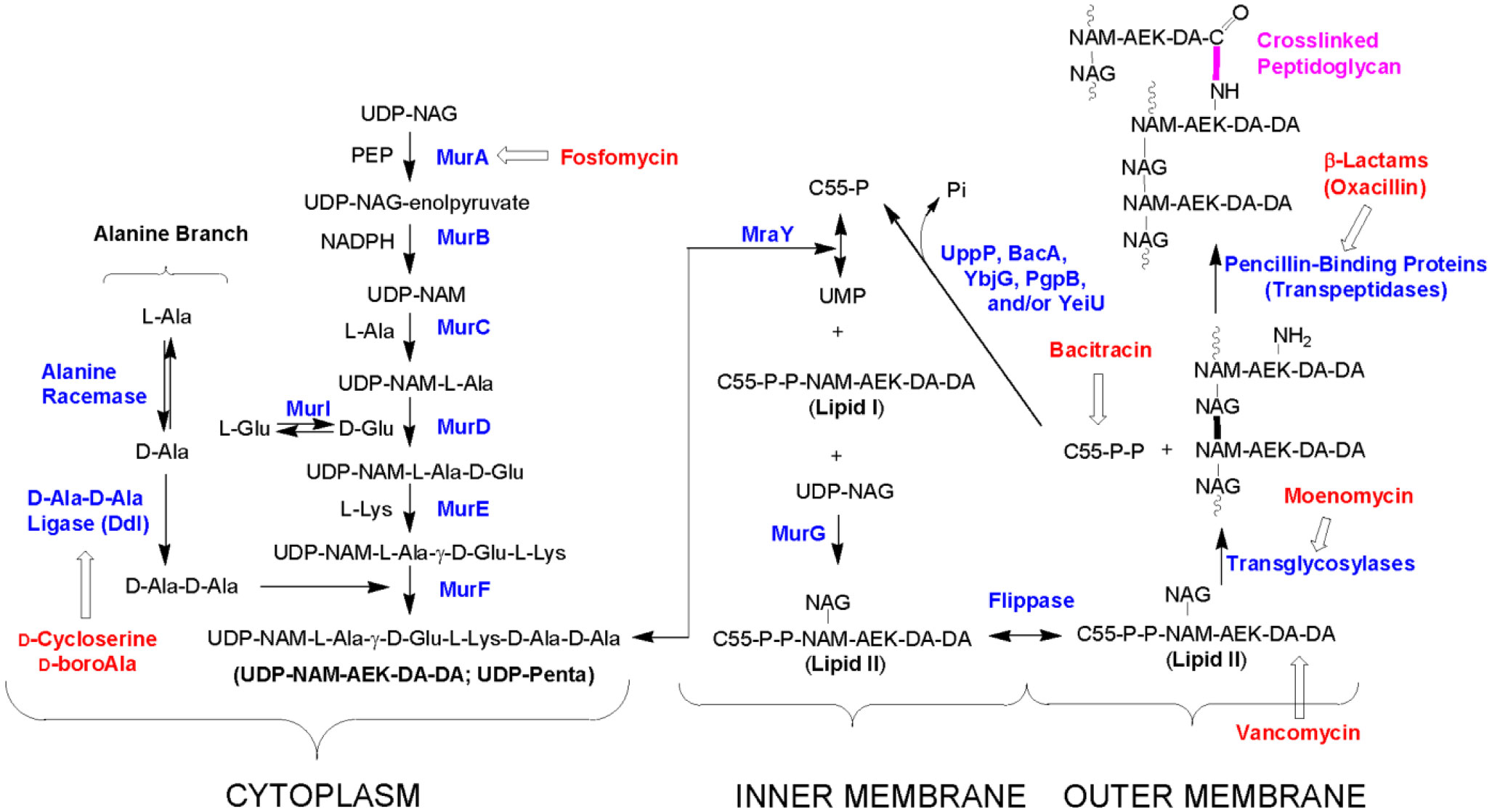
Teo AC, Roper DI (2015) Core Steps of Membrane-Bound Peptidoglycan Biosynthesis: Recent Advances, Insight and Opportunities. Antibiotics (Basel) 4: 495-520. doi: 10.3390/antibiotics4040495
|
| [167] |
Vemula H, Ayon NJ, Burton A, et al. (2017) Antibiotic Effects on Methicillin-Resistant Staphylococcus aureus Cytoplasmic Peptidoglycan Intermediate Levels and Evidence for Potential Metabolite Level Regulatory Loops. Antimicrob Agents Chemother 61: e02253-02216. doi: 10.1128/AAC.02253-16
|
| [168] |
Stranden AM, Ehlert K, Labischinski H, et al. (1997) Cell wall monoglycine cross-bridges and methicillin hypersusceptibility in a femAB null mutant of methicillin-resistant Staphylococcus aureus. J Bacteriol 179: 9-16. doi: 10.1128/JB.179.1.9-16.1997
|
| [169] |
Vemula H, Ayon NJ, Gutheil WG (2016) Cytoplasmic peptidoglycan intermediate levels in Staphylococcus aureus. Biochimie 121: 72-78. doi: 10.1016/j.biochi.2015.11.017
|
| [170] |
Lam H, Oh DC, Cava F, et al. (2009) D-amino acids govern stationary phase cell wall remodeling in bacteria. Science 325: 1552-1555. doi: 10.1126/science.1178123
|
| [171] |
Alvarez L, Aliashkevich A, de Pedro MA, et al. (2018) Bacterial secretion of D-arginine controls environmental microbial biodiversity. Isme J 12: 438-450. doi: 10.1038/ismej.2017.176
|
| [172] |
de Pedro MA, Quintela JC, Holtje JV, et al. (1997) Murein segregation in Escherichia coli. J Bacteriol 179: 2823-2834. doi: 10.1128/JB.179.9.2823-2834.1997
|
| [173] | Wu H, Xue E, Zhi N, et al. (2020) d-Methionine and d-Phenylalanine Improve Lactococcus lactis F44 Acid Resistance and Nisin Yield by Governing Cell Wall Remodeling. Appl Environ Microbiol 86. |
| [174] |
Hu H, Emerson J, Aronson AI (2007) Factors involved in the germination and inactivation of Bacillus anthracis spores in murine primary macrophages. FEMS Microbiol Lett 272: 245-250. doi: 10.1111/j.1574-6968.2007.00766.x
|
| [175] |
Reynolds PE, Courvalin P (2005) Vancomycin resistance in enterococci due to synthesis of precursors terminating in D-alanyl-D-serine. Antimicrob Agents Chemother 49: 21-25. doi: 10.1128/AAC.49.1.21-25.2005
|
| [176] |
Hall-Stoodley L, Costerton JW, Stoodley P (2004) Bacterial biofilms: from the Natural environment to infectious diseases. Nat Rev Microbiol 2: 95-108. doi: 10.1038/nrmicro821
|
| [177] |
Kostakioti M, Hadjifrangiskou M, Hultgren SJ (2013) Bacterial biofilms: development, dispersal, and therapeutic strategies in the dawn of the postantibiotic era. Cold Spring Harbor Perspect Med 3: a010306. doi: 10.1101/cshperspect.a010306
|
| [178] |
Kolodkin-Gal I, Romero D, Cao S, et al. (2010) D-amino acids trigger biofilm disassembly. Science 328: 627-629. doi: 10.1126/science.1188628
|
| [179] |
Hochbaum AI, Kolodkin-Gal I, Foulston L, et al. (2011) Inhibitory effects of D-amino acids on Staphylococcus aureus biofilm development. J Bacteriol 193: 5616-5622. doi: 10.1128/JB.05534-11
|
| [180] |
Ramon-Perez ML, Diaz-Cedillo F, Ibarra JA, et al. (2014) D-Amino acids inhibit biofilm formation in Staphylococcus epidermidis strains from ocular infections. J Med Microbiol 63: 1369-1376. doi: 10.1099/jmm.0.075796-0
|
| [181] |
Sarkar S, Pires MM (2015) d-Amino acids do not inhibit biofilm formation in Staphylococcus aureus. PLoS One 10: e0117613. doi: 10.1371/journal.pone.0117613
|
| [182] | Bucher T, Kartvelishvily E, Kolodkin-Gal I (2016) Methodologies for Studying B. subtilis Biofilms as a Model for Characterizing Small Molecule Biofilm Inhibitors. J Vis Exp 116: 54612. |
| [183] |
Jia R, Li Y, Al-Mahamedh HH, et al. (2017) Enhanced Biocide Treatments with D-amino Acid Mixtures against a Biofilm Consortium from a Water Cooling Tower. Front Microbiol 8: 1538. doi: 10.3389/fmicb.2017.01538
|
| [184] | Li H, Anuwongcharoen N, Malik AA, et al. (2016) Roles of d-Amino Acids on the Bioactivity of Host Defense Peptides. Int J Mol Sci 17. |
| [185] |
Ghssein G, Brutesco C, Ouerdane L, et al. (2016) Biosynthesis of a broad-spectrum nicotianamine-like metallophore in Staphylococcus aureus. Science 352: 1105-1109. doi: 10.1126/science.aaf1018
|
| [186] | Strydom DJ, Andersen TT, Apostol I, et al. (1993) Cysteine and Tryptophan Amino Acid Analysis of ABRF92-AAA. Techniques in Protein Chemistry IV Academic Press, 279-288. |
| [187] |
Martínez-Rodríguez S, Martínez-Gómez AI, Rodríguez-Vico F, et al. (2010) Natural Occurrence and Industrial Applications of d-Amino Acids: An Overview. Chem Biodiversity 7: 1531-1548. doi: 10.1002/cbdv.200900245
|
| [188] |
Cartus AT (2012) 12 - d-Amino acids and cross-linked amino acids as food contaminants. Chemical Contaminants and Residues in Food Woodhead Publishing, 286-319. doi: 10.1533/9780857095794.2.286
|
| [189] | Stokes JL, Gunness M, Dwyer IM, et al. (1945) Microbiological Methods for the Determination of Amino Acids. J Biol Chem 163: 159-168. |
| [190] |
Archibald RM (1946) Enzymatic methods in amino acid analysis. Ann N Y Acad Sci 47: 181-186. doi: 10.1111/j.1749-6632.1946.tb31712.x
|
| [191] |
Dale T, Court WE (1981) Improved separation of amino acids by thin-layer chromatography. Chromatographia 14: 617-620. doi: 10.1007/BF02291097
|
| [192] |
Ali M, Abdul M, Gaber E-D (2012) Amino acid and vitamin determinations by TLC/HPTLC: review of the current state. Open Chem 10: 731-750. doi: 10.2478/s11532-012-0019-0
|
| [193] |
Mayadunne R, Nguyen T-T, Marriott PJ (2005) Amino acid analysis by using comprehensive two-dimensional gas chromatography. Anal Bioanal Chem 382: 836-847. doi: 10.1007/s00216-005-3083-x
|
| [194] |
E. Otter D (2012) Standardised methods for amino acid analysis of food. Br J Nutr 108: S230-S237. doi: 10.1017/S0007114512002486
|
| [195] |
Ilisz I, Aranyi A, Pataj Z, et al. (2012) Recent advances in the direct and indirect liquid chromatographic enantioseparation of amino acids and related compounds: A review. J Pharm Biomed Anal 69: 28-41. doi: 10.1016/j.jpba.2012.01.020
|
| [196] |
Ilisz I, Péter A, Lindner W (2016) State-of-the-art enantioseparations of natural and unnatural amino acids by high-performance liquid chromatography. TrAC, Trends Anal Chem 81: 11-22. doi: 10.1016/j.trac.2016.01.016
|
| [197] |
Ayon NJ, Sharma AD, Gutheil WG (2019) LC-MS/MS-Based Separation and Quantification of Marfey's Reagent Derivatized Proteinogenic Amino Acid DL-Stereoisomers. J Am Soc Mass Spectrom 30: 448-458. doi: 10.1007/s13361-018-2093-9
|
| [198] |
Kim BY, Yang J, Gong M, et al. (2009) Multidimensional separation of chiral amino acid mixtures in a multilayered three-dimensional hybrid microfluidic/nanofluidic device. Anal Chem 81: 2715-2722. doi: 10.1021/ac802630p
|
| [199] |
Poinsot V, Carpene MA, Bouajila J, et al. (2012) Recent advances in amino acid analysis by capillary electrophoresis. Electrophoresis 33: 14-35. doi: 10.1002/elps.201100360
|
| [200] |
Creamer JS, Mora MF, Willis PA (2017) Enhanced Resolution of Chiral Amino Acids with Capillary Electrophoresis for Biosignature Detection in Extraterrestrial Samples. Anal Chem 89: 1329-1337. doi: 10.1021/acs.analchem.6b04338
|
| [201] |
Waldhier MC, Gruber MA, Dettmer K, et al. (2009) Capillary electrophoresis and column chromatography in biomedical chiral amino acid analysis. Anal Bioanal Chem 394: 695-706. doi: 10.1007/s00216-009-2792-y
|
| [202] |
Giuffrida A, Maccarrone G, Cucinotta V, et al. (2014) Recent advances in chiral separation of amino acids using capillary electromigration techniques. J Chromatogr A 1363: 41-50. doi: 10.1016/j.chroma.2014.08.041
|
| [203] |
Moini M, Schultz CL, Mahmood H (2003) CE/Electrospray Ionization-MS Analysis of Underivatized d/l-Amino Acids and Several Small Neurotransmitters at Attomole Levels through the Use of 18-Crown-6-tetracarboxylic Acid as a Complexation Reagent/Background Electrolyte. Anal Chem 75: 6282-6287. doi: 10.1021/ac034708i
|
| [204] |
Wu Y, Zhai Y, Zhang Y, et al. (2014) Chiral separation and determination of amino acids in real samples by LE-MEKC using Cu(II)-l-proline as chiral selector. J Chromatogr B 965: 254-259. doi: 10.1016/j.jchromb.2014.07.001
|
| [205] | Hirs CH, Stein WH, Moore S (1954) The amino acid composition of ribonuclease. J Biol Chem 211: 941-950. |
| [206] |
Tsugita A, Scheffler J-J (1982) A Rapid Method for Acid Hydrolysis of Protein with a Mixture of Trifluoroacetic Acid and Hydrochloric Acid. Eur J Biochem 124: 585-588. doi: 10.1111/j.1432-1033.1982.tb06634.x
|
| [207] |
Kabaha K, Taralp A, Cakmak I, et al. (2011) Accelerated hydrolysis method to estimate the amino acid content of wheat (Triticum durum Desf.) flour using microwave irradiation. J Agric Food Chem 59: 2958-2965. doi: 10.1021/jf103678c
|
| [208] |
Sharer JD, De Biase I, Matern D, et al. (2018) Laboratory analysis of amino acids, 2018 revision: a technical standard of the American College of Medical Genetics and Genomics (ACMG). Genet Med 20: 1499-1507. doi: 10.1038/s41436-018-0328-6
|
| [209] |
Miyazawa T, Minowa H, Imagawa K, et al. (1997) Enantiomeric Separation of Non-Protein Amino Acids by Chiral Ligand-Exchange High-Performance Liquid Chromatography. Anal Lett 30: 867-882. doi: 10.1080/00032719708006430
|
| [210] |
Petritis K, Valleix A, Elfakir C, et al. (2001) Simultaneous analysis of underivatized chiral amino acids by liquid chromatography–ionspray tandem mass spectrometry using a teicoplanin chiral stationary phase. J Chromatogr A 913: 331-340. doi: 10.1016/S0021-9673(00)01268-1
|
| [211] |
Miyoshi Y, Koga R, Oyama T, et al. (2012) HPLC analysis of naturally occurring free d-amino acids in mammals. J Pharm Biomed Anal 69: 42-49. doi: 10.1016/j.jpba.2012.01.041
|
| [212] |
Cohen SA, Michaud DP (1993) Synthesis of a fluorescent derivatizing reagent, 6-aminoquinolyl-N-hydroxysuccinimidyl carbamate, and its application for the analysis of hydrolysate amino acids via high-performance liquid chromatography. Anal Biochem 211: 279-287. doi: 10.1006/abio.1993.1270
|
| [213] |
Bhushan R, Brückner H (2011) Use of Marfey's reagent and analogs for chiral amino acid analysis: Assessment and applications to natural products and biological systems. J Chromatogr B 879: 3148-3161. doi: 10.1016/j.jchromb.2011.05.058
|
| [214] |
Vemula H, Kitase Y, Ayon NJ, et al. (2017) Gaussian and linear deconvolution of LC-MS/MS chromatograms of the eight aminobutyric acid isomers. Anal Biochem 516: 75-85. doi: 10.1016/j.ab.2016.10.017
|
| [215] |
Langrock T, Czihal P, Hoffmann R (2006) Amino acid analysis by hydrophilic interaction chromatography coupled on-line to electrospray ionization mass spectrometry. Amino Acids 30: 291-297. doi: 10.1007/s00726-005-0300-z
|
| [216] |
Armstrong M, Jonscher K, Reisdorph NA (2007) Analysis of 25 underivatized amino acids in human plasma using ion-pairing reversed-phase liquid chromatography/time-of-flight mass spectrometry. Rapid Commun Mass Spectrom 21: 2717-2726. doi: 10.1002/rcm.3124
|
| [217] |
Piraud M, Vianey-Saban C, Petritis K, et al. (2005) Ion-pairing reversed-phase liquid chromatography/electrospray ionization mass spectrometric analysis of 76 underivatized amino acids of biological interest: a new tool for the diagnosis of inherited disorders of amino acid metabolism. Rapid Commun Mass Spectrom 19: 1587-1602. doi: 10.1002/rcm.1957
|
| [218] |
Le A, Ng A, Kwan T, et al. (2014) A rapid, sensitive method for quantitative analysis of underivatized amino acids by liquid chromatography-tandem mass spectrometry (LC-MS/MS). J Chromatogr B Analyt Technol Biomed Life Sci 944: 166-174. doi: 10.1016/j.jchromb.2013.11.017
|
| [219] |
Fujii K, Ikai Y, Oka H, et al. (1997) A Nonempirical Method Using LC/MS for Determination of the Absolute Configuration of Constituent Amino Acids in a Peptide: Combination of Marfey's Method with Mass Spectrometry and Its Practical Application. Anal Chem 69: 5146-5151. doi: 10.1021/ac970289b
|
| [220] |
Ferré S, González-Ruiz V, Guillarme D, et al. (2019) Analytical strategies for the determination of amino acids: Past, present and future trends. J Chromatogr B 1132: 121819. doi: 10.1016/j.jchromb.2019.121819
|
| [221] |
Stokvis E, Rosing H, Beijnen JH (2005) Stable isotopically labeled internal standards in quantitative bioanalysis using liquid chromatography/mass spectrometry: necessity or not? Rapid Commun Mass Spectrom 19: 401-407. doi: 10.1002/rcm.1790
|
| [222] |
Barrado E, Rodriguez JA, Castrillejo Y (2009) Determination of primary amino acids in wines by high performance liquid magneto-chromatography. Talanta 78: 672-675. doi: 10.1016/j.talanta.2008.12.023
|
| [223] |
Eto S, Yamaguchi M, Bounoshita M, et al. (2011) High-throughput comprehensive analysis of d- and l-amino acids using ultra-high performance liquid chromatography with a circular dichroism (CD) detector and its application to food samples. J Chromatogr B 879: 3317-3325. doi: 10.1016/j.jchromb.2011.07.025
|
| [224] |
Pérez-Míguez R, Bruyneel B, Castro-Puyana M, et al. (2019) Chiral Discrimination of DL-Amino Acids by Trapped Ion Mobility Spectrometry after Derivatization with (+)-1-(9-Fluorenyl)ethyl Chloroformate. Anal Chem 91: 3277-3285. doi: 10.1021/acs.analchem.8b03661
|
| [225] |
Schurig V (2002) Chiral separations using gas chromatography. TrAC, Trends Anal Chem 21: 647-661. doi: 10.1016/S0165-9936(02)00808-7
|
| [226] |
Erbe T, Brückner H (2000) Chromatographic determination of amino acid enantiomers in beers and raw materials used for their manufacture. J Chromatogr A 881: 81-91. doi: 10.1016/S0021-9673(00)00255-7
|
| [227] |
Pätzold R, Brückner H (2006) Gas chromatographic determination and mechanism of formation of D-amino acids occurring in fermented and roasted cocoa beans, cocoa powder, chocolate and cocoa shell. Amino Acids 31: 63. doi: 10.1007/s00726-006-0330-1
|
| [228] |
Gu L, Jones AD, Last RL (2007) LC−MS/MS Assay for Protein Amino Acids and Metabolically Related Compounds for Large-Scale Screening of Metabolic Phenotypes. Anal Chem 79: 8067-8075. doi: 10.1021/ac070938b
|
| [229] |
Kaspar H, Dettmer K, Gronwald W, et al. (2009) Advances in amino acid analysis. Anal Bioanal Chem 393: 445-452. doi: 10.1007/s00216-008-2421-1
|
| [230] |
Schurig V (2011) Gas chromatographic enantioseparation of derivatized α-amino acids on chiral stationary phases—Past and present. J Chromatogr B 879: 3122-3140. doi: 10.1016/j.jchromb.2011.04.005
|
| [231] |
Szökő É, Vincze I, Tábi T (2016) Chiral separations for d-amino acid analysis in biological samples. J Pharm Biomed Anal 130: 100-109. doi: 10.1016/j.jpba.2016.06.054
|
| [232] |
Konya Y, Bamba T, Fukusaki E (2016) Extra-facile chiral separation of amino acid enantiomers by LC-TOFMS analysis. J Biosci Bioeng 121: 349-353. doi: 10.1016/j.jbiosc.2015.06.017
|
| [233] |
Nakano Y, Konya Y, Taniguchi M, et al. (2017) Development of a liquid chromatography-tandem mass spectrometry method for quantitative analysis of trace d-amino acids. J Biosci Bioeng 123: 134-138. doi: 10.1016/j.jbiosc.2016.07.008
|
| [234] |
Konya Y, Taniguchi M, Fukusaki E (2017) Novel high-throughput and widely-targeted liquid chromatography–time of flight mass spectrometry method for d-amino acids in foods. J Biosci Bioeng 123: 126-133. doi: 10.1016/j.jbiosc.2016.07.009
|
| [235] |
Dell'mour M, Jaitz L, Oburger E, et al. (2010) Hydrophilic interaction LC combined with electrospray MS for highly sensitive analysis of underivatized amino acids in rhizosphere research. J Sep Sci 33: 911-922. doi: 10.1002/jssc.200900743
|
| [236] |
Horak J, Lämmerhofer M (2019) Stereoselective separation of underivatized and 6-aminoquinolyl-N-hydroxysuccinimidyl carbamate derivatized amino acids using zwitterionic quinine and quinidine type stationary phases by liquid chromatography–High resolution mass spectrometry. J Chromatogr A 1596: 69-78. doi: 10.1016/j.chroma.2019.02.060
|
| [237] |
Bäurer S, Ferri M, Carotti A, et al. (2020) Mixed-mode chromatography characteristics of chiralpak ZWIX(+) and ZWIX(−) and elucidation of their chromatographic orthogonality for LC × LC application. Anal Chim Acta 1093: 168-179. doi: 10.1016/j.aca.2019.09.068
|
| [238] |
Han M, Xie M, Han J, et al. (2018) Development and validation of a rapid, selective, and sensitive LC–MS/MS method for simultaneous determination of d- and l-amino acids in human serum: application to the study of hepatocellular carcinoma. Anal Bioanal Chem 410: 2517-2531. doi: 10.1007/s00216-018-0883-3
|
| [239] |
Kinoshita K, Jingu S, Yamaguchi J-i (2013) A surrogate analyte method to determine d-serine in mouse brain using liquid chromatography–tandem mass spectrometry. Anal Biochem 432: 124-130. doi: 10.1016/j.ab.2012.09.035
|
| [240] |
Pesek JJ, Matyska MT, Fischer SM, et al. (2008) Analysis of hydrophilic metabolites by high-performance liquid chromatography–mass spectrometry using a silica hydride-based stationary phase. J Chromatogr A 1204: 48-55. doi: 10.1016/j.chroma.2008.07.077
|
| [241] |
Raimbault A, Dorebska M, West C (2019) A chiral unified chromatography–mass spectrometry method to analyze free amino acids. Anal Bioanal Chem 411: 4909-4917. doi: 10.1007/s00216-019-01783-5
|
| [242] |
Raimbault A, Noireau A, West C (2020) Analysis of free amino acids with unified chromatography-mass spectrometry—application to food supplements. J Chromatogr A 1616: 460772. doi: 10.1016/j.chroma.2019.460772
|
| [243] |
Miller L, Yue L (2020) Chiral separation of underivatized amino acids in supercritical fluid chromatography with chiral crown ether derived column. Chirality 32: 981-989. doi: 10.1002/chir.23204
|
| [244] |
Sánchez-Hernández L, Bernal JL, Nozal MJd, et al. (2016) Chiral analysis of aromatic amino acids in food supplements using subcritical fluid chromatography and Chirobiotic T2 column. J Supercrit Fluids 107: 519-525. doi: 10.1016/j.supflu.2015.06.027
|
| [245] |
Oyama T, Negishi E, Onigahara H, et al. (2015) Design and synthesis of a novel pre-column derivatization reagent with a 6-methoxy-4-quinolone moiety for fluorescence and tandem mass spectrometric detection and its application to chiral amino acid analysis. J Pharm Biomed Anal 116: 71-79. doi: 10.1016/j.jpba.2015.05.039
|
| [246] |
Pietrogrande MC, Basaglia G (2010) Enantiomeric resolution of biomarkers in space analysis: Chemical derivatization and signal processing for gas chromatography–mass spectrometry analysis of chiral amino acids. J Chromatogr A 1217: 1126-1133. doi: 10.1016/j.chroma.2009.09.055
|
| [247] |
Bhushan R, Brückner H (2004) Marfey's reagent for chiral amino acid analysis: a review. Amino Acids 27: 231-247. doi: 10.1007/s00726-004-0118-0
|
| [248] |
Ilisz I, Berkecz R, Péter A (2008) Application of chiral derivatizing agents in the high-performance liquid chromatographic separation of amino acid enantiomers: A review. J Pharm Biomed Anal 47: 1-15. doi: 10.1016/j.jpba.2007.12.013
|
| [249] |
Tanwar S, Bhushan R (2015) Enantioresolution of Amino Acids: A Decade's Perspective, Prospects and Challenges. Chromatographia 78: 1113-1134. doi: 10.1007/s10337-015-2933-8
|
| [250] |
Kotthaus AF, Altenbach HJ (2011) A new chiral derivatizing agent for the HPLC separation of α-amino acids on a standard reverse-phase column. Amino Acids 40: 527-532. doi: 10.1007/s00726-010-0665-5
|
| [251] |
Péter A, Vékes E, Török G (2000) Application of (S)-N-(4-nitrophenoxycarbonyl) phenylalanine methoxyethyl ester as a new chiral derivatizing agent for proteinogenic amino acid analysis by high-performance liquid chromatography. Chromatographia 52: 821-826. doi: 10.1007/BF02491012
|
| [252] |
Olajos E, Peter A, Casimir R, et al. (2001) HPLC enantioseparation of phenylalanine analogs by application of (S)-N-(4-nitrophenoxycarbonyl)phenylalanine methoxyethyl ester as a new chiral derivatizing agent. Chromatographia 54: 77-82. doi: 10.1007/BF02491837
|
| [253] |
Visser WF, Verhoeven-Duif NM, Ophoff R, et al. (2011) A sensitive and simple ultra-high-performance-liquid chromatography–tandem mass spectrometry based method for the quantification of d-amino acids in body fluids. J Chromatogr A 1218: 7130-7136. doi: 10.1016/j.chroma.2011.07.087
|
| [254] |
Reischl RJ, Lindner W (2012) Methoxyquinoline labeling—A new strategy for the enantioseparation of all chiral proteinogenic amino acids in 1-dimensional liquid chromatography using fluorescence and tandem mass spectrometric detection. J Chromatogr A 1269: 262-269. doi: 10.1016/j.chroma.2012.07.046
|
| [255] |
Reischl RJ, Hartmanova L, Carrozzo M, et al. (2011) Chemoselective and enantioselective analysis of proteinogenic amino acids utilizing N-derivatization and 1-D enantioselective anion-exchange chromatography in combination with tandem mass spectrometric detection. J Chromatogr A 1218: 8379-8387. doi: 10.1016/j.chroma.2011.09.046
|
| [256] |
Harada M, Karakawa S, Yamada N, et al. (2019) Biaryl axially chiral derivatizing agent for simultaneous separation and sensitive detection of proteinogenic amino acid enantiomers using liquid chromatography–tandem mass spectrometry. J Chromatogr A 1593: 91-101. doi: 10.1016/j.chroma.2019.01.075
|
| [257] |
Manica DP, Lapos JA, Daniel Jones A, et al. (2003) Analysis of the stability of amino acids derivatized with naphthalene-2,3-dicarboxaldehyde using high-performance liquid chromatography and mass spectrometry. Anal Biochem 322: 68-78. doi: 10.1016/j.ab.2003.07.002
|
| [258] |
Karakawa S, Shimbo K, Yamada N, et al. (2015) Simultaneous analysis of d-alanine, d-aspartic acid, and d-serine using chiral high-performance liquid chromatography-tandem mass spectrometry and its application to the rat plasma and tissues. J Pharm Biomed Anal 115: 123-129. doi: 10.1016/j.jpba.2015.05.024
|
| [259] |
Müller C, Fonseca JR, Rock TM, et al. (2014) Enantioseparation and selective detection of D-amino acids by ultra-high-performance liquid chromatography/mass spectrometry in analysis of complex biological samples. J Chromatogr A 1324: 109-114. doi: 10.1016/j.chroma.2013.11.026
|
| [260] |
Zampolli MG, Basaglia G, Dondi F, et al. (2007) Gas chromatography–mass spectrometry analysis of amino acid enantiomers as methyl chloroformate derivatives: Application to space analysis. J Chromatogr A 1150: 162-172. doi: 10.1016/j.chroma.2006.12.033
|
| [261] |
Menestrina F, Osorio Grisales J, Castells CB (2016) Chiral analysis of derivatized amino acids from kefir by gas chromatography. Microchem J 128: 267-273. doi: 10.1016/j.microc.2016.05.007
|
| [262] |
Bertrand M, Chabin A, Brack A, et al. (2008) Separation of amino acid enantiomers VIA chiral derivatization and non-chiral gas chromatography. J Chromatogr A 1180: 131-137. doi: 10.1016/j.chroma.2007.12.004
|
| [263] |
Zampolli M, Meunier D, Sternberg R, et al. (2006) GC-MS analysis of amino acid enantiomers as their N(O,S)-perfluoroacyl perfluoroalkyl esters: Application to space analysis. Chirality 18: 279-295. doi: 10.1002/chir.20241
|
| [264] |
Pätzold R, Schieber A, Brückner H (2005) Gas chromatographic quantification of free D-amino acids in higher vertebrates. Biomed Chromatogr 19: 466-473. doi: 10.1002/bmc.515
|
| [265] |
Lorenzo MP, Dudzik D, Varas E, et al. (2015) Optimization and validation of a chiral GC–MS method for the determination of free d-amino acids ratio in human urine: Application to a Gestational Diabetes Mellitus study. J Pharm Biomed Anal 107: 480-487. doi: 10.1016/j.jpba.2015.01.015
|
| [266] |
Lajkó G, Ilisz I, Tóth G, et al. (2015) Application of Cinchona alkaloid-based zwitterionic chiral stationary phases in supercritical fluid chromatography for the enantioseparation of Nα-protected proteinogenic amino acids. J Chromatogr A 1415: 134-145. doi: 10.1016/j.chroma.2015.08.058
|
| [267] |
Fleischer H, Thurow K (2013) Fast mass spectrometry-based enantiomeric excess determination of proteinogenic amino acids. Amino Acids 44: 1039-1051. doi: 10.1007/s00726-012-1439-z
|
| [268] |
Ianni F, Sardella R, Lisanti A, et al. (2015) Achiral–chiral two-dimensional chromatography of free amino acids in milk: A promising tool for detecting different levels of mastitis in cows. J Pharm Biomed Anal 116: 40-46. doi: 10.1016/j.jpba.2014.12.041
|
| [269] |
Woiwode U, Neubauer S, Lindner W, et al. (2018) Enantioselective multiple heartcut two-dimensional ultra-high-performance liquid chromatography method with a Coreshell chiral stationary phase in the second dimension for analysis of all proteinogenic amino acids in a single run. J Chromatogr A 1562: 69-77. doi: 10.1016/j.chroma.2018.05.062
|
| [270] |
Miyoshi Y, Nagano M, Ishigo S, et al. (2014) Chiral amino acid analysis of Japanese traditional Kurozu and the developmental changes during earthenware jar fermentation processes. J Chromatogr B 966: 187-192. doi: 10.1016/j.jchromb.2014.01.034
|
| [271] |
Ishii C, Akita T, Mita M, et al. (2018) Development of an online two-dimensional high-performance liquid chromatographic system in combination with tandem mass spectrometric detection for enantiomeric analysis of free amino acids in human physiological fluid. J Chromatogr A 1570: 91-98. doi: 10.1016/j.chroma.2018.07.076
|
| [272] |
Myrgorodska I, Meinert C, Martins Z, et al. (2016) Quantitative enantioseparation of amino acids by comprehensive two-dimensional gas chromatography applied to non-terrestrial samples. J Chromatogr A 1433: 131-136. doi: 10.1016/j.chroma.2016.01.014
|

Figures(5) / Tables(7)

Navid J. Ayon. Features, roles and chiral analyses of proteinogenic amino acids[J]. AIMS Molecular Science, 2020, 7(3): 229-268. doi: 10.3934/molsci.2020011


 DownLoad: