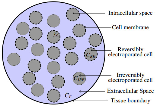
In order to overcome the obstruction of cell membranes in the path of drug delivery to diseased cells, the applications of electric pulses of adequate potency are designated as electroporation. In the present study, a mathematical model of drug delivery into the electroporated tissue is advocated, which deals with both reversibly and irreversibly electroporated cells. This mathematical formulation is manifested through a set of differential equations, which are solved analytically, and numerically, according to the complexity, with a pertinent set of initial and boundary conditions. The time-dependent mass transfer coefficient in terms of pores is used to find the drug concentrations through reversibly and irreversibly electroporated cells as well as extracellular space. The effects of permeability of drug, electric field and pulse period on drug concentrations in extracellular and intracellular regions are discussed. The threshold value of an electric field ($ E > 100 $ V cm$ ^{-1} $) to initiate drug uptake is identified in this study. Special emphasis is also put on two cases of electroporation, drug dynamics during ongoing electroporation and drug dynamics after the electric pulse period is over. Furthermore, all the simulated results and graphical portrayals are discussed in detail to have a transparent vision in comprehending the underlying physical and physiological phenomena. This model could be useful to various clinical experiments for drug delivery in the targeted tissue by controlling the model parameters depending on the tissue condition.
Citation: Nilay Mondal, Koyel Chakravarty, D. C. Dalal. A mathematical model of drug dynamics in an electroporated tissue[J]. Mathematical Biosciences and Engineering, 2021, 18(6): 8641-8660. doi: 10.3934/mbe.2021428
In order to overcome the obstruction of cell membranes in the path of drug delivery to diseased cells, the applications of electric pulses of adequate potency are designated as electroporation. In the present study, a mathematical model of drug delivery into the electroporated tissue is advocated, which deals with both reversibly and irreversibly electroporated cells. This mathematical formulation is manifested through a set of differential equations, which are solved analytically, and numerically, according to the complexity, with a pertinent set of initial and boundary conditions. The time-dependent mass transfer coefficient in terms of pores is used to find the drug concentrations through reversibly and irreversibly electroporated cells as well as extracellular space. The effects of permeability of drug, electric field and pulse period on drug concentrations in extracellular and intracellular regions are discussed. The threshold value of an electric field ($ E > 100 $ V cm$ ^{-1} $) to initiate drug uptake is identified in this study. Special emphasis is also put on two cases of electroporation, drug dynamics during ongoing electroporation and drug dynamics after the electric pulse period is over. Furthermore, all the simulated results and graphical portrayals are discussed in detail to have a transparent vision in comprehending the underlying physical and physiological phenomena. This model could be useful to various clinical experiments for drug delivery in the targeted tissue by controlling the model parameters depending on the tissue condition.

| [1] |
k K. A. DeBruin, W. Krassowska, Modeling electroporation in a single cell. i. effects of field strength and rest potential, Biophys. J., 77 (1999), 1213–1224. doi: 10.1016/S0006-3495(99)76973-0

|
| [2] |
R. V. Davalos, B. Rubinsky, L. M. Mir, Theoretical analysis of the thermal effects during in vivo tissue electroporation, Bioelectrochemistry, 61 (2003), 99–107. doi: 10.1016/j.bioelechem.2003.07.001
|
| [3] |
W. Krassowska, P. D. Filev, Modeling electroporation in a single cell, Biophys. J., 92 (2007), 404–417. doi: 10.1529/biophysj.106.094235
|
| [4] |
T. Kotnik, L. Rems, M. Tarek, D. Miklavčič, Membrane electroporation and electropermeabilization: Mechanisms and models, Annu. Rev. Biophys., 48 (2019), 63–91. doi: 10.1146/annurev-biophys-052118-115451
|
| [5] |
J. Dermol-Černe, E. Pirc, D. Miklavčič, Mechanistic view of skin electroporation–models and dosimetry for successful applications: an expert review, Expert. Opin. Drug. Deliv., 17 (2020), 689–704. doi: 10.1080/17425247.2020.1745772
|
| [6] |
S. Mahnič-Kalamiza, E. Vorobiev, D. Miklavčič, Electroporation in food processing and biorefinery, J. Membr. Biol., 247 (2014), 1279–1304. doi: 10.1007/s00232-014-9737-x
|
| [7] |
T. B. Napotnik, M. Reberšek, P. T. Vernier, B. Mali, D. Miklavčič, Effects of high voltage nanosecond electric pulses on eukaryotic cells (in vitro): A systematic review, Bioelectrochemistry, 110 (2016), 1–12. doi: 10.1016/j.bioelechem.2016.02.011
|
| [8] |
M. Pavlin, D. Miklavčič, Effective conductivity of a suspension of permeabilized cells: A theoretical analysis, Biophys. J., 85 (2003), 719–729. doi: 10.1016/S0006-3495(03)74515-9
|
| [9] |
J. Dermol-Černe, D. Miklavčič, From cell to tissue properties-modeling skin electroporation with pore and local transport region formation, IEEE Trans. Biomed. Eng., 65 (2018), 458–468. doi: 10.1109/TBME.2017.2773126
|
| [10] | P. A. Garcia, R. V. Davalos, D. Miklavčič, A numerical investigation of the electric and thermal cell kill distributions in electroporation-based therapies in tissue, PLoS One, 9 (2014), 1–12. |
| [11] |
Y. Granot, B. Rubinsky, Mass transfer model for drug delivery in tissue cells with reversible electroporation, Int. J. Heat Mass Transf., 51 (2008), 5610–5616. doi: 10.1016/j.ijheatmasstransfer.2008.04.041
|
| [12] |
M. Pavlin, S. M. Kandu, M. Rebersek, G. Pucihar, F. X. Hart, R. Mag-jareviccacute, et al., Effect of cell electroporation on the conductivity of a cell suspension, Biophys. J., 88 (2005), 4378–4390. doi: 10.1529/biophysj.104.048975
|
| [13] |
R. V. Davalos, L. M. Mir, B. Rubinsky, Tissue ablation with irreversible electroporation, Ann. Biomed. Eng., 33 (2005), 223–231. doi: 10.1007/s10439-005-8981-8
|
| [14] |
B. Rubinsky, Irreversible electroporation in medicine, Technol. Cancer Res. Treat., 6 (2007), 255–260. doi: 10.1177/153303460700600401
|
| [15] |
C. Jiang, R. Davalos, J. Bischof, A review of basic to clinical studies of irreversible electroporation therapy, IEEE Trans. Biomed. Eng., 62 (2015), 4–20. doi: 10.1109/TBME.2014.2367543
|
| [16] |
D. Miklavčič, G. Serša, E. Brecelj, J. Gehl, D. Soden, G. Bianchi, et al., Electrochemotherapy: technological advancements for efficient electroporation-based treatment of internal tumors, Med. Biol. Eng. Comput., 50 (2012), 1213–25. doi: 10.1007/s11517-012-0991-8
|
| [17] | J. Dermol-Černe, J. Vidmar, J. Ščančar, K. Uršič, G. Serša, D. Miklavčič, Connecting the in vitro and in vivo experiments in electrochemotherapy - a feasibility study modeling cisplatin transport in mouse melanoma using the dual-porosity model, J. Control. Release, 286 (2018), 33–45. |
| [18] |
C. Rosazza, S. H. Meglic, A. Zumbusch, M. P. Rols, D. Miklavčič, Gene electrotransfer: A mechanistic perspective, Curr. Gene Ther., 16 (2016), 98–129. doi: 10.2174/1566523216666160331130040
|
| [19] |
B. Zorec, S. Becker, M. Rebersek, D. Miklavčič, N. Pavselj, Skin electroporation for transdermal drug delivery: The influence of the order of different square wave electric pulses, Int. J. Pharm., 457 (2013), 214–223. doi: 10.1016/j.ijpharm.2013.09.020
|
| [20] |
S. Čorović, L. M. Mir, D. Miklavčič, In vivo muscle electroporation threshold determination: Realistic numerical models and in vivo experiments, J. Membr. Biol., 245 (2012), 509–520. doi: 10.1007/s00232-012-9432-8
|
| [21] |
G. Pucihar, J. Krmelj, M. Reberšek, T. B. Napotnik, D. Miklavčič, Equivalent pulse parameters for electroporation, IEEE Trans. Biomed. Eng., 58 (2011), 3279–3288. doi: 10.1109/TBME.2011.2167232
|
| [22] |
S. Satkauskas, M. Bureau, M. Puc, A. Mahfoudi, D. Scherman, D. Miklavčič, et al., Mechnisms of in vivo dna electrotransfer: Respective contributions of cell electropermeabilization and dna electrophoresis, Mol. Ther., 5 (2002), 133–140. doi: 10.1006/mthe.2002.0526
|
| [23] |
J. C. Weaver, Electroporation of biological membranes from multicellular to nano scales, IEEE Trans. Dielectr. Electr. Insul., 10 (2003), 754–768. doi: 10.1109/TDEI.2003.1237325
|
| [24] |
S. Mahnič-Kalamiza, D. Miklavčič, E. Vorobiev, Dual-porosity model of solute diffusion in biological tissue modified by electroporation, Biochem. Biophys. Acta. Biomembr., 1838 (2014), 1950–1966. doi: 10.1016/j.bbamem.2014.03.004
|
| [25] |
S. Mahnič-Kalamiza, D. Miklavčič, E. Vorobiev, Dual-porosity model of mass transport in electroporated biological tissue: Simulations and experimental work for model validation, Innov. Food Sci. Emerg. Technol., 29 (2015), 41–54. doi: 10.1016/j.ifset.2014.09.011
|
| [26] |
B. Boyd, S. Becker, Macroscopic modeling of in vivo drug transport in electroporated tissue, J. Biomech. Eng., 138 (2016), 031008–11. doi: 10.1115/1.4032380
|
| [27] |
F. Argus, B. Boyd, S. M. Becker, Electroporation of tissue and cells: A three-equation model of drug delivery, Comput. Biol. Med., 84 (2017), 226–234. doi: 10.1016/j.compbiomed.2017.04.001
|
| [28] |
E. Goldberg, A. Soba, D. Gandía, M. L. Fernández, C. Suárez, Coupled mathematical modeling of cisplatin electroporation, Bioelectrochemistry, 140 (2021), 107788. doi: 10.1016/j.bioelechem.2021.107788
|
| [29] |
D. Sel, D. Cukjati, D. Batiuskaite, T. Slivnik, L. M. Mir, D. Miklavčič, Sequential finite element model of tissue electropermeabilization, IEEE Trans. Biomed. Eng., 52 (2005), 816–827. doi: 10.1109/TBME.2005.845212
|
| [30] |
R. W. Glaser, S. L. Leikin, L. V. Chernomordik, V. F. Pastushenko, A. I. Sokirko, Reversible electrical breakdown of lipid bilayers: formation and evolution of pores, Biochem. Biophys. Acta. Biomembr., 940 (1988), 275–287. doi: 10.1016/0005-2736(88)90202-7
|
| [31] | J. C. Neu, W. Krassowska, Asymptotic model of electroporation, Phys. Rev. E, 59 (1999), 3471–3482. |
| [32] |
M. Pavlin, Miklavčič, Theoretical and experimental analysis of conductivity, ion diffusion and molecular transport during cell electroporation-relation between short-lived and long-lived pores, Bioelectrochemistry, 74 (2008), 38–46. doi: 10.1016/j.bioelechem.2008.04.016
|


Figures(13) / Tables(1)

Nilay Mondal, Koyel Chakravarty, D. C. Dalal. A mathematical model of drug dynamics in an electroporated tissue[J]. Mathematical Biosciences and Engineering, 2021, 18(6): 8641-8660. doi: 10.3934/mbe.2021428


 DownLoad: