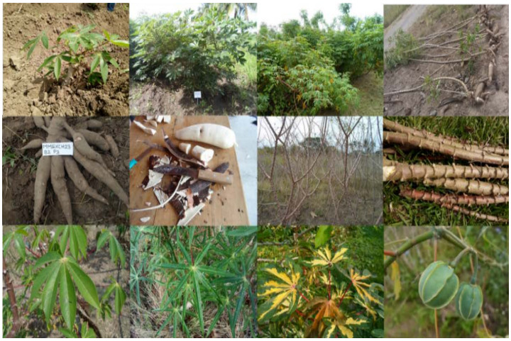
Cassava (Manihot esculenta Crantz) has garnered global attention due to its importance as a crucial raw material for ethanol and other derivative production. Nonetheless, its agroindustry generates a substantial amount of residues. We examined the potential utilization of co–products from both agricultural and industrial sectors concerning starch extraction processes. A total of 319 million tons of fresh cassava roots are globally produced, yielding up to 55% of agricultural co–products during harvesting. For every ton of starch extracted, 2.5 tons of bagasse, along with 100 to 300 kg of peel per ton of fresh processed cassava, and 17.4 m3 of residual liquid tributaries are generated. Consequently, both solid agricultural biomass and solid/liquid residues could be directed towards cogenerating bioenergy such as bioethanol, biobutanol, biodiesel, bio–oil, charcoal, and other bioproducts. In conclusion, the conversion of cassava agroindustrial co–products into food and non–food products with high added value could be promoted, thus fostering a circular economy to enhance profitability, sustainability, and crop promotion.
Citation: Pablo Andrés–Meza, Noé Aguilar–Rivera, Isaac Meneses–Márquez, José Luis Del Rosario–Arellano, Gloria Ivette Bolio–López, Otto Raúl Leyva–Ovalle. Cassava cultivation; current and potential use of agroindustrial co–products[J]. AIMS Environmental Science, 2024, 11(2): 248-278. doi: 10.3934/environsci.2024012
Cassava (Manihot esculenta Crantz) has garnered global attention due to its importance as a crucial raw material for ethanol and other derivative production. Nonetheless, its agroindustry generates a substantial amount of residues. We examined the potential utilization of co–products from both agricultural and industrial sectors concerning starch extraction processes. A total of 319 million tons of fresh cassava roots are globally produced, yielding up to 55% of agricultural co–products during harvesting. For every ton of starch extracted, 2.5 tons of bagasse, along with 100 to 300 kg of peel per ton of fresh processed cassava, and 17.4 m3 of residual liquid tributaries are generated. Consequently, both solid agricultural biomass and solid/liquid residues could be directed towards cogenerating bioenergy such as bioethanol, biobutanol, biodiesel, bio–oil, charcoal, and other bioproducts. In conclusion, the conversion of cassava agroindustrial co–products into food and non–food products with high added value could be promoted, thus fostering a circular economy to enhance profitability, sustainability, and crop promotion.

| [1] | Jiang D, Wang Q, Ding F, et al. (2019) Potential marginal land resources of cassava worldwide: A data–driven analysis. Renew. and Sustain. Energy Rev 104: 167–173. https://doi.org/10.1016/j.rser.2019.01.024 |
| [2] | FAO (Food and Agriculture Organization of the United Nations) (2018) Food Outlook–Biannual Report on Global Food Markets–November 2018. Available from: https://reliefweb.int/report/world/food-outlook-biannual-report-global-food-markets-november-2018 |
| [3] |
Esuma W, Nanyonjo AR, Miiro R, et al. (2019) Men and women's perception of yellow–root cassava among rural farmers in eastern Uganda. Agri Food Secur 8: 1–9. https://doi.org/10.1186/s40066-019-0253-1 doi: 10.1186/s40066-019-0253-1

|
| [4] | Okoruwa VO, Abass AB, Akin–olagunju OA, et al. (2020) Does institution type affect access to finance for cassava actors in Nigeria? J of Agric and Food Res 2(October 2019): 100023. https://doi.org/10.1016/j.jafr.2020.100023 |
| [5] |
Kihara J, Bolo P, Kinyua M, et al. (2020) Micronutrient deficiencies in African soils and the human nutritional nexus: opportunities with staple crops. Environ Geochem and Health 42: 3015–3033. https://doi.org/10.1007/s10653-019-00499-w doi: 10.1007/s10653-019-00499-w
|
| [6] | FAOStat (2023) Data, production, crops. Rome (Ita): Food and Agriculture Organisation. Available from: http://www.fao.org/faostat/en/#data/QC. 15 |
| [7] |
Fernando NML, Amaraweera APSM, Gunawardane, OHP, et al. (2022) Sustainable biorefinery approach for cassava: A Review. Eng: J of the Inst of Eng Sri Lanka 2: 71–88. http://doi.org/10.4038/engineer.v55i2.7510 doi: 10.4038/engineer.v55i2.7510
|
| [8] | Ayetigbo O, Latif S, Abass A, et al. (2018). Comparing characteristics of root, flour and starch of biofortified yellow–flesh and white–flesh cassava variants, and sustainability considerations: A review. Sustainability (Switzerland). 10: 1–32. https://doi.org/10.3390/su10093089 |
| [9] |
Olukanni DO, Olatunji TO (2018) Cassava waste management and biogas generation potential in selected local government areas in Ogun State, Nigeria. Recycl 3: 1–12. https://doi.org/10.3390/recycling3040058 doi: 10.3390/recycling3040058
|
| [10] |
Lerdlattaporn R, Phalakornkula C, Trakulvichean S, et al. (2021) Implementing circular economy concept by converting cassava pulp and wastewater to biogas for sustainable production in starch industry. Sustain Environ Res 31: 1–12. https://doi.org/10.1186/s42834-021-00093-9 doi: 10.1186/s42834-021-00093-9
|
| [11] | Bradu P, Biswas A, Nair C, et al. (2023) Recent advances in green technology and Industrial Revolution 4.0 for a sustainable future. Environ Sci Pollution Res 30: 124488–124519. https://doi.org/10.1007/s11356-022-20024-4 |
| [12] | OCDE/FAO (2019) OCDE–FAO Perspectivas Agrícolas 2019–2028, OECD Publishing, París/Organización de las Naciones Unidas para la Alimentación y la Agricultura (FAO), Roma. https://doi.org/10.1787/7b2e8ba3-es |
| [13] |
Martínez DG, Feiden A, Bariccatti R, et al. (2018) Ethanol production from waste of cassava processing. Appl Sci 8: 1–8. https://doi.org/10.3390/app8112158 doi: 10.3390/app8112158
|
| [14] | Chatellard L, Marone A, Carrère H, et al. (2017) Trends and challenges in biohydrogen production from agricultural waste. In: Singh A, Rathore D. Biohydrogen production: sustainability of current technology and future perspective. Springer, New Delhi. p. 69–95. https://doi.org/10.1007/978-81-322-3577-4_4 |
| [15] |
Sivamani S, Chandrasekaran AP, Balajii M, et al. (2018) Evaluation of the potential of cassava–based residues for biofuels production. Rev Environ Sci Biotech 17: 553–570. https://doi.org/10.1007/s11157-018-9475-0 doi: 10.1007/s11157-018-9475-0
|
| [16] | Gogoi N, Sarma B, Mondal SC, et al. (2019) Use of biochar in sustainable agriculture. In: Farooq, M., and Pisante, M. editors. Innovations in Sustainable Agriculture. Springer, Cham, p. 501–528. https://doi.org/10.1007/978-3-030-23169-9_16 |
| [17] | Aruwajoye GS, Faloye FD, Kana EG (2020a) Process optimisation of enzymatic saccharification of soaking assisted and thermal pretreated cassava peels waste for bioethanol production. Waste Biomass Valor 11: 2409–2420 https://doi.org/10.1007/s12649-018-00562-0 |
| [18] |
Pason P, Tachaapaikoon C, Panichnumsin P, et al. (2020) One–step biohydrogen production from cassava pulp using novel enrichment of anaerobic thermophilic bacteria community. Biocatal Agric Biotechnol 27: 1–6. https://doi.org/10.1016/j.bcab.2020.101658 doi: 10.1016/j.bcab.2020.101658
|
| [19] |
Aduba CC, Ndukwe JK, Onyejiaka CK, et al. (2023) Integrated valorization of cassava wastes for production of bioelectricity, biogas and biofertilizer. Waste Biomass Valori 14: 4003–4019. https://doi.org/10.1007/s12649-023-02126-3 doi: 10.1007/s12649-023-02126-3
|
| [20] |
Xiong X, Iris KM, Tsang DC, et al. (2019) Value-added chemicals from food supply chain wastes: State-of-the-art review and future prospects. Chem Eng J 375: 121983. https://doi.org/10.1016/j.cej.2019.121983 doi: 10.1016/j.cej.2019.121983
|
| [21] |
Rodrigues CG, Silva VDM, Loyola ACDF, et al. (2022) Characterization and identification of bioactive compounds in agro-food waste flours. Quím N 45: 403–409. http://dx.doi.org/10.21577/0100-4042.20170853 doi: 10.21577/0100-4042.20170853
|
| [22] |
Ratnadewi AAI, Santoso AB, Sulistyaningsih E (2016) Application of cassava peel and waste as raw materials for xylooligosaccharide production using endoxylanase from Bacillus subtilis of soil termite abdomen. Procedia Chem 18: 31–38. https://doi.org/10.1016/j.proche.2016.01.007 doi: 10.1016/j.proche.2016.01.007
|
| [23] |
AMAO JA, Omojasola PF, Barooah M (2019) Isolation and characterization of some exopolysaccharide producing bacteria from cassava peel heaps. Sci Afr 4: 1–11. https://doi.org/10.1016/j.sciaf.2019.e00093 doi: 10.1016/j.sciaf.2019.e00093
|
| [24] |
John T, Salihu A, Onyike E (2020) Assessment of cassava peels as renewable substrate for production of poly–γ–glutamic acid by Bacillus subtilis. Environ Sustain 3: 179–186. https://doi.org/10.1007/s42398-020-00102-4 doi: 10.1007/s42398-020-00102-4
|
| [25] | Vishnu D, Dhandapani B, Mahadevan S (2020) Recent advances in organic acid production from microbial sources by utilizing agricultural by–products as substrates for industrial applications. In: Jerold M, Arockiasamy S, Sivasubramanian V. The Handbook of Environmental Chemistry. Springer, Berlin, Heidelberg. p. 67–87. https://doi.org/10.1007/698_2020_577 |
| [26] |
Acosta AHA, Barraza YCA, Albis AAR (2017) Adsorción de cromo (VI) utilizando cáscara de yuca (Manihot esculenta) como biosorbente: Estudio cinético. Ing y Desarro 35: 58–79. http://dx.doi.org/10.14482/inde.35.1.8943 doi: 10.14482/inde.35.1.8943
|
| [27] |
Albis AA, López AJ, Romero MC (2017) Remoción de azul de metileno de soluciones acuosas utilizando cáscara de yuca (Manihot esculenta) modificada con ácido fosfórico. Prospectiva 15: 60–73. https://doi.org/10.15665/rp.v15i2.777 doi: 10.15665/rp.v15i2.777
|
| [28] |
Santos LN, Porto CE, Bulla MK, et al. (2021) Peach palm and cassava wastes as biosorbents of tartrazine yellow dye and their application in industrial effluent. Scientia Plena 17: 1–19. https://doi.org/10.14808/sci.plena.2021.054201 doi: 10.14808/sci.plena.2021.054201
|
| [29] |
Salgaonkar BB, Mani K, Bragança JM (2019) Sustainable bioconversion of cassava waste to poly(3–hydroxybutyrate–co–3–hydroxyvalerate) by Halogeometricum borinquense Strain E3. J Polym Environ 27: 299–308. https://doi.org/10.1007/s10924-018-1346-9 doi: 10.1007/s10924-018-1346-9
|
| [30] |
Keller M, Ambrosio E, de Oliveira VM et al. (2020). Polyurethane foams synthesis with cassava waste for biodiesel removal from water bodies. Bioresour Technol Rep 10: 1–5. https://doi.org/10.1016/j.biteb.2020.100396 doi: 10.1016/j.biteb.2020.100396
|
| [31] | Sharma HK, Kaushal P (2016) Introduction to tropical roots and tubers. In: Sharma HK, Njintang NY, Singhal RS, Kaushal P. Tropical roots and tubers: production, processing and technology. John Wiley & Sons. p. 1–33. https://doi.org/10.1002/9781118992739.ch1 |
| [32] |
Olsen KM (2004) SNPs, SSRs and inferences on cassava's origin. Plant Mol Biol 56: 517–526. https://doi.org/10.1007/s11103-004-5043-9 doi: 10.1007/s11103-004-5043-9
|
| [33] |
Léotard G, Duputié A, Kjellberg F, et al. (2009) Phylogeography and the origin of cassava: new insights from the northern rim of the Amazonian basin. Mol Phylogenetics and Evol 53: 329–334. https://doi.org/10.1016/j.ympev.2009.05.003 doi: 10.1016/j.ympev.2009.05.003
|
| [34] | Tovar E, Bocanegra JL, Villafañe C, et al. (2016). Diversity and genetic structure of cassava landraces and their wild relatives (Manihot spp.) in Colombia revealed by simple sequence repeats. Plant Genet Resour: Charact and Util 14: 200–210. https://doi.org/10.1017/S1479262115000246 |
| [35] | Ogbonna AC, Braatz de AL., Mueller LA, et al. (2021) Comprehensive genotyping of a Brazilian cassava (Manihot esculenta Crantz) germplasm bank: insights into diversification and domestication. Theoretical App Genet 134: 1343–1362. https://doi.org/10.1007/s00122-021-03775-5 |
| [36] |
Watling J, Shock MP, Mongelo GZ, et al. (2018) Direct archaeological evidence for Southwestern Amazonia as an early plant domestication and food production centre. PLoS ONE 13: 28. https://doi.org/https://doi.org/10.1371/journal.pone.0199868 doi: 10.1371/journal.pone.0199868
|
| [37] |
Carvalho LJCB, Schaal BA (2001) Assessing genetic diversity in the cassava (Manihot esculenta Crantz) germplasm collection in Brazil using PCR–based markers. Euphytica 120: 133–142. https://doi.org/10.1023/A:1017548930235 doi: 10.1023/A:1017548930235
|
| [38] |
Isendahl C (2011) The Domestication and early spread of manioc (Manihot esculenta Crantz): A Brief Synthesis. Latin American Antiquity 22: 452–468. https://doi.org/10.7183/1045-6635.22.4.452 doi: 10.7183/1045-6635.22.4.452
|
| [39] |
Simon MF, Reis TS, Mendoza FJM, et al. (2020) Conservation assessment of cassava wild relatives in central Brazil. Biodiver Conserv 29: 1589–1612. https://doi.org/10.1007/s10531-018-1626-7 doi: 10.1007/s10531-018-1626-7
|
| [40] |
Nassar N (2000) Cytogenetics and evolution of cassava (Manihot esculenta Crantz). Genet and Mol Biol 23: 1003–1014. https://doi.org/10.1590/S1415-47572000000400046 doi: 10.1590/S1415-47572000000400046
|
| [41] |
Pope KO, Pohl MED, Jones JC, et al. (2001) Origin and environmental setting of ancient agriculture in the lowlands of Mesoamerica. Sci 292: 1370–1373. https://doi.org/10.1126/science.292.5520.1370 doi: 10.1126/science.292.5520.1370
|
| [42] | Acosta OG (2005) Asentamiento y sistemas agrícolas en los márgenes del Tonalá bases para el estudio de la paleosubsistencia olmeca en La Venta, Tabasco (ca. 1500-500 aC). Dialogo Antropog 3: 57–72. |
| [43] | Cagnato C, Ponce JM (2018) Ancient Maya manioc (Manihot esculenta Crantz) consumption: Starch grain evidence from late to terminal classic (8th-9th century CE) occupation at La Corona, northwestern Petén, Guatemala. J of Archaeol Sci: Rep 16(December 2017): 276–286. https://doi.org/10.1016/j.jasrep.2017.09.035 |
| [44] | Carter SE, Fresco LO, Jones PG, et al. (1993) Introduction and diffusion of cassava in Africa. Ibadan: ⅡTA, Research Guide 49. Training Program, International Institute of Tropical Agriculture (ⅡTA). Ibadan, Nigeria. 34 p. https://www.researchgate.net/publication/40207427_Introduction_and_diffusion_of_cassava_in_Africa |
| [45] | Jones WO (1959) Manioc in Africa. Michigan (EE. UU.): Stanford Univ. Press, Oxford Univ. Press. 315 p. |
| [46] | Cock JH (2019) Cassava: new potential for a neglected crop. New York (EE. UU.): CRC Press, 208 p. |
| [47] | Onwueme IC (2002) Cassava in Asia and the Pacific. In: Hillocks RJ, Thresh JM, Bellotti AC. Cassava: Biology, production and utilization. Cab 55–65. |
| [48] | Lim TK (2016) Manihot esculenta. In: Lim TK. Edible medicinal and non–medicinal plants. Springer, Dordrecht. p. 308–353. https://doi.org/10.1007/978-94-017-7276-1_17 |
| [49] | Aguilar-Rivera N (2024) Life cycle assessment of valorization of root and tuber crop wastes for bio-commodities and biofuels: Cassava as a case study. In: Ray RC editor. Roots, tubers, and bulb crop wastes: Management by biorefinery approaches. Springer, Singapore 333–350 https://doi.org/10.1007/978-981-99-8266-0_15 |
| [50] |
Anyanwu CN, Ibeto CN, Ezeoha SL, et al. (2015) Sustainability of cassava (Manihot esculenta Crantz) as industrial feedstock, energy and food crop in Nigeria. Renew Energy 81: 745–752. https://doi.org/10.1016/j.renene.2015.03.075 doi: 10.1016/j.renene.2015.03.075
|
| [51] |
Ikuemonisan ES, Mafimisebi TE, Ajibefun I, et al. (2020) Cassava production in Nigeria: trends, instability and decomposition analysis (1970–2018). Heliyon 6: e05089. https://doi.org/10.1016/j.heliyon.2020.e05089 doi: 10.1016/j.heliyon.2020.e05089
|
| [52] | Tokunaga H, Tamon B, Ishitani M, Ito K, et al. (2018). Sustainable management of invasive cassava pests in Vietnam, Cambodia, and Thailand. In: Kokubun M, Asanuma S. Crop Production under Stressful Conditions: Application of Cutting–edge Science and Technology in Developing Countries. p. 131–157. https://doi.org/10.1007/978-981-10-7308-3 |
| [53] | OEC (The Observatory of Economic Complexity) (2020) Cassava. https://oec.world/en |
| [54] | GSOVN (General Statistics Office of Vietnam) (2019) Agriculture, Forestry and Fishing. Vietnam. Available from: https://www.gso.gov.vn/en/agriculture-forestry-and-fishery/ |
| [55] | ACIAR (Australian Centre for International Agricultural Research) (2023) A tale of two diseases: Cassava and COVID–19. https://www.aciar.gov.au/media-search/blogs/a-tale-two-diseases-cassava-and-covid-19 |
| [56] | Ceballos H, Hershey CH (2017) Cassava (Manihot esculenta Crantz). In: Campos H, Caligari PDS. Genetic Improv of Tropical Crops 129–180. https://doi.org/10.1007/978-3-319-59819-2 |
| [57] |
de Souza FD, Rodrigues dos STP, Mazetti FA, et al. (2019) Harvest time optimization leads to the production of native cassava starches with different properties. Int J of Biol Macromol 132: 710–721. https://doi.org/10.1016/j.ijbiomac.2019.03.245 doi: 10.1016/j.ijbiomac.2019.03.245
|
| [58] | Vilpoux O, de Oliveira GD, Pascoli CM (2017) Cassava cultivation in Latin America. Burleigh Dodds Science Publishing. 1–26. https://doi.org/10.19103/as.2016.0014.07 |
| [59] |
Andrade CI, Andrade LRS, Bharagava RN, et al. (2021) Valorization of cassava residues for biogas production in Brazil based on the circular economy: An updated and comprehensive review. Clean Eng Technol 4: 100196. https://doi.org/10.1016/j.clet.2021.100196 doi: 10.1016/j.clet.2021.100196
|
| [60] | Thiele G, Friedmann M, Polar V, et al. (2022) Overview. In: Thiele G, Friedmann M, Campos H, Polar V, Bentley JW. editors. Root, Tuber and Banana Food System Innovations. Springer Cham 3–28. https://doi.org/10.1007/978-3-030-92022-7 |
| [61] |
Dah-Sol K, Fumiko I (2023) Nutritional composition of cassava (Manihot esculenta) and its application to elder-friendly food based on enzyme treatment. Int J of Food Prop 26: 1311–1323. https://doi.org/10.1080/10942912.2023.2213410 doi: 10.1080/10942912.2023.2213410
|
| [62] | Panghal A, Munezero C, Sharma P, et al. (2019) Cassava toxicity, detoxification and its food applications: a review. Toxin Reviews. https://doi.org/10.1080/15569543.2018.1560334 |
| [63] |
Ospina MA, Pizarro M, Tran T, et al. (2021) Cyanogenic, carotenoids and protein composition in leaves and roots across seven diverse population found in the world cassava germplasm collection at CIAT, Colombia. Int J Food Sci Technol 56: 1343–1353. https://doi.org/10.1111/ijfs.14888 doi: 10.1111/ijfs.14888
|
| [64] | FAO WHO (2019) Codex Committee on Contaminats in Foods. In: Organisation, FAA, United, WHOOT. & NATIONS U. editors. Discussion paper on the establishment of MLS for HCN in Cassava and Cassava-based Products and Occurrence of Mycotoxins in these Products. 1–4. Rome, Italy, FAO. |
| [65] |
Boakye PB, Parkes EY, Harrison OA, et al. (2020) Proximate composition, cyanide content, and carotenoid retention after boiling of provitamin A–rich cassava grown in Ghana. Foods 9: 1800. https://doi.org/10.3390/foods9121800 doi: 10.3390/foods9121800
|
| [66] |
Herminingrum S (2019) The genealogy of traditional Javanese cassava–based foods. J Ethn Foods 6: 1–16. https://doi.org/10.1186/s42779-019-0015-5 doi: 10.1186/s42779-019-0015-5
|
| [67] |
da Silva Santos BR, Silva EFR, Minho LAC, et al. (2020) Evaluation of the nutritional composition in effect of processing cassava leaves (Manihot esculenta) using multivariate analysis techniques. Microchem J 152: 104271. https://doi.org/10.1016/j.microc.2019.104271 doi: 10.1016/j.microc.2019.104271
|
| [68] | Chiwona-Karltun L, Brimer L, Jackson J. (2022) Improving safety of cassava products. In: Thiele G, Friedmann M, Campos H, Polar V, Bentley JW. editors. Root, Tuber and Banana Food System Innovations. Springer Cham, p. 241–258. https://doi.org/10.1007/978-3-030-92022-7 |
| [69] | Tambalo FMZ, Capuno RBA, Estrellana CD, et al. (2023) Effect of processing on the antinutrient and protein contents of cassava leaves from selected varieties. Philippine J of Sci 152: 561–570. |
| [70] |
Wadhwa M, Singh H, Kumar B, et al. (2021) In vitro evaluation of short duration cassava varieties as llivestock feed. Indian J of Anim Sci 91: 965-970. https://doi.org/10.56093/ijans.v91i11.118142 doi: 10.56093/ijans.v91i11.118142
|
| [71] |
Leguizamón AJ, Rompato KM, Hoyos RE, et al. (2021) Nutritional evaluation of three varieties of cassava leaves (Manihot esculenta Crantz) grown in Formosa, Argentina. J Food Compos Anal 101: 103986. https://doi.org/10.1016/j.jfca.2021.103986 doi: 10.1016/j.jfca.2021.103986
|
| [72] | Mohidin SRNSP, Moshawih S, Hermansyah A, et al. (2023) Cassava (Manihot esculenta Crantz): A systematic review for the pharmacological activities, traditional uses, nutritional values, and phytochemistry. J Evid–Based Integr Med 28: 2515690X231206227. |
| [73] | Ryan PJ, Riechman SE, Fluckey JD, et al. (2021) Interorgan metabolism of amino acids in human health and disease. In: Wu G. editor. Amino Acids in Nutrition and Health, Advances in Experimental Medicine and Biology. Springer, Cham 1332: 129–149. https://doi.org/10.1007/978-3-030-74180-8_8 |
| [74] |
Byju G, Suja G (2020) Mineral nutrition of cassava. Adv Agron 159: 169–235. https://doi.org/10.1016/bs.agron.2019.08.005 doi: 10.1016/bs.agron.2019.08.005
|
| [75] | Laxminarayana K, Mishra S, Soumya, S (2016) Good agricultural practices in tropical root and tuber crops. In: Sharma HK, Kaushal P. Tropical roots and tubers: production, processing and technology. p. 183–224. https://doi.org/10.1002/9781118060858.ch3 |
| [76] |
Morgante CV, Nunes SLP, Chaves ARDM, et al. (2020) Genetic and physiological analysis of early drought response in Manihot esculenta and its wild relative. Acta physiologiae plant 42: 1–11. https://doi.org/10.1007/s11738-019-3005-8 doi: 10.1007/s11738-019-3005-8
|
| [77] | Boundy-Mills K, Karuna N, Garay LA, et al. (2019) Conversion of cassava leaf to bioavailable, high-protein yeast cell biomass. J Sci Food Agric 99: 3034–3044. DOI10.1002/jsfa.9517 |
| [78] | USDA (U.S. Department of Agriculture) (2019) Food data central, Cassava, raw. https://fdc.nal.usda.gov/fdc-app.html#/food-details/169985/nutrients |
| [79] | Mardina P, Irawan C, Putra MD, et al. (2021) Bioethanol production from cassava peel treated with sulfonated carbon catalyzed hydrolysis. Jurnal Kimia Sains dan Aplikasi 24: 1–8. https://doi.0rg/io.i47io/jksa.24.1.1-9 |
| [80] |
Chaiareekitwat S, Latif S, Mahayothee B, et al. (2022) Protein composition, chlorophyll, carotenoids, and cyanide content of cassava leaves (Manihot esculenta Crantz) as influenced by cultivar, plant age, and leaf position. Food Chem 372: 131–173. https://doi.org/10.1016/j.foodchem.2021.131173 doi: 10.1016/j.foodchem.2021.131173
|
| [81] |
Gundersen E, Christiansen AHC, Jørgensen K, et al. (2022) Production of leaf protein concentrates from cassava: Protein distribution and anti-nutritional factors in biorefining fractions. J Clean Prod 379: 134730. https://doi.org/10.1016/j.jclepro.2022.134730 doi: 10.1016/j.jclepro.2022.134730
|
| [82] |
Fanelli NS, Torres-Mendoza LJ, Abelilla JJ, et al. (2023) Chemical composition of cassava-based feed ingredients from South-East Asia. Anim Biosci 36: 908–919. https://doi.org/10.5713/ab.22.0360 doi: 10.5713/ab.22.0360
|
| [83] |
Munyahali W, Pypers P, Swennen R, et al. (2017) Responses of cassava growth and yield to leaf harvesting frequency and NPK fertilizer in South Kiv, Democratic Republic of Congo. F Crops Res 214: 194–201. https://doi.org/10.1016/j.fcr.2017.09.018 doi: 10.1016/j.fcr.2017.09.018
|
| [84] |
Otun S, Escrich A, Achilonu I, et al. (2023) The future of cassava in the era of biotechnology in Southern Africa. Crit Re Biotechnol 43: 594–612. https://doi.org/10.1080/07388551.2022.2048791 doi: 10.1080/07388551.2022.2048791
|
| [85] | Saraiva LL, da Silva LCA, da Silva SV (2019) Effect of harvesting times on agronomic characteristics of industrial cassava genotypes. Revista Brasileira de Ciências Agrárias. 14: 1–6. https://doi.org/10.5039/agraria.v14i2a5647 |
| [86] |
Sukara E, Hartati S, Ragamustari SK (2020). State of the art of Indonesian agriculture and the introduction of innovation for added value of cassava. Plant Biotechnol Rep 14: 207–212. https://doi.org/10.1007/s11816-020-00605-w doi: 10.1007/s11816-020-00605-w
|
| [87] |
Veiga JPS, Valle TL, Feltran JC, et al. (2016). Characterization and productivity of cassava waste and its use as an energy source. Renew Energy 93: 691–699. https://doi.org/10.1016/j.renene.2016.02.078 doi: 10.1016/j.renene.2016.02.078
|
| [88] |
Ozoegwu CG, Eze C, Onwosi CO, et al. (2017) Biomass and bioenergy potential of cassava waste in Nigeria: Estimations based partly on rural–level garri processing case studies. Renew and Sustain Energy Rev 72: 625–638. https://doi.org/10.1016/j.rser.2017.01.031 doi: 10.1016/j.rser.2017.01.031
|
| [89] | Howeler R, Lutaladio N, Thomas G (2013) Save and grow cassava: a guide to sustainable production intensification. Available from: http://www.fao.org/3/i3278e/i3278e.pdf |
| [90] | Lismeri L, Anggraini M, Sudarno A. et al (2019) Characterization and analysis of cassava stems as potential biomass for bio–oil production via electromagnetic–assisted catalytic liquefaction. Adv Eng Res 202: 292–298. http://repository.lppm.unila.ac.id/16247/1/ICBS%20pdf.pdf |
| [91] | Akogun OA, Waheed MA, Ismaila SO, et al. (2020) Co-briquetting characteristics of cassava peel with sawdust at different torrefaction pretreatment conditions. Energy Sources Part A Recovery Utilization Environ Eff 1–19. https://doi.org/10.1080/15567036.2020.1752333 |
| [92] |
Mbinda W, Mukami A (2022) Breeding for postharvest physiological deterioration in cassava: problems and strategies. CABI Agric Biosci 3: 30. https://doi.org/10.1186/s43170-022-00097-4 doi: 10.1186/s43170-022-00097-4
|
| [93] |
Amelework AB, Bairu MW, Maema O, et al. (2021) Adoption and promotion of resilient crops for climate risk mitigation and import substitution: A case analysis of cassava for South African agriculture. Front Sustain Food Syst 5: 617783. https://doi.org/10.3389/fsufs.2021.617783 doi: 10.3389/fsufs.2021.617783
|
| [94] |
Kringel DH, El Halal SLM, Zavareze EDR, et al. (2020) Methods for the extraction of roots, tubers, pulses, pseudocereals, and other unconventional starches sources: a review. Starch-Stärke 72: 1900234. https://doi.org/10.1002/star.201900234 doi: 10.1002/star.201900234
|
| [95] |
Guangyu D, Xueting W, Bochao Z, et al. (2021) The transformation and outcome of traditional cassava starch processing in Guangxi, China. Environ Techn 42: 3278–3287. DOI: 10.1080/09593330.2020.1725647 doi: 10.1080/09593330.2020.1725647
|
| [96] |
Salla DA, Furlaneto FP, Cabello C, et al. (2010) Energetic analysis of the ethanol production systems of cassava (Manihot esculenta Crantz). Revista Brasileira de Engenharia Agrícola e Ambiental 14: 444–448. https://doi.org/10.1590/S1415-43662010000400015 doi: 10.1590/S1415-43662010000400015
|
| [97] |
Santos SA, Lopes SY, Araújo KR, et al. (2017) Waste bio–refineries for the cassava starch industry: New trends and review of alternatives. Renew Sustain Energy Rev 73: 1265–1275. https://doi.org/10.1016/j.rser.2017.02.007 doi: 10.1016/j.rser.2017.02.007
|
| [98] |
Tan X, Gu B, Li X, et al. (2017). Effect of growth period on the multi–scale structure and physicochemical properties of cassava starch. Int J Biol Macromol 101: 9–15. https://doi.org/10.1016/j.ijbiomac.2017.03.031 doi: 10.1016/j.ijbiomac.2017.03.031
|
| [99] |
Buddhakulsomsiri J, Parthanadee P, Pannakkong W (2018) Prediction models of starch content in fresh cassava roots for a tapioca starch manufacturer in Thailand. Comput Electron Agric 154: 296–303. https://doi.org/10.1016/j.compag.2018.09.016 doi: 10.1016/j.compag.2018.09.016
|
| [100] |
Tappiban P, Sraphet S, Srisawad N, et al. (2020) Effects of cassava variety and growth location on starch fine structure and physicochemical properties. Food Hydrocoll 108: 106074. https://doi.org/10.1016/j.foodhyd.2020.106074 doi: 10.1016/j.foodhyd.2020.106074
|
| [101] | García–Mogollón C, Salcedo–Mendoza J, Alvis–Bermúdez A (2018) Optimum conditions for the leaching step in the extraction of cassava starch. Biotecnología en el Sect Agropecu y Agroind 16: 62–67. http://www.scielo.org.co/scielo.php?script = sci_arttext & pid = S1692-35612018000100062 |
| [102] |
Zhang X, Guo D, Blennow A, et al. (2021) Mineral nutrients and crop starch quality. Trends Food Sci Technol 114: 148–157. https://doi.org/10.1016/j.tifs.2021.05.016 doi: 10.1016/j.tifs.2021.05.016
|
| [103] | Agama–Acevedo E, Flores–Silva PC, Bello–Perez LA (2019) Cereal starch production for food applications. In: Silva CMTP, Schmiele M. editors. Starches for Food Application, Chemical, Technological and Health Properties 71–102. https://doi.org/10.1016/B978-0-12-809440-2.00003-4 |
| [104] |
Devi A, Bajar S, Sihag P, et al. (2023) A panoramic view of technological landscape for bioethanol production from various generations of feedstocks. Bioeng 14: 81–112. https://doi.org/10.1080/21655979.2022.2095702 doi: 10.1080/21655979.2022.2095702
|
| [105] | IFBB (Institute for Bioplastics and Biocomposites) (2019) Biopolymers facts and statistics, Production capacities, processing routes, feedstock, land and water use. https://www.ifbb-hannover.de/en/facts-and-statistics.html |
| [106] |
Del Rosario–Arellano JL, Bolio–López GI, Valadez–González A, et al. (2021) Exploration of cassava clones for the development of biocomposite films. AIMS Mater Sci 9: 85–104. doi: 10.3934/matersci.2022006 doi: 10.3934/matersci.2022006
|
| [107] |
Sunmonu M, Sanusi M, Lawal H (2021) Effect of different processing conditions on quality of cassava. Croatian J Food Sci Technol 13: 69–77. https://doi.org/10.17508/CJFST.2021.13.1.09 doi: 10.17508/CJFST.2021.13.1.09
|
| [108] |
Adewale P, Yancheshmeh MS, Lam E (2022) Starch modification for non-food, industrial applications: Market intelligence and critical review. Carbohydr Polym 291: 119590. https://doi.org/10.1016/j.carbpol.2022.119590 doi: 10.1016/j.carbpol.2022.119590
|
| [109] |
Del Rosario–Arellano JL, Meneses–Márquez I, Leyva–Ovalle OR, et al. (2020) Morphoagronomic and industrial performance of cassava (Manihot esculenta Crantz) germplasm for the production of starch and solid byproducts. AIMS Agric Food 5: 617–634. doi: 10.3934/agrfood.2020.4.617 doi: 10.3934/agrfood.2020.4.617
|
| [110] |
Trakulvichean S, Chaiprasert P, Otmakhova J, et al. (2017) Integrated economic and environmental assessment of biogas and bioethanol production from cassava cellulosic waste. Waste Biomass Valor 10: 691–700. https://doi.org/10.1007/s12649-017-0076-x doi: 10.1007/s12649-017-0076-x
|
| [111] | Giau VV, Van TT. Le LT, et al. (2023) Application of linear programming for cassava starch production optimization in Vietnam within a circular economy framework toward zero emission. Environ Eng Res 28: 220214. https://doi.org/10.4491/eer.2022.214 |
| [112] |
de Carvalho JC, Borghetti IA, Cartas LC, et al. (2018) Biorefinery integration of microalgae production into cassava processing industry: Potential and perspectives. Bioresour Technol 247(September 2017): 1165–1172. https://doi.org/10.1016/j.biortech.2017.09.213 doi: 10.1016/j.biortech.2017.09.213
|
| [113] | Okunade DA, Adekalu KO (2013) Physico–chemical analysis of contaminated water resources due to cassava wastewater effluent disposal. Eur Int J Sci Technol 2: 75–84. https://www.researchgate.net/publication/263328306_Physico-chemical_analysis_of_contaminated_water_resources_due_to_cassava_wastewater_effluent_disposal |
| [114] |
Zhang M, Xie L, Yin Z, et al. (2016) Biorefinery approach for cassava–based industrial wastes: current status and opportunities. Bioresour Technol 215: 50–62. https://doi.org/10.1016/j.biortech.2016.04.026 doi: 10.1016/j.biortech.2016.04.026
|
| [115] |
Raza QUA, Bashir MA, Rehim A, et al. (2021) Sugarcane industrial byproducts as challenges to environmental safety and their remedies: A review. Water 13: 3495. https://doi.org/10.3390/w13243495 doi: 10.3390/w13243495
|
| [116] | Jennings DL (2019) Starch crops. In: CRC Handbook of plant Science in Agriculture 137–144. CRC press. |
| [117] | Benesi IR, Labuschagne MT, Dixon AG, et al. (2004) Stability of native starch quality parameters, starch extraction and root dry matter of cassava genotypes in different environments. J Sci Food Agric 84: 1381–1388. https://doi.org/10.1002/jsfa.1734 |
| [118] |
Liang S, Gliniewicz K, Mendes-Soares H, et al. (2015) Comparative analysis of microbial community of novel lactic acid fermentation inoculated with different undefined mixed cultures. Bioresour Technol 179: 268–274. https://doi.org/10.1016/j.biortech.2014.12.032. doi: 10.1016/j.biortech.2014.12.032
|
| [119] | Mangla AK, Chawla V, Singh G (2017) Review paper on high temperature corrosion and its control in coal fired boilers. Int J Latest Trends Eng Technol (Special Issue–AFTMME): 088–092. https://www.ijltet.org/pdfviewer.php?id = 925 & j_id = 4238. |
| [120] |
Vargas YA, Peréz LI (2018) Aprovechamiento de residuos agroindustriales en el mejoramiento de la calidad del ambiente. Revista Facultad de Ciencias Básicas 1: 59–72. https://doi.org/10.18359/rfcb.3108 doi: 10.18359/rfcb.3108
|
| [121] | Cao X, Tong J, Ding M, et al. (2019) Physicochemical properties of starch in relation to rheological properties of wheat dough (Triticum aestivum L.). Food chem 297: 125000. https://doi.org/10.1016/j.foodchem.2019.125000 |
| [122] |
Park J, O0h SK, Chun, HJ, et al. (2020) Structural and physicochemical properties of native starches and non-digestible starch residues from Korean rice cultivars with different amylose contents. Food Hydrocoll 102: 105544. https://doi.org/10.1016/j.foodhyd.2019.105544 doi: 10.1016/j.foodhyd.2019.105544
|
| [123] |
Awoyale AA, Lokhat D, Eloka-Eboka AC (2021) Experimental characterization of selected Nigerian lignocellulosic biomasses in bioethanol production. Int J Ambient Energy 42: 1343–1351. https://doi.org/10.1080/01430750.2019.1594375 doi: 10.1080/01430750.2019.1594375
|
| [124] |
Biel W, Jaroszewska A, Stankowski S, et al. (2021) Comparison of yield, chemical composition and farinograph properties of common and ancient wheat grains. Eur Food Res Technol 247: 1525–1538. https://doi.org/10.1007/s00217-021-03729-7 doi: 10.1007/s00217-021-03729-7
|
| [125] |
Pineda-Gómez P, González NM, Contreras-Jiménez B, et al. (2021) Physicochemical characterisation of starches from six potato cultivars native to the Colombian andean region. Potato Res 64: 21–39. https://doi.org/10.1007/s11540-020-09462-0 doi: 10.1007/s11540-020-09462-0
|
| [126] |
Singh SP, Jawaid M, Chandrasekar M, et al. (2021) Sugarcane wastes into commercial products: Processing methods, production optimization and challenges. J Clean Prod 328: 129453. https://doi.org/10.1016/j.jclepro.2021.129453 doi: 10.1016/j.jclepro.2021.129453
|
| [127] |
Ghaffar Y, Ashraf W, Akhtar N, et al. (2022) Estimation of statistical parameters in candidate wheat genotypes for yield-related traits. J King Saud University-Sci 34: 102364. https://doi.org/10.1016/j.jksus.2022.102364 doi: 10.1016/j.jksus.2022.102364
|
| [128] | Martínez RD, Cirilo AG, Cerrudo AA, et al. (2022) Environment affects starch composition and kernel hardness in temperate maize. J Sci Food Agric 102: 5488–5494. DOI10.1002/jsfa.1190 |
| [129] | Yossa R, Ahmad FN. Kumari J, et al. (2022) Apparent digestibility coefficients of banana peel, cassava peel, cocoa husk, copra waste, and sugarcane bagasse in the GIFT strain of Nile tilapia (Oreochromis niloticus). J Appl Aquaculture 34: 734–754. https://doi.org/10.1080/10454438.2021.1890304 |
| [130] |
Liu H, Lin X, Li X, et al. (2023) Haplotype variations of sucrose phosphate synthase B gene among sugarcane accessions with different sucrose content. BMC Genom 24: 1–12. https://doi.org/10.1186/s12864-023-09139-1 doi: 10.1186/s12864-023-09139-1
|
| [131] |
Thuppahige VTW, Moghaddam L, Welsh ZG (2023) Investigation of critical properties of Cassava (Manihot esculenta) peel and bagasse as starch-rich fibrous agro-industrial wastes for biodegradable food packaging. Food Chem 422: 136200. https://doi.org/10.1016/j.foodchem.2023.136200 doi: 10.1016/j.foodchem.2023.136200
|
| [132] |
Kovač M, Ravnjak B, Šubarić D, et al. (2024) Isolation and characterization of starch from different potato cultivars grown in Croatia. Applied Sci 14: 909. https://doi.org/10.3390/app14020909 doi: 10.3390/app14020909
|
| [133] |
Barros FFC, Ponezi AN, Pastore GM (2008) Production of biosurfactant by Bacillus subtilis LB5a on a pilot scale using cassava wastewater as substrate. J Ind Microbiol Biotech 35: 1071–1078. https://doi.org/10.1007/s10295-008-0385-y doi: 10.1007/s10295-008-0385-y
|
| [134] |
Fleck L, Tavares MH, Eyng E, et al. (2017) Optimization of anaerobic treatment of cassava processing wastewater. Engenharia Agríc 37: 574–590. https://doi.org/10.1590/1809-4430-Eng.Agric.v37n3p574-590/2017 doi: 10.1590/1809-4430-Eng.Agric.v37n3p574-590/2017
|
| [135] | Santos RJE, da Silva SAM, Martini M, et al. (2019) Rhodotorula glutinis cultivation on cassava wastewater for carotenoids and fatty acids generation. Biocatal Agric Biotechnol. 22: 101419. https://doi.org/10.1016/j.bcab.2019.101419 |
| [136] | Muniz, MJ., Santos TT., Ronchesel RM, et al. (2022) Chlorella sorokiniana as bioremediator of wastewater: Nutrient removal, biomass production, and potential profit. Bioresour Technol Rep 17: 100933. https://doi.org/10.1016/j.biteb.2021.100933 |
| [137] |
Padi RK, Chimphango A (2020) Commercial viability of integrated waste treatment in cassava starch industries for targeted resource recoveries. J Clean Prod 265: 1–33. https://doi.org/10.1016/j.jclepro.2020.121619 doi: 10.1016/j.jclepro.2020.121619
|
| [138] |
Li M, Zhou H, Zi X, et al. (2024) Feeding value assessment of five varieties whole-plant cassava in tropical China. Fermentation 10: 45. https://doi.org/10.3390/fermentation10010045 doi: 10.3390/fermentation10010045
|
| [139] |
Parmar A, Sturm B, Hensel O (2017) Crops that feed the world: Production and improvement of cassava for food, feed, and industrial uses. Food Secur 9: 907–927. https://doi.org/10.1007/s12571-017-0717-8 doi: 10.1007/s12571-017-0717-8
|
| [140] |
Kombate K, Dansi D, Dossou–Aminon I, et al. (2017) Diversity of cassava (Manihot esculenta Crantz) cultivars in the traditional agriculture of Togo. Int J Curr Res Biosci Plant Biol 4: 98–113. https://doi.org/10.20546/ijcrbp.2017.406.012 doi: 10.20546/ijcrbp.2017.406.012
|
| [141] |
Quadros FGS, Gomide IS (2021) Aspectos socioeconômicos e ambientais da produção de farinha de mandioca na comunidade quilombola Amazônica do Cuxiú, Bonito/PA. Nat Conserv 14: 55–61. http://doi.org/10.6008/CBPC2318-2881.2021.001.0006 doi: 10.6008/CBPC2318-2881.2021.001.0006
|
| [142] | Achi CG, Coker AO, Sridhar MKC (2018) Cassava processing wastes: options and potentials for resource recovery in Nigeria. In: Ghosh S. Utilization and management of bioresources. Springer, Singapore. p. 77–89 https://doi.org/10.1007/978-981-10-5349-8_8 |
| [143] |
Silva PA, Pires AJ, Pina DDS, et al. (2022) Cassava wastewater can be safely used in the diet of feedlot lambs. Anim Prod Sci 62: 601–609. https://doi.org/10.1071/AN20214 doi: 10.1071/AN20214
|
| [144] |
Hassan ZM, Manyelo TG, Selaledi L, et al. (2020) The effects of tannins in monogastric animals with special reference to alternative feed ingredients. Mol 25: 4680. https://doi.org/10.3390/molecules25204680. doi: 10.3390/molecules25204680
|
| [145] | Apata DF, Babalola TO (2012) The use of cassava, sweet potato and cocoyam, and their by–products by non–ruminants. Int J Food Sci Nutr Eng 2: 54–62. https://www.researchgate.net/publication/233532871_The_Use_of_Cassava_Sweet_Potato_and_Cocoyam_and_Their_By-Products_by_Non_-_Ruminants |
| [146] | Okike I, Wigboldus S, Samireddipalle A, et al. (2022) Turning waste to wealth: harnessing the potential of cassava peels for nutritious animal feed. In: Thiele G, Friedmann M, Campos H, Polar V, Bentley JW. editors. Root, Tuber and Banana Food System Innovations. Springer Cham, 173–206. https://doi.org/10.1007/978-3-030-92022-7 |
| [147] |
Marin ME, Zajul M, Goldman M, et al. (2020) Effects of solid–state fermentation and the potential use of cassava by–products as fermented food. Waste Biomass Valor 11: 1289–1299 https://doi.org/10.1007/s12649-018-0479-3 doi: 10.1007/s12649-018-0479-3
|
| [148] |
de Souza AP, da Silva PGP, de Souza AS, et al. (2020) Changes in biochemical composition of cassava and beet residues during solid state bioprocess with Pleurotus ostreatus. Biocatal Agric Biotechnol 26: 1–8. https://doi.org/10.1016/j.bcab.2020.101641 doi: 10.1016/j.bcab.2020.101641
|
| [149] |
Morm S, Lunpha A, Pilajun R, et al. (2023) Gas kinetics, rumen characteristics, and in vitro degradability of varied levels of dried and fresh cassava leaf top fermented with cassava pulp. Tropical Anim Sci J 46: 105–111. https://doi.org/10.5398/tasj.2023.46.1.105 doi: 10.5398/tasj.2023.46.1.105
|
| [150] | Contino–Esquijerosa Y, Herrera–González R, Ojeda–García F, et al. (2017) Evaluación del comportamiento productivo en cerdos en crecimiento alimentados con una dieta no convencional. Pastos y Forrajes 40: 152–157. http://scielo.sld.cu/scielo.php?pid = S0864-03942017000200009 & script = sci_arttext & tlng = pt |
| [151] |
Williams GA, Akinola OS, Adeleye TM, et al. (2023) Processed cassava peel–leaf blends: effect on performance, carcass yield, organ weights and ileal microflora of growing pigs. Anim Prod Sci 63: 751–760. https://doi.org/10.1071/AN22101 doi: 10.1071/AN22101
|
| [152] | Adiaha MS (2017) Potential of cassava peel as a biotechnical nutrient carrier for organic fertilizer production to increase crop production and soil fertility. World Sci News 72: 103–107. https://www.infona.pl/resource/bwmeta1.element.psjd-7cbacb46-5426-45a3-9103-e81f1933267b |
| [153] |
Syamala C, Kuzhivilayil SJ, Nair MM, et al. (2017) Management of cassava starch factory solid waste (thippi) through composting to a nutrient-rich organic manure. Commun Soil Sci Plant Anal 48: 595–607. http://dx.doi.org/10.1080/00103624.2016.1243700 doi: 10.1080/00103624.2016.1243700
|
| [154] |
Makinde EA, Salau AW (2017) Fortified cassava peel compost amendment for Amaranthus: influence on plant growth, nutrients uptake and on soil nutrient changes. J Plant Nutr 40: 645–655. https://doi.org/10.1080/01904167.2016.1245328 doi: 10.1080/01904167.2016.1245328
|
| [155] | Nguefack J, Onguene D, Lekagne JD, et al. (2022) Effect of aqueous extract of clove basil (Ocimum gratissimum L.) and soil amendment with cassava peels compost on nutrients, pesticide residues, yield and antioxidant properties of sweet pepper (Capsicum annuum L.). Sci Hortic 295: 110872. https://doi.org/10.1016/j.scienta.2021.110872 |
| [156] |
Oo AN, Iwai CB, Saenjan P (2015) Soil properties and maize growth in saline and nonsaline soils using cassava–industrial waste compost and vermicompost with or without earthworms. L Degrad Dev 26: 300–310. https://doi.org/10.1002/ldr.2208 doi: 10.1002/ldr.2208
|
| [157] |
Bezerra MGDS, da Silva GG, Difante GDS, et al. (2019) Chemical attributes of soil under cassava wastewater application in Marandugrass cultivation. Revista Brasileira de Engenharia Agrícola e Ambiental 23: 579–585. https://doi.org/10.1590/1807-1929/agriambi.v23n8p579-585 doi: 10.1590/1807-1929/agriambi.v23n8p579-585
|
| [158] |
Ghimire A, Frunzo L, Pirozzi F, et al. (2015) A review on dark fermentative biohydrogen production from organic biomass: process parameters and use of by-products. Applied Energy 144: 73–95. DOI: 10.1016/j.apenergy.2015.01.045 doi: 10.1016/j.apenergy.2015.01.045
|
| [159] |
Zanatta ER, Reinehr TO, Awadallak JA, et al. (2016) Kinetic studies of thermal decomposition of sugarcane bagasse and cassava bagasse. J Therm Anal Calorim 125: 437–445. https://doi.org/10.1007/s10973-016-5378-x doi: 10.1007/s10973-016-5378-x
|
| [160] |
Cruz G, Rodríguez ADLP, da Silva DF, et al. (2020) Physical–chemical characterization and thermal behavior of cassava harvest waste for application in thermochemical processes. J Therm Anal Calorim 143: 1–12. https://doi.org/10.1007/s10973-020-09330-6 doi: 10.1007/s10973-020-09330-6
|
| [161] | Aruwajoye GS, Sewsynker–Sukai Y, Kana EG (2020b) Valorisation of cassava peels through simultaneous saccharification and ethanol production: Effect of prehydrolysis time, kinetic assessment and preliminary scale up. Fuel 278: 118351. https://doi.org/10.1016/j.fuel.2020.118351 |
| [162] |
García-Velásquez CA, Daza L, Cardona CA (2020) Economic and energy valorization of cassava stalks as feedstock for ethanol and electricity production. BioEnergy Res 13: 810–823. https://doi.org/10.1007/s12155-020-10098-8 doi: 10.1007/s12155-020-10098-8
|
| [163] | Kumar B, Bhardwaj N, Agrawal K, et al. (2020) Bioethanol production: generation–based comparative status measurements. In: Srivastava, N., Srivastava, M., Mishra, P., Gupta, V. editors. Biofuel production technologies: critical analysis for sustainability. Clean Energy Prod Technol Springer, Singapore. p. 155–201. https://doi.org/10.1007/978-981-13-8637-4_7 |
| [164] |
Jusakulvijit P, Bezama A, Thrän D (2021) Availability and assessment of potential agricultural residues for the regional development of second-generation bioethanol in Thailand. Waste Biomass Valor 12: 6091–6118. https://doi.org/10.1007/s12649-021-01424-y doi: 10.1007/s12649-021-01424-y
|
| [165] | Adeleke KM, Itabiyi OE, Ilori OO (2018) Temperature effect on the product yield from pyrolysis of cassava peels. Int J Sci Eng Res 9: 953. https://www.ijser.org/researchpaper/Temperature-Effect-on-the-Products-Yield-from-Pyrolysis-of-Cassava-Peels.pdf |
| [166] |
Budzianowski WM (2017) High–value low–volume bioproducts coupled to bioenergies with potential to enhance business development of sustainable biorefineries. Renew Sustain Energy Rev 70(December 2016): 793–804. https://doi.org/10.1016/j.rser.2016.11.260 doi: 10.1016/j.rser.2016.11.260
|
| [167] | Rodrigues ADLP, Sousa AVS, Braz CEM, et al. (2018) Physical–chemical and thermal characterization of cassava harvest residues for application in combustion and pyrolysis processes. In: X Congreso Nacional de Engenharia Mecánica, 20 al 24 de mayo 2018. https://scholar.google.com.mx/scholar?hl = es & as_sdt = 0%2C5 & as_vis = 1 & q = Physical-chemical+and+thermal+characterization+of+cassava+harvest+residues+ & btnG = |
| [168] | Ray RC (2024) Roots, Tubers, and Bulb Crop Wastes: Management by Biorefinery Approaches Springer Singapore 1: 1–374. https://doi.org/10.1007/978-981-99-8266-0 |
| [169] |
Escaramboni B, Núñez EGF, Carvalho AFA, et al. (2018) Ethanol biosynthesis by fast hydrolysis of cassava bagasse using fungal amylases produced in optimized conditions. Ind Crops Prod 112: 368–377. https://doi.org/10.1016/j.indcrop.2017.12.004 doi: 10.1016/j.indcrop.2017.12.004
|
| [170] | Ayutthaya PPN, Charoenrat T, Krusong W, et al. (2019) Repeated cultures of Saccharomyces cerevisiae SC90 to tolerate inhibitors generated during cassava processing waste hydrolysis for bioethanol production. 3 Biotech 9: 1–13. https://doi.org/10.1007/s13205-019-1607-x |
| [171] |
Polachini TC, Fachin L, Betiol L, et al. (2016) Water adsorption isotherms and thermodynamic properties of cassava bagasse. Thermochim Acta 632: 79–85. https://doi.org/10.1016/j.tca.2016.03.032 doi: 10.1016/j.tca.2016.03.032
|
| [172] |
Hasselmann VI, Lisboa MGL, Pereira FS, et al. (2018) Cassava pulp enzymatic hydrolysate as a promising feedstock for ethanol production. Braz Arch Biol Technol 61: 1–10. https://doi.org/10.1590/1678-4324-2018161214 doi: 10.1590/1678-4324-2018161214
|
| [173] |
Huang J, Du Y, Bao T, et al. (2019) Production of n–butanol from cassava bagasse hydrolysate by engineered Clostridium tyrobutyricum overexpressing adhE2: kinetics and cost analysis. Bioresour Technol 292: 1–7. 121969. https://doi.org/10.1016/j.biortech.2019.121969 doi: 10.1016/j.biortech.2019.121969
|
| [174] |
Madadi M, Wang Y, Xu C, et al. (2021) Using Amaranthus green proteins as universal biosurfactant and biosorbent for effective enzymatic degradation of diverse lignocellulose residues and efficient multiple trace metals remediation of farming lands. J Hazard Mater 406: 124727. https://doi.org/10.1016/j.jhazmat.2020.124727 doi: 10.1016/j.jhazmat.2020.124727
|
| [175] |
Lyu H, Zhang J, Zhou J, et al. (2019) The byproduct-organic acids strengthened pretreatment of cassava straw: Optimization and kinetic study. Bioresource techn 290: 121756. https://doi.org/10.1016/j.biortech.2019.121756 doi: 10.1016/j.biortech.2019.121756
|
| [176] | Olaniyan AM, Olawale TT, Alabi KP, et al. (2017) Design, construction and testing of a biogas reactor for production of biogas using cassava peel and cow dung as biomass. Arid Zone J Eng Technol Environ 13: 478–488. https://www.researchgate.net/publication/320842514_Design_Construction_and_Testing_of_a_Biogas_Reactor_for_Production_of_Biogas_using_Cassava_Peel_and_Cow_Dung_as_Biomass |
| [177] |
Varongchayakul S, Songkasiri W, Chaiprasert P (2021) Optimization of cassava pulp pretreatment by liquid hot water for biomethane production. BioEner Res 14: 1312–1327. https://doi.org/10.1007/s12155-020-10238-0 doi: 10.1007/s12155-020-10238-0
|
| [178] |
Kabir G, Hameed BH (2017) Recent progress on catalytic pyrolysis of lignocellulosic biomass to high–grade bio–oil and bio–chemicals. Renew and Sustain Energy Rev 70(December 2016): 945–967. https://doi.org/10.1016/j.rser.2016.12.001 doi: 10.1016/j.rser.2016.12.001
|
| [179] |
Rueangsan K, Suwapaet N, Pattiya A (2018) Bio–oil production by fast pyrolysis of cassava residues in a free–fall reactor using liquid media–assisted condensation. Energy Sour Part A: Recovery Utilization Environ Eff 40: 615–622. https://doi.org/10.1080/15567036.2018.1440874 doi: 10.1080/15567036.2018.1440874
|
| [180] |
Wu J, Yang J, Huang G, et al. (2020) Hydrothermal carbonization synthesis of cassava slag biochar with excellent adsorption performance for Rhodamine B. J Clean Prod 251: 119717. https://doi.org/10.1016/j.jclepro.2019.119717 doi: 10.1016/j.jclepro.2019.119717
|
| [181] | OECD/FAO (2022) OECD-FAO Agricultural Outlook 2022-2031, OECD Publishing, Paris, https://doi.org/10.1787/f1b0b29c-en |
| [182] | Ohimain EI, Silas–Olu DI, Zipamoh JT (2013) Biowastes generation by small scale cassava processing centres in Wilberforce Island, Bayelsa State, Nigeria. Gr J of Environ Manag and Public Saf 2: 51–59. https://www.researchgate.net/publication/236247754_Biowastes_Generation_by_Small_Scale_Cassava_Processing_Centres_in_Wilberforce_Island_Bayelsa_State_Nigeria |
| [183] | Niyomvong N, Boondaeng A (2019) Ethanol production from cassava stem using Saccharomyces cerevisiae TISTR 5339 through simultaneous saccharification and fermentation. Agric Nat Resour 53: 667–673. https://li01.tci-thaijo.org/index.php/anres/article/view/232624 |
| [184] |
Ajala AS, Adeoye AO, Olaniyan SA, et al. (2020) A study on effect of fermentation conditions on citric acid production from cassava peels. Sci Afr 8: 1–6. https://doi.org/10.1016/j.sciaf.2020.e00396 doi: 10.1016/j.sciaf.2020.e00396
|
| [185] |
Rogoski W, Pereira GN, Cesca K, et al. (2023) An overview on pretreatments for the production of cassava peels-based xyloligosaccharides: State of art and challenges. Waste Biomass Valorization 14: 2115–2131. https://doi.org/10.1007/s12649-023-02044-4 doi: 10.1007/s12649-023-02044-4
|
| [186] |
He CW, Wei JH, Zeng LY, et al. (2020) Triterpenoids and flavonoids from cassava leaves. Chem Nat Compd 56: 331–333. https://doi.org/10.1007/s10600-020-03022-1 doi: 10.1007/s10600-020-03022-1
|
| [187] |
Abotbina W, Sapuan SM, Sultan MTH, et al. (2022) Extraction, characterization, and comparison of properties of cassava bagasse and black seed fibers. J Nat Fibers 19: 14525–14538. https://doi.org/10.1080/15440478.2022.2068103 doi: 10.1080/15440478.2022.2068103
|
| [188] |
Aisien FA, Amenaghawon AN, Bienose KC (2015) Particle boards produced from cassava stalks: Evaluation of physical and mechanical properties. S Afr J Sci 111: 1–4. http://dx.doi.org/10.17159/sajs.2015/20140042 doi: 10.17159/sajs.2015/20140042
|
| [189] |
Qiu Y, Wang F, Ma X, et al. (2023) Carbon quantum dots derived from cassava stems via acid/alkali-assisted hydrothermal carbonization: formation, mechanism and application in drug release. Ind Crops Prod 204: 117243. https://doi.org/10.1016/j.indcrop.2023.11724 doi: 10.1016/j.indcrop.2023.11724
|
| [190] |
Aisien FA, Amenaghawon AN, Onyekezine FD (2013) Roofing sheets produced from cassava stalks and corn cobs: evaluation of physical and mechanical properties. Int J Sci Res Knowl 1: 521–527. http://dx.doi.org/10.12983/ijsrk-2013-p521-527 doi: 10.12983/ijsrk-2013-p521-527
|
| [191] | Bokanisereme UF, Okechukwu PN (2013) Anti–inflammatory, analgesic and anti–pyretic activity of cassava leaves extract. Asian J Pharm Clin Res 6: 89–92. https://www.academia.edu/es/50486819/Anti_Inflammatory_Analgesic_and_Anti_Pyretic_Activity_of_Cassava_Leaves_Extract |
| [192] | Abiaziem CV, Ojelade IA (2019) Cassava peel wax: its extraction and characterization. J Chem Bio Phys Sci 9: 316–322. http://eprints.federalpolyilaro.edu.ng/345/ |
| [193] |
Zhang C, Ali KRA, Wei H, et al. (2022) Rapid and mass production of biopesticide Trichoderma Brev T069 from cassava peels using newly established solid-state fermentation bioreactor system. J Environ manag 313: 114981. https://doi.org/10.1016/j.jenvman.2022.114981 doi: 10.1016/j.jenvman.2022.114981
|
| [194] | Attahdaniel EB, Enwerem PO, Lawrence PG, et al. (2020) Green synthesis and characterization of sodium cyanide from cassava (Manihot esculenta Crantz). FUW Trends Sci Technol J 5: 247–251. |
| [195] |
Roza L, Fauzia V, Rahman MYA, (2020) ZnO nanorods decorated with carbon nanodots and its metal doping as efficient photocatalyst for degradation of methyl blue solution. Optical Mater 109: 110360. https://doi.org/10.1016/j.optmat.2020.110360 doi: 10.1016/j.optmat.2020.110360
|
| [196] | Adebisi JA, Agunsoye JO, Bello SA, et al. (2019) Extraction of silica from sugarcane bagasse, cassava periderm and maize stalk: Proximate analysis and physico-chemical properties of wastes. Waste Biomass Valor 10: 617–629. https://doi.org/10.1007/s12649-017-0089-5 |
| [197] |
Sopapan P, Laopaiboon R, Laopaiboon J, et al. (2020) Study of bagasse and cassava rhizome effects on the physical, mechanical and structural properties of soda–lime borate glasses. SN Appl Sci 2: 1–10. https://doi.org/10.1007/s42452-020-2721-4 doi: 10.1007/s42452-020-2721-4
|
| [198] | Diabor E (2017) Isolation and characterization of cassava fibre for tissue engineering scaffold application[doctoral dissertation]. Legon (Ghana): University of Ghana, College of Basic and Applied Sciences, Department of Biomedical engineering. 139 p. https://inis.iaea.org/search/search.aspx?orig_q = RN: 52026537 |


Figures(5) / Tables(4)

Pablo Andrés–Meza, Noé Aguilar–Rivera, Isaac Meneses–Márquez, José Luis Del Rosario–Arellano, Gloria Ivette Bolio–López, Otto Raúl Leyva–Ovalle. Cassava cultivation; current and potential use of agroindustrial co–products[J]. AIMS Environmental Science, 2024, 11(2): 248-278. doi: 10.3934/environsci.2024012


 DownLoad: