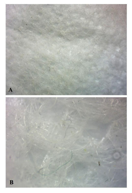
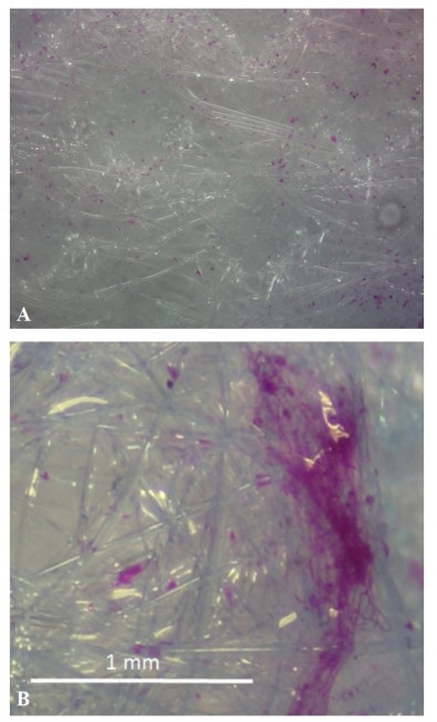
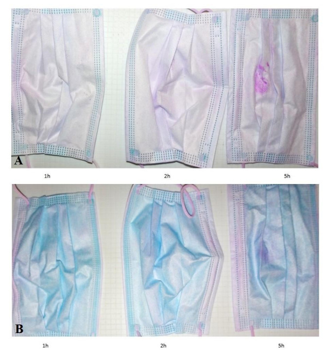
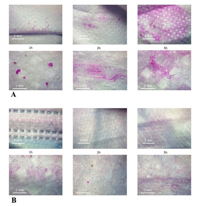
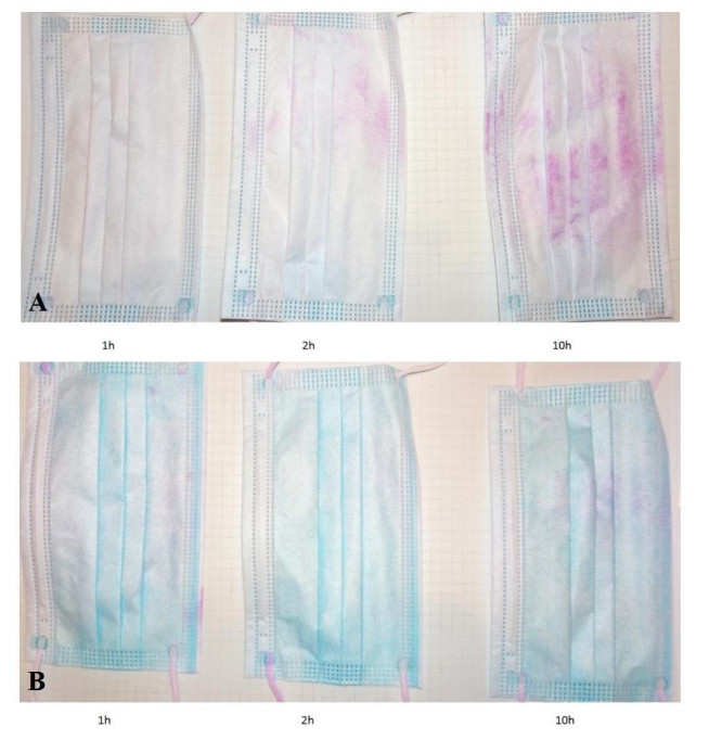
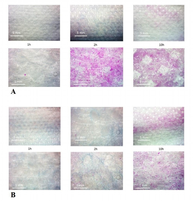
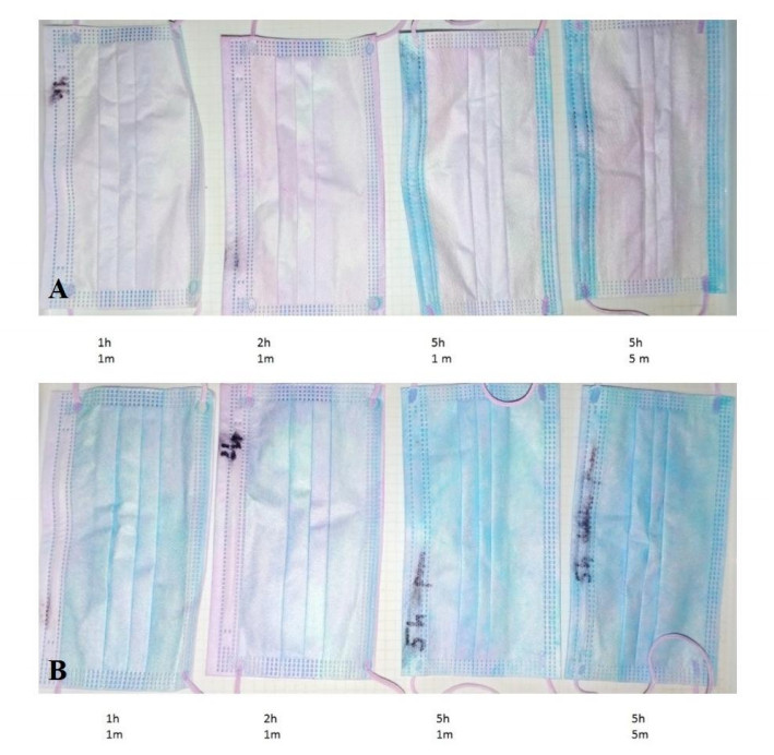
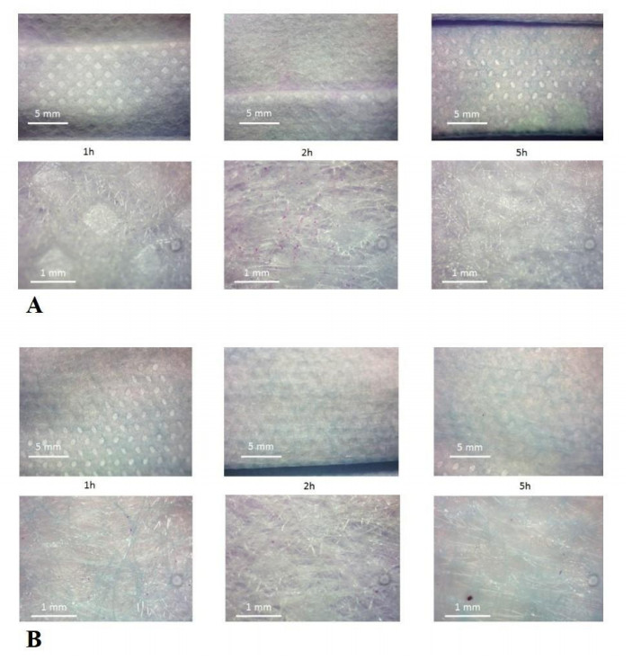
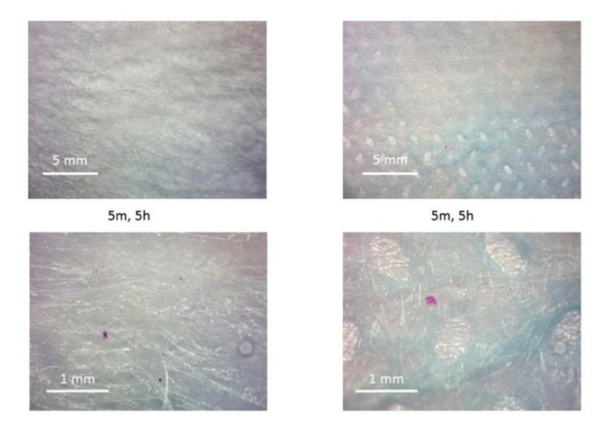
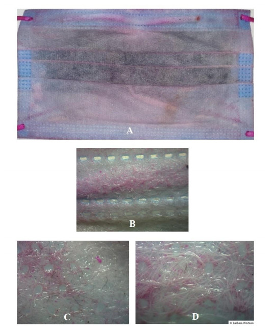
Unworn masks and masks provided to us after having been worn conformable to law (mandatory wearing of masks) served as test objects. In order to identify the distribution of living microorganisms on the surface of a mask dependent on exposure time and distance from the human face we conducted a staining study using the bengal rose method. The regular deposition of living microorganisms on artificial mask surfaces was more intense in the areas close to the mouth and nose. A time dependent accumulation was larger on the inside in comparison to the outside of the mask, even if the mask was not worn but only left in the room. The most interesting finding was the ability of microorganisms to penetrate all layers of the mask. We therefore conclude that masks are a suitable substrate for the cultivation of germs, even when not worn. Colonisation increases with human use and with time.
Citation: Kai Kisielinski, Barbara Wojtasik. Suitability of Rose Bengal sodium salt staining for visualisation of face mask contamination by living organisms[J]. AIMS Environmental Science, 2022, 9(2): 218-231. doi: 10.3934/environsci.2022015
Unworn masks and masks provided to us after having been worn conformable to law (mandatory wearing of masks) served as test objects. In order to identify the distribution of living microorganisms on the surface of a mask dependent on exposure time and distance from the human face we conducted a staining study using the bengal rose method. The regular deposition of living microorganisms on artificial mask surfaces was more intense in the areas close to the mouth and nose. A time dependent accumulation was larger on the inside in comparison to the outside of the mask, even if the mask was not worn but only left in the room. The most interesting finding was the ability of microorganisms to penetrate all layers of the mask. We therefore conclude that masks are a suitable substrate for the cultivation of germs, even when not worn. Colonisation increases with human use and with time.

| [1] |
Delanghe L, Cauwenberghs E, Spacova I, et al. (2021) Cotton and Surgical Face Masks in Community Settings: Bacterial Contamination and Face Mask Hygiene. Front Med (Lausanne) 8: 732047. doi:10.3389/fmed.2021.732047 doi: 10.3389/fmed.2021.732047

|
| [2] |
Sachdev R, Garg K, Singh G, et al. (2020) Is safeguard compromised? Surgical mouth mask harboring hazardous microorganisms in dental practice. J Family Med Prim Care 9: 759-763. doi:10.4103/jfmpc.jfmpc_1039_19 doi: 10.4103/jfmpc.jfmpc_1039_19
|
| [3] | Walton WR (1952) Techniques for recognition of living foraminifera. Cushman Found Foram Res Contr 3: 56-60. |
| [4] | Wojtasik B (2021) Method for assessment of biological corrosion of porous structures, including concrete, in particular hydrotechnical concrete. Polish Patent Office |
| [5] |
Wojtasik B, Zbawicka M, Grabarczyk L, et al. (2019) The lethal effect of hydrotechnical concrete on freshwater Bivalvia. Limnological Review. 19: 137-145. doi:10.2478/limre-2019-0012 doi: 10.2478/limre-2019-0012
|
| [6] |
Wojtasik B, Zbawicka M, Grabarczyk L, et al. (2021) Flow cytometric approach to evaluate the impact of hydro-technical concrete compounds' release to the freshwater microbiome. Environ Monit Assess 193: 698. doi:10.1007/s10661-021-09481-5 doi: 10.1007/s10661-021-09481-5
|
| [7] |
Kisielinski K, Giboni P, Prescher A, et al. (2021) Is a Mask That Covers the Mouth and Nose Free from Undesirable Side Effects in Everyday Use and Free of Potential Hazards? Int J Environ Res Public Health 18: 4344. doi:10.3390/ijerph18084344 doi: 10.3390/ijerph18084344
|
| [8] |
Monalisa AC, Padma KB, Manjunath K, et al. (2017) Microbial Contamination of the Mouth Masks Used by Post-Graduate Students in a Private Dental Institution: An In-Vitro Study. IOSR J Dent Med Sci 16: 61-67. doi:10.9790/0853-1605046167 doi: 10.9790/0853-1605046167
|
| [9] |
Luksamijarulkul P, Aiempradit N, Vatanasomboon P (2014) Microbial Contamination on Used Surgical Masks among Hospital Personnel and Microbial Air Quality in their Working Wards: A Hospital in Bangkok. Oman Med J 29: 346-350. doi:10.5001/omj.2014.92 doi: 10.5001/omj.2014.92
|
| [10] |
Zhiqing L, Yongyun C, Wenxiang C, et al. (2018) Surgical masks as source of bacterial contamination during operative procedures. J Orthop Translat 14: 57-62. doi:10.1016/j.jot.2018.06.002 doi: 10.1016/j.jot.2018.06.002
|
| [11] |
Rawal A (2020) Multi-layered masks to combat COVID-19. Indian J Med Res 152: 9-11. doi:10.4103/ijmr.IJMR_2709_20 doi: 10.4103/ijmr.IJMR_2709_20
|
| [12] | Organization WH. (2020) WHO - Advice on the use of masks in the context of COVID-19: interim guidance, 5 June 2020. Published online 2020. https://apps.who.int/iris/handle/10665/332293 |
Figures(10)

Kai Kisielinski, Barbara Wojtasik. Suitability of Rose Bengal sodium salt staining for visualisation of face mask contamination by living organisms[J]. AIMS Environmental Science, 2022, 9(2): 218-231. doi: 10.3934/environsci.2022015


 DownLoad: