
Citation: A. Sydney Gladman, Manuel Garcia-Leiner, Alexis F. Sauer-Budge. Emerging polymeric materials in additive manufacturing for use in biomedical applications[J]. AIMS Bioengineering, 2019, 6(1): 1-20. doi: 10.3934/bioeng.2019.1.1
[1] | Ana Letícia Braz, Ifty Ahmed . Manufacturing processes for polymeric micro and nanoparticles and their biomedical applications. AIMS Bioengineering, 2017, 4(1): 46-72. doi: 10.3934/bioeng.2017.1.46 |
[2] | Sven Sölmann, Anke Rattenholl, Hannah Blattner, Guido Ehrmann, Frank Gudermann, Dirk Lütkemeyer, Andrea Ehrmann . Mammalian cell adhesion on different 3D printed polymers with varying sterilization methods and acidic treatment. AIMS Bioengineering, 2021, 8(1): 25-35. doi: 10.3934/bioeng.2021004 |
[3] | Segun Akinola, Leelakrishna Reddy . Nanoscale antenna systems: Transforming wireless communications and biomedical applications. AIMS Bioengineering, 2023, 10(3): 300-330. doi: 10.3934/bioeng.2023019 |
[4] | Valeria Miranda-Arizmendi, Diana Fimbres-Olivarria, Anselmo Miranda-Baeza, Agustín Rascón-Chu, Jorge Marquez-Escalante, Jaime Lizardi-Mendoza, Mayra A. Méndez-Encinas, Elizabeth Carvajal-Millan . Sulfated polysaccharides from marine diatoms: Insight into molecular characteristics and biological activity. AIMS Bioengineering, 2024, 11(1): 110-129. doi: 10.3934/bioeng.2024007 |
[5] | E. Langford, C.A. Griffiths . The mechanical strength of additive manufactured intraosseous transcutaneous amputation prosthesis, known as the ITAP. AIMS Bioengineering, 2018, 5(3): 133-150. doi: 10.3934/bioeng.2018.3.133 |
[6] | Rachel Emerine, Shih-Feng Chou . Fast delivery of melatonin from electrospun blend polyvinyl alcohol and polyethylene oxide (PVA/PEO) fibers. AIMS Bioengineering, 2022, 9(2): 178-196. doi: 10.3934/bioeng.2022013 |
[7] | Daniel Calle, Duygu Yilmaz, Sebastian Cerdan, Armagan Kocer . Drug delivery from engineered organisms and nanocarriers as monitored by multimodal imaging technologies. AIMS Bioengineering, 2017, 4(2): 198-222. doi: 10.3934/bioeng.2017.2.198 |
[8] | Induni N. Weerarathna, Anurag Luharia . Exploring the nexus of biomedical science and robots for enhanced clinical outcomes—a literature review. AIMS Bioengineering, 2024, 11(1): 1-17. doi: 10.3934/bioeng.2024001 |
[9] | Amit Aherwar, Amit K Singh, Amar Patnaik . Current and future biocompatibility aspects of biomaterials for hip prosthesis. AIMS Bioengineering, 2016, 3(1): 23-43. doi: 10.3934/bioeng.2016.1.23 |
[10] | Dominik Gaweł, Paweł Główka, Michał Nowak, Tomasz Kotwicki . Visualization of the medical imaging data in 3D space using portable format. AIMS Bioengineering, 2016, 3(2): 176-187. doi: 10.3934/bioeng.2016.2.176 |
In recent years, the medical device community steadily adopted innovative uses of additive manufacturing (AM), also known as 3D printing (3DP), throughout the entire product lifecycle. Originally used only for rapid manufacture of visual aids or low-grade prototypes, advances in the quality and types of available materials and printing resolution have enabled medical device manufacturers opportunities to use AM as their final manufacturing methodology. In fact, as of December 4, 2017, the United States Food and Drug Administration (FDA) has cleared over 100 AM devices [1]. Some recent examples are listed in Table 1. Many of the approved devices address orthopedic specialties with joint replacements and bone grafts; however, devices for dentistry, general & plastic surgery, and neurology are also included.
The current devices capitalize on two key advantages offered by AM: the ability to fabricate complex porosities and patient-specific geometries. Being able to control the porosity of a material has significant implications for the improvement of implant osseointegration. For example, Stryker has commercialized a 3DP titanium alloy surface with defined porosity that enhances bone ingrowth (see an example product in Figure 1) [2],[3]. Stryker describes this ‘Tritanium’ surface as consisting of ‘random interconnected architecture with rugged, irregular pore sizes and shapes that are designed to mimic cancellous bone’ [4]. This unique surface can only be manufactured by AM.
The FDA approved the first 3DP drug, Spritam, in 2015 [5]. Spritam is formulated using the ZipDose Technology, which is a proprietary platform that produces formulations that rapidly disintegrate within seconds with a sip of liquid. The method starts by depositing a powder blend in a single layer, which is then bound together with a binding fluid. The process is repeated several times to yield a highly porous structure which enables the rapid dissolution [6].
Company Name | Device Name | 510(k) number | 510k Review Panel | Clearance Date | Material |
Additive Orthopaedics | Patient Specific 3D Printed Bone Segments | K180239 | Orthopedic | 5/16/2018 | Medical Grade Titanium Alloy (Ti-6Al-4V) |
Stryker | Intervertebral Fusion Device with Bone Graft | K1734776 | Orthopedic | 1/18/2018 | Titanium Alloy (Ti-6Al-4V) |
Clariance | Idys TLIF 3DTi Cages | K172465 | Orthopedic | 12/15/2017 | Titanium Alloy (Ti-6Al-4V) |
Medicrea International | IMPIX 3D Print Cages | K163595 | Orthopedic | 11/13/2017 | Titanium Alloy (Ti-6Al-4V) |
RTI Surgical | Fortilink Interbody Fusion with TETRAfuse 3D Technology | K172343 | Orthopedic | 10/23/2017 | Radiolucent Polyetherketoneketone (PEKK) |
Medtronic Sofamor Danek | ArTiC-L 3D Ti Spinal System with TiONIC Tech | K171689 | Orthopedic | 10/5/2017 | Titanium |
Medacta International | 3DMetal Tibial Cones | K170149 | Orthopedic | 8/18/2017 | Titanium Alloy |
Materialise | TruMatch CMF Titanium 3D Printed Implant System | K170272 | Dental | 8/8/2017 | Commercially Pure Titanium |
HT Medical | NeoFuse Ti3D PLIF//TLIF/Cervical Interbody | K170318 | Orthopedic | 7/12/2017 | Titanium Grade 23 |
Additive Orthopaedics | Bunion System | K163593 | Orthopedic | 6/20/2017 | Medical Grade Titanium Alloy (Ti-6Al-4V) |
MicroPort Orthopedics | BIOFOAM AM | K170288 | Orthopedic | 6/15/2017 | Commercially Pure Titanium |
RTI Surgical | Fortilink-C with TETRAfuse 3D Technology | K163673 | Orthopedic | 5/23/2017 | Radiolucent Polyetherketoneketone (PEKK) |
SI-Bone | iFuse Implant System - iFuse-3D implant | K162733 | Orthopedic | 3/10/2017 | Medical Grade Titanium Alloy (Ti-6Al-4V) |
Additive Orthopaedics | Hammertoe Correction System | K160264 | Orthopedic | 6/1/2016 | Medical Grade Titanium Alloy (Ti-6Al-4V) |
Renovis Surgical Technologies | Anterior Lumbar Interbody Fusion System | K142095 | Orthopedic | 10/15/2014 | Titanium |
Oxford Performance Materials | Osteofab Patient Specific Facial Device | K133809 | General & Plastic Surgery | 7/28/2014 | Polyetherketoneketone (PEKK) |
Renovis Surgical Technologies | Tesera Trabecular Technology (T3) Acetabular Shell System | K132312 | Orthopedic | 4/11/2014 | Titanium Alloy (Ti-6Al-4V) |
Stryker Orthopaedics | Triathlon Tritanium Metal-Backed Patella | K132624 | Orthopedic | 11/26/2013 | Titanium |
Tissue Regeneration Systems | TRS PCL Cranial Bone Void Filler | K123633 | Neurology | 8/16/2013 | Polycaprolactone (PCL) |
Another class of devices that leverage the unique capabilities of AM are those that are printed on-demand in patient-specific geometries. Devices in this class include custom implants for cranial surgery, dentistry, and maxillofacial surgery, molds for making prosthetics, and surgical guides [8]. Patient-specific implants are designed from magnetic resonance imaging (MRI) or computed tomography (CT) scans of the patient's bone or joint, which enables better alignment and fit to the patient's unique anatomy [9]. Studies specifically assessing whether patient-specific instrumentation improves patient outcomes have demonstrated significant improvements in the selection of the optimal implant and reduction in misaligned implants [10]. Additionally, the ability to design customized implants will continue to have a strong impact on the reconstruction of traumatic injuries and in the treatment of other complex disease processes [11].
Of the FDA approved devices, the majority are made of titanium, but some are manufactured from polymers such as polyetherketoneketone (PEKK) and polycaprolactone (PCL). Due to the variability of polymer formulations, the approval process can be more involved for polymeric devices. Nonetheless, many biomedical applications require materials with properties that can only be met with polymers. Thus, to date, the impact of AM on the medical device industry has been in part hindered by the lack of materials which meet the demands of biocompatibility and appropriate structural and rheological properties. Significant effort has been expended to meet this need with polymeric materials and this review focuses on these recent developments.
Although a wide range of AM methods are available, developments in polymer printing are focused in three main approaches: 1) powder bed fusion processes such as Selective Laser Sintering (SLS), 2) deposition-on-demand processes which include extrusion-based technologies, such as Fused Deposition Modeling (FDM) and direct ink-write printing [12], and inkjet or drop-wise deposition methods, and 3) photopolymer-based printing methods, such as stereolithography (SLA) and Continuous Liquid Interface Production (CLIP). These, have proven to be successful in incorporating a variety of polymers as raw materials. AM methods and instrumentation are the subject of numerous reviews [13]–[15], and thus we will not elaborate further on this topic.
When considering AM material choices for biomedical applications, the requirements include appropriate mechanical properties, temperature stability and chemical resistance for sterilization, radiolucency, and biocompatibility and/or biodegradability. ‘Hard’ polymers, such as engineering thermoplastics and thermosets, are good choices for structural implants, rigid medical devices, and dental applications. ‘Soft’ polymers include hydrogels and elastomers. The former is of specific interest to the tissue engineering field and the latter in the development of surgical models and phantoms. First, we will discuss the traditional ‘hard’ polymers, which have already begun making inroads to the marketplace. Second, we will highlight the emerging applications related to ‘soft’ polymers.
While a variety of traditional thermoplastics and thermosets are commercially available for use in AM (e.g., nylon, ABS, epoxides) [16], a few key polymers have found use in the emerging biomedical space. Biodegradable polymers are of interest for implantable devices that initially require a structural element, but as the natural tissue grows to fill in the repair site, the structural element is no longer needed. Non-biodegradable polymers have use in applications where structural support in needed for the lifetime of the implant. Table 2 lists some examples of hard polymers that are used in FDA approved devices and have been adapted for use in AM methods.
Many biodegradable polymers are polyesters that are degraded in the human body by hydrolysis of their ester linkages. Examples of such synthetic polymers that are used in biomedical devices and drug delivery platforms include polylactic acid (PLA), polycaprolactone (PCL), poly (glycolic acid) (PGA) and polydioxanone (PDO). Each of these polymers have different mechanical properties and degradation rates [17], which influence a product manufacturer's material selection process. The polymer properties can be further tuned by blending different polymers together.
PLA is arguably the most extensively used biodegradable polymer as it is derived from renewable resources, has good thermal processability, requires less energy to produce than petroleum-based polymers, and is biocompatible [18]. PLA's degradation products are neither toxic nor carcinogenic and do not interfere with tissue healing [19]. PLA has a relatively slow degradation rate with in vivo lifetimes normally in the range of 9–15 months [20], but can be up to 3 years [21], although degradation rate is a function of multiple factors, including degree of crystallinity, molecular weight, molecular weight distribution, morphology, water diffusion rate into the polymer, and stereoisomeric content. While PLA has many attractive qualities, it is also a very brittle material with less than 10% elongation at break, is relatively hydrophobic and lacks reactive side-chain groups limiting the ability to execute surface and bulk modifications.
PCL was first synthesized in the early 1930s, and is therefore one of the earliest polymers [22]. It is hydrophobic, semicrystalline, is easily processed at low temperatures, and is biocompatible. It degrades in vivo by both bulk and surface erosion mechanisms. PCL also has a relatively long lifetime in vivo with standard degradation times in the 9–12 month range [20], and has been widely used in long-term implants and controlled drug release applications [23]. To tune the degradation time and other properties, manufacturers can blend or copolymerize PCL with other polymers [24], for example as is done by Ethicon in their Monocryl resorbable sutures that is a co-polymer of glycolide and ε-caprolactone [25]. PGA is the simplest linear aliphatic polyester and was first patented in 1954 [26]. Due to its relatively high hydrolytic instability, PGA degrades in vivo by a bulk erosion mechanism with loss of mechanical properties in 1–2 months [20]. As previously mentioned, it is often combined with other polymers in biomedical applications [24].
PDO is a biodegradable polymer with repeating ether-ester units. Compared to the polyesters described above, PDO has been investigated less even though Ethicon introduced it to the marketplace in 1981 [27],[28]. PDO has several unique properties, including shape memory. PDO is best known for its clinical use as a suture, but the shape memory property hinders knot tying. However, this feature is very useful in some applications to provide rebound and kink resistance, such as in vascular conduits [29] and as a structural component in a surgical meshes [30]. PDO is assumed to degrade via bulk erosion, and suture absorption rate is in the range of 180 to 210 days [31].
Polymer | Abbreviation | Type | Structure | Examples of use in FDA approved devices |
Polylactic acid | PLA | Biodegradable polyester | SmartNail bone fixation nail (ConMed) [32] | |
Polycaprolactone | PCL | Biodegradable polyester | Monocryl sutures (Ethicon) [25] | |
Poly(glycolic acid) | PGA | Biodegradable polyester | Monocryl sutures (Ethicon) [25] | |
Polydioxanone | PDO | Biodegradable polyester | Ventrio Hernia Patch (BD Bard) [30] | |
Polyetheretherketone | PEEK | Thermoplastic | Rampart O Interbody Fusion (Spineology) [33] | |
Polyetherketoneketone | PEKK | Thermoplastic | Fortilink™-C Interbody Fusion (RTI Surgical) [34] |
Of particular interest in the category of non-biodegradable polymers are the family of polyaryletherketones (PAEKs). Among the PAEK family, polyetheretherketone (PEEK) is without a doubt the most commercially successful polymer, introduced in a multitude of applications that directly benefit from its unique physical properties. PEEK is a semi-crystalline thermoplastic with a linear, highly aromatic molecular backbone including ether and ketone linkages [35]–[37]. PEEK exhibits unparalleled properties compared to most other plastic resins, displaying excellent mechanical properties over a wide range of environmental conditions. PEEK products offer a high elastic modulus and ultimate strength coupled with post-yield ductility, and excellent creep resistance. PEEK is used as an alternative to metals in a growing number of applications [35]–[37]. Moreover, PEEK's unique properties have allowed for the development of implantable grades that are fully recognized as biocompatible and bioinert. Compared to metals, PEEK exhibits lower density, lower thermal and electrical conductivity, greater corrosion resistance, and compatibility with X-rays and magnetic fields often used in diagnostic imaging [38]–[40]. PEEK resins exhibit excellent solvent resistance, thermal oxidative stability, and radiation resistance, and can be sterilized by steam, ethylene oxide, and gamma radiation.
Another important member of the PAEK family is poly(etherketoneketone) (PEKK). Just like PEEK, PEKK is a semi-crystalline thermoplastic polymer with a linear, highly aromatic molecular structure including ether and ketone linkages. PEKK's chemical structure and synthetic route offer unique abilities to produce polymers with a wide range of thermal properties, superior mechanical properties, chemical resistance and low flammability. PEKK polymers differ from the general class of PAEKs in that the incorporation of the second ketone group to the polymer backbone leads to two different isomeric forms, included as repeating units along the polymer chain. A 1,4 addition, through a para-phenylene radical (terephthalic, T) provides linearity and chain stiffness to the polymer. In contrast, a 1,3 addition, through a meta-phenylene radical (isophthalic, I) increases the degree of freedom of the chain, reducing linearity and affecting the overall rate of crystallization. The ratio of these isomeric units (T/I) is often used to control the physical and chemical properties of PEKK resins, favoring precise manipulation of key parameters, including among others very high melting points and glass transition temperatures, a wide range of crystallization rates and degrees of crystallinity [41],[42]. These properties enable PEKK devices to be compatible with autoclave sterilization.
Unlike PEEK, in terms of crystal structure, PEKK resins are polymorphic and are able to crystallize in various crystalline unit cells, depending on the crystallization route. The crystal structure, polymorphism and morphology of PEKK has been reported in the literature [43]–[45]. Recent studies of the morphological changes observed in PEKK materials when subjected to specific heat treatments and processing conditions have provided useful information of particular interest for the introduction of PEKK resins in AM processes [41],[42]. In addition, PEKK can be tuned such that it has mechanically similar properties to cortical bone and thus is attractive for bone replacement implants [46]. In some specific cases, these approaches have led to commercially successful grades introduced into biomedical applications, particularly for patient-specific implants [47],[48].
Photocurable resins, which are thermosetting in nature, are another class of hard polymers employed in photopolymer-based printing methods such as SLA and CLIP, which have found particular use in dental applications (see Section 2.3.2). There are a number of commercial proprietary materials (e.g., polyurethane-based, cyanate ester-based, and acrylate-based) and printers used in this field [49]–[51].
Biomedical implants, which can be permanent or transient solutions for repair or replacement of biological tissues, are one of the most commonly investigated applications for AM in the biomedical space. Here, tissue engineering scaffolds can be fabricated with open network structures that enable structural function as well as integration with host tissue, and is most commonly employed for mesenchymal tissue (e.g., bone and cartilage) [56]. FDM-based processes have utilized PCL materials to fabricate bone and cartilage tissue scaffolds [57],[58], and there is continued research in scaffolds comprised of natural biological materials as will be discussed below. 3D powder binding with PLA and PLGA has been used to fabricate scaffolds for tissue engineering and drug delivery [59]. For the case of PEEK, high temperature laser sintering studies focused on the effect of build orientation have been recently reported with a focus on the introduction of PEEK to cranial implants and the healthcare sector in general [60],[61]. Importantly, patient-specific imaging data can be utilized to resolve the specific geometry needed for the implant, which can be imported into the 3D printing software to create the exact morphology necessary for the implant [62]. In an exciting example of the impact that polymer-based AM can have on biomedical implants, researchers recently successfully printed a tracheal split for at least four children suffering from tracheobronchomalacia since 2013 (Figure 2a) [52],[63]. There are a number of technical and regulatory considerations to ensure that 3D-printed implants have a path to commercial adoption [64]. While PDO is poised to be used in AM with resins available [65], no published examples of 3DP biomedical devices were found.
The dental market has been one of the earliest adopters of AM technology in the biomedical industry (Figure 3). Silicone and wax based materials are currently used to 3D print molds for various dental elements, and in some cases acrylates and other materials are used for the direct printing of teeth or jaw implants, prosthetics, and other applications including modeling and drilling guides for facial surgeries and dental instruments [66]. In orthodontics, dental alignment services currently produce millions of custom aligners per year using SLA-based technologies (Figure 2b). A variety of commercial printers and materials are available for use in the dental industry [49]–[51] with continued development of advanced polymeric and composite resins.
AM can also supplement the medical design product development process by aiding in low cost device verification by replacing testing in animal and/or cadaver models, for example in the case of surgical tools [68], with 3D printed anatomical models. Besides the obvious limitations of cost, biosafety, availability and ease of use concerns with testing on cadavers, the conventional models do not usually have the specific pathologies that the medical device is meant to address. Thus, AM anatomical models are increasingly being used [69],[70]. Moreover, expected anatomical variations, e.g., small, medium, and large or straight and tortuous lumens can easily be printed. These models, particularly patient-specific models, are also being applied to surgical preparation for complex anatomies. Numerous examples have been published demonstrating improved outcomes of patients when physicians have been trained on 3D-printed models from patient CT-scans vs. preparation by studying the images alone [54],[71]–[75]. These models may incorporate a variety of commodity-grade AM polymeric materials as they do not require biocompatibility or biodegradability (Figure 2c). Some polymers such as polyurethane can produce either hard polymeric structures or soft polymeric structures based on the specific chemistry and degree of cross-linking, and as such can be considered in either category depending upon the formulation.
The trend in prosthetic arms and hands has moved towards systems that include additional degrees of actuation. AM has been applied to address various hardware design challenges, patient-specific customization, and facile repairs [76]. Additionally, efforts are being made to increase accessibility of prosthetic designs in low-resource settings for the ∼40 million amputees by providing guidelines for non-assembly based designs [77]. As AM moves into the consumer space, open-source designs for customizable prosthetics are being collected in centralized repositories [55]. Many different types of polymeric materials and printing techniques have been utilized for this application (Figure 2d).
Unlike many of the hard materials utilized in AM of biomedical applications, which build upon commercial success of traditional AM methods and materials, the use of soft materials such as hydrogels in this space is a nascent development requiring further research and development before commercial adoption. This is partly due to some of the unique applications such as drug delivery and tissue engineering, which require more complex performance and regulatory considerations.
Soft polymers used in biomedical AM applications can be categorized as natural, synthetic, or hybrid materials. Natural soft polymers are those commonly associated with the extracellular matrix such as collagen, hyaluronic acid, fibrin/fibrinogen, and mixtures thereof [78]. Such natural polymers are highly favored due to their inherent biocompatibility and bioactivity, but often suffer from limitations in processability and cost. A subset of natural polymers used in biomedical AM are not native to humans, which include cellulose, silk, alginate, chitosan, and agarose, as well as gelatin, which is a denatured form of collagen [78]. Researchers often employ blends or composites of these materials to capitalize on the enhancements in mechanical performance and biofunctionality when combined [79]. Almost all polymers in this category are biodegradable and thus are intended to serve as transient implants or delivery vehicles.
Synthetic soft polymers for use in biomedical AM include polyethylene glycol (PEG), acrylate-based hydrogels, elastomers, and hydrogels with unique thermal properties such as the poloxamer Pluronic F127 [80]. Such materials have finer control in chemistry, structure, and physical properties such as rheological flow, but often lack biocompatibility and degradation pathways. Elastomers (e.g., polyurethanes and silicones) are most commonly used as tissue phantoms or surgical models due to their mechanical and tactile resemblance to native tissue (See Section 3.2.3), but are less commonly considered for implantables due to their lack of biodegradability. Synthetic hydrogels may find use in 3D-printed contact lenses [81] or pharmaceutical delivery applications (See Section 3.2.2). Pluronic F127 (a poly(ethylene oxide)–poly(propylene oxide) copolymer) has found unique use as a templating material for bioprinting, where the polymer can be applied in specific patterns and encapsulated with another material of choice [82]–[84]. Upon exposure to refrigeration temperature, the micellar structure of the Pluronic F127 disassembles, liquefying the gel. With application of a light suction, the Pluronic F127 can be removed to reveal open structures, most notably serving as vascularization paths for the printed scaffold or tissue construct or as templates for other tubular tissue structures such as renal proximal tubules [82]–[84].
Hybrid soft polymers, or those which contain both natural and synthetic moieties, find themselves at the intersection of synthetic and natural polymer attributes. Natural polymers like gelatin and hyaluronic acid suffer from processing and stability challenges in their native form [85] and thus rheological properties must be tuned to enable extrusion-based printing. Gelatin is a thermally reversible gel, which returns to the liquid state at body temperature, making it difficult to maintain 3D-printed conformation at physiological conditions [86] in its natural state. Hyaluronic acid is a linear polymer, which forms a loose physical entanglement that provides minimal structural integrity in solution [87]. A common strategy to improve the structural integrity of such materials involves the reaction of free amine groups with methacrylic anhydride to produce methacrylated compounds (i.e., gelatin methacrylol or GelMA, methacrylated hyaluronic acid or MeHA) [88],[89]. The methacrylation enables the hydrogels to be covalently crosslinked via free radicals to form stable network gels, which retain their native enzymatic degradation capabilities, but have greater control over rheological and mechanical properties. Both GelMA and MeHA have found use in biomedical AM applications [84],[90],[91].
Another form of hybrid soft polymers for medical AM applications involves the conjugation of bioactive moieties to synthetic polymer backbones. The most notable example of this type is the conjugation of arginylglycylaspartic acid (RGD) peptide to synthetic polymer backbones such as PEG or acrylated-PEGs, although a variety of bioactive additives have been considered [92]. Here, the peptide enables the synthetic polymer to adhere to the cell membrane, thus promoting cellular adhesion and proliferation on the surface of printed scaffolds and structures. This technique allows for the fine control over molecular weight, network structure, and rheology with synthetic hydrogel backbones while integrating cellular response.
Class of Soft Polymer | Example polymers | Example structure |
Natural polymer | hyaluronic acid | |
alginate | ||
Synthetic polymer | Pluronic F127 | |
silicone | ||
Hybrid soft polymers | acrylate-PEG-RGD | |
GelMA |
One of the applications that would have the most disruptive impact on the biomedical field is in AM-driven tissue engineering and bioprinting, where cell-laden or cell-free scaffolds and tissue constructs could enable functional implants and devices for disease modeling, and tissue/organ repair and replacement [93]. While this field has yet to penetrate the commercial biomedical market, the research and development in this space in the last few decades has expanded substantially. The most common methods employed in bioprinting are extrusion-based printing (e.g., direct-write), photopolymer-based printing such as SLA, electrohydrodynamic printing or droplet-based printing methods such as inkjet printing [94]–[96]. A variety of polymeric materials, including natural, synthetic, and natural-synthetic hybrids have been utilized in each of these methods, with specific materials highlighted above. In each case, if live cell printing is being conducted, careful attention to processing parameters that may affect cell viability and function must be considered, such as light and temperature exposure and shear stresses [97].
One notable advancement in AM-based tissue engineering and bioprinting is the incorporation of vascularization. The tissue engineering field has suffered for decades with the inability to incorporate functional vasculature, which prevent tissue constructs from reaching substantial thicknesses necessary for use as implants or viable tissue models. By the use of sacrificial templates or direct printing of hollow tubes, researchers have enabled the patterning of vascular structures, which can reveal open vascular networks capable of lining with native vascular cells such as endothelial cells [84],[98]. This advancement has enabled tissue constructs to be printed upwards of a centimeter in thickness with actively perfusable vasculature, enabling viability greater than one month (Figure 4a) [83].
Drug delivery applications are seeing a growing interest in the biomedical AM community [99]. The first FDA-approved 3D-printed drug, Spritam, was approved in 2015 for treatment of epilepsy [100]. The powder-bed based printing method (ZipDose Technology [6]) enabled a rapidly disintegrating tablet to be produced, marketed for patients with difficulty swallowing tablets [101]. While bioresorbable materials such as PLA and PCL have been employed, hydrophilic, water-soluble polymers such as PEG and cellulose have also been investigated as drug-delivery platforms via AM [102]. Printed stimuli-responsive hydrogels have garnered interest for drug-delivery systems due to their ability to release pharmaceutical agents in response to a specific environmental trigger (Figure 4b) [103]–[105]. These AM drug-delivery strategies enable unique geometries as well as graded and layered structures with control over composition and release kinetics that may enable ‘personalized’ medicine.[106]
Tissue phantoms and surgical models are currently attainable applications for soft (and in some cases hard) polymers as they are intended to be models that do not intend to be implanted or therapeutic in nature. Instead, tissue phantoms enable validation of disease modeling and assistance with diagnosis and treatment plans for injuries and disease states. They may mimic tissue properties in tactile or visual representations, or mimic ultrasound or CT imaging representations [107]. Such materials must have the aforementioned characteristics of native tissue with precise control over geometry. Elastomers (e.g., polyurethane elastomers and polydimethylsiloxane (PDMS)) are commonly employed for this application, where AM can enable fine control over spatial resolution [107]–[109]. Multimaterial or gradient printing can be employed to enable printed structures with localized regions of relatively stiff or soft materials for mimicking the tactile properties of a given tissue, and can also vary the color and translucency for visual inspection by medical professionals (Figure 4c) [110].
Biomedical applications will continue to benefit from the additive manufacturing of polymeric materials. Hard polymers, which have seen early adoption by the medical community, will continue to be utilized due to their emerging success in FDA-approved implantable devices and their market penetration in the dental community. Research in soft polymers for biomedical AM applications is accelerating, and with continued development, clinical breakthroughs are anticipated particularly in the area of tissue engineering, bioprinting, and personalized medicine. Both types of polymers contribute to the successful implementation of realistic surgical models or phantoms due to AM's unmatched ability to produce complex models with fine geometrical features, and in some cases, tunable mechanical properties. Nascent breakthroughs in additive manufacturing include holographic printing [111] and 4D printing (i.e., printed stimuli-responsive structures capable of changing over time) [112]. The former may enable exceedingly fast printing speeds for the fabrication of large biomedical devices and implants, while the latter produces responsive shape-changing geometries that may impact drug delivery or ‘smart’ implantables. There remains a need for continued development and characterization of polymeric materials suitable for AM methods for use in these biomedical applications, and with time, many believe that a 3D printer will become a standard outfit in the clinic to assist medical professionals in every aspect of their care and treatment.
[1] | Gottlieb S (2017) Ushering in new era of 3D printing of medical products; provides guidance to manufacturers of medical devices. United States Food and Drug Administration (FDA). Available from : https://www.fda.gov/newsevents/newsroom/pressannouncements/ucm587547.htm. |
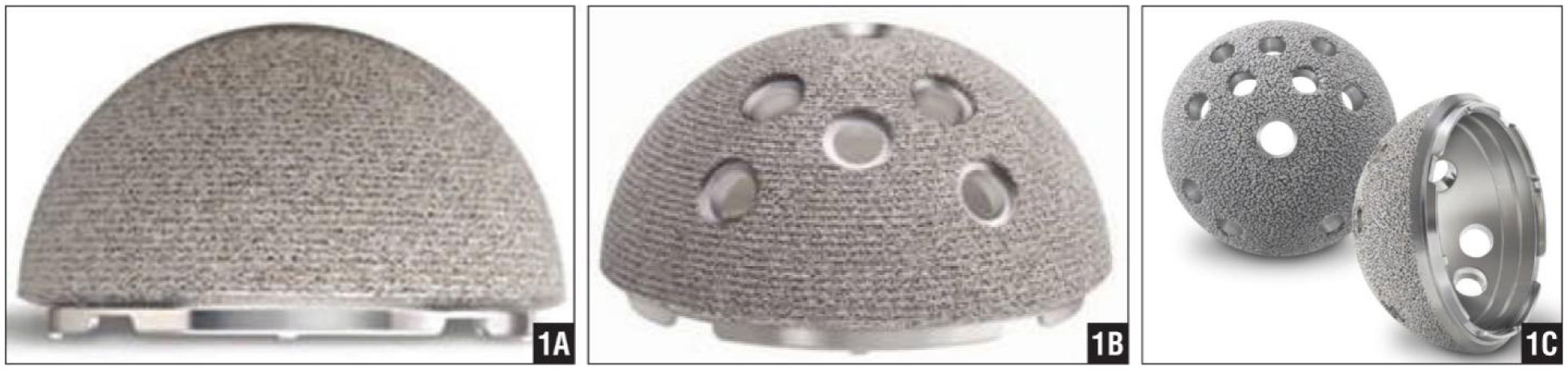
[2] | Frenkel SR, Jaffe WL, Dimaano F, et al. (2004) Bone response to a novel highly porous surface in a canine implantable chamber. J Biomed Mater Res B Appl Biomater 71: 387–391. |
[3] | United States Food and Drug Administration (FDA) (2013) K123486 triathalon tritanium tibial baseplate. Available from: https://www.accessdata.fda.gov/scripts/cdrh/cfdocs/cfpmn/pmn.cfm?ID=K123486. |
[4] | Stryker Tritanium. In-growth technology built to fuse. Available from: http://www.stryker.com/builttofuse/#. |
[5] |
Alhnan MA, Okwuosa TC, Sadia M, et al. (2016) Emergence of 3D printed dosage forms: Opportunities and challenges. Pharm Res 33: 1817–1832. doi: 10.1007/s11095-016-1933-1
![]() |
[6] | Aprecia Pharmaceuticals. What is ZipDose technology? Available from: https://www.spritam.com/#/hcp/zipdose-technology/what-is-zipdose-technology. |
[7] | De Jesus C (2016) New Tritanium spinal implant fully integrates with the human body, Available from : https://futurism.com/3d-printed-tritanium-spinal-implant-simulates-actual-bone. |
[8] |
Tack P, Victor J, Gemmel P, et al. (2016) 3D-printing techniques in a medical setting: a systematic literature review. Biomed Eng Online 15: 115. doi: 10.1186/s12938-016-0236-4
![]() |
[9] |
Haglin JM, Eltorai AE, Gil JA, et al. (2016) Patient-specific orthopaedic implants. Orthop Surg 8: 417–424. doi: 10.1111/os.12282
![]() |
[10] |
Hendel MD, Bryan JA, Barsoum WK, et al. (2012) Comparison of patient-specific instruments with standard surgical instruments in determining glenoid component position: A randomized prospective clinical trial. J Bone Joint Surg Am 94: 2167–2175. doi: 10.2106/JBJS.K.01209
![]() |
[11] |
Bauermeister AJ, Zuriarrain A, Newman MI (2016) Three-dimensional printing in plastic and reconstructive surgery: A systematic review. Ann Plast Surg 77: 569–576. doi: 10.1097/SAP.0000000000000671
![]() |
[12] |
Lewis JA (2006) Direct ink writing of 3D functional materials. Adv Funct Mater 16: 2193–2204. doi: 10.1002/adfm.200600434
![]() |
[13] | Wong KV, Hernandez A (2012) A review of additive manufacturing. ISRN Mech Eng 2012: 10. |
[14] |
Bikas H, Stavropoulos P, Chryssolouris G (2016) Additive manufacturing methods and modelling approaches: A critical review. Int J Adv Manuf Technol 83: 389–405. doi: 10.1007/s00170-015-7576-2
![]() |
[15] |
Ngo TD, Kashani A, Imbalzano G, et al. (2018) Additive manufacturing (3D printing): A review of materials, methods, applications and challenges. Compos Part B: Eng 143: 172–196. doi: 10.1016/j.compositesb.2018.02.012
![]() |
[16] | Stratasys Ltd. our materials. Available from: https://www.stratasys.com/materials/search. |
[17] |
Lyu S, Untereker D (2009) Degradability of polymers for implantable biomedical devices. Int J Mol Sci 10: 4033–4065. doi: 10.3390/ijms10094033
![]() |
[18] |
Farah S, Anderson DG, Langer R (2016) Physical and mechanical properties of PLA, and their functions in widespread applications - A comprehensive review. Adv Drug Deliv Rev 107: 367–392. doi: 10.1016/j.addr.2016.06.012
![]() |
[19] |
Gupta B, Revagade N, Hilborn J (2007) Poly(lactic acid) fiber: An overview. Prog Poly Sci 32: 455–482. doi: 10.1016/j.progpolymsci.2007.01.005
![]() |
[20] |
Hutmacher DW (2000) Scaffolds in tissue engineering bone and cartilage. Biomaterials 21: 2529–2543. doi: 10.1016/S0142-9612(00)00121-6
![]() |
[21] |
Rasal RM, Janorkar AV, Hirt DE (2010) Poly (lactic acid) modifications. Prog Poly Sci 35: 338–356. doi: 10.1016/j.progpolymsci.2009.12.003
![]() |
[22] | Natta FJv, Hill JW, Carothers WH (1934) Studies of polymerization and ring formation. XXIII.1 ε-Caprolactone and its polymers. J Am Chem Soc 56: 455–457. |
[23] |
Woodruff MA, Hutmacher DW (2010) The return of a forgotten polymer-Polycaprolactone in the 21st century. Prog Poly Sci 35: 1217–1256. doi: 10.1016/j.progpolymsci.2010.04.002
![]() |
[24] |
Athanasiou KA, Agrawal CM, Barber FA, et al. (1998) Orthopaedic applications for PLA-PGA biodegradable polymers. Arthroscopy 14: 726–737. doi: 10.1016/S0749-8063(98)70099-4
![]() |
[25] | Ethicon Monocryl (poliglecaprone 25) suture. (2018) Available from: https://www.ethicon.com/emea/products/wound-closure/absorbable-sutures/monocryl-poliglecaprone-25-suture. |
[26] | Lowe CE (1954) Preparation of high molecular weight polyhydroxyacetic ester. USA Patent Office. US2668162A. |
[27] |
Goonoo N, Jeetah R, Bhaw-Luximon A, et al. (2015) Polydioxanone-based bio-materials for tissue engineering and drug/gene delivery applications. Eur J Pharm Biopharm 97: 371–391. doi: 10.1016/j.ejpb.2015.05.024
![]() |
[28] | Bartholomew RS (1981) PDS (polydioxanone suture): A new synthetic absorbable suture in cataract surgery. A preliminary study. Ophthalmologica 183: 81–85. |
[29] |
Boland ED, Coleman BD, Barnes CP, et al. (2005) Electrospinning polydioxanone for biomedical applications. Acta Biomater 1: 115–123. doi: 10.1016/j.actbio.2004.09.003
![]() |
[30] | Bard Ventrio Hernia Patch. (2018) Available from: https://www.crbard.com/Davol/en-US/products/Ventrio-Hernia-Patch. |
[31] | Gierek M, Kusnierz K, Lampe P, et al. (2018) Absorbable sutures in general surgery - review, available materials, and optimum choices. Pol Przegl Chir 90: 34–37. |
[32] | ConMed SmartNail Implant. Available from: http://www.conmed.com/en/products/orthopedics/knee/cartilage/cartilage-repair/smartnail-implant. |
[33] | Spineology Rampart O Interbody Fusion. Available from: https://www.spineology.com/united-states/our-products/rampart-o. |
[34] | FDA (2017) K163673 510K Summary Fortilink-C with TETRAfuse 3D Technology, available from: https://www.accessdata.fda.gov/cdrh_docs/pdf16/K163673.pdf. |
[35] |
Herold F, Schneller A (1992) High-performance polymers. Adv Mater 4: 143–152. doi: 10.1002/adma.19920040304
![]() |
[36] |
Nguyen HX, Ishida H (1987) Poly(aryl-ether-ether-ketone) and its advanced composites - a review. Polym Composite 8: 57–73. doi: 10.1002/pc.750080202
![]() |
[37] | Fink JK (2008) High performance polymers. Norwich, NY: William Andrew, Inc. |
[38] | Kemmish D (2010) Update on the technology and applications of polyaryletherketones. Shawbury, UK: Smithers Rapra Publishing. |
[39] |
Kurtz SM, Devine JN (2007) PEEK biomaterials in trauma, orthopedic, and spinal implants. Biomaterials 28: 4845–4869. doi: 10.1016/j.biomaterials.2007.07.013
![]() |
[40] | Kurtz SM (2012) PEEK Biomaterials Handbook. Amsterdam: Elsevier, Inc. |
[41] | Garcia-Leiner M, Clay B, Ricou P (2014) High performance polymers in Selective Laser Sintering processes: Understanding structure and property; San Francisco, CA. ACS Fall 2014 National Meeting. |
[42] | Bertelo CA, Garcia-Leiner MA, DeCarmine A, et al. (2017) Heat treated polymer powders, USA, US 9587107. |
[43] |
Cheng SZD, Ho RM, Hsiao BS, et al. (1996) Polymorphism and crystal structure identification in poly(aryl ether ketone ketone)s. Macromol Chem Phys 197: 185–213. doi: 10.1002/macp.1996.021970115
![]() |
[44] |
Gardner KH, Hsiao BS, Matheson RR, et al. (1992) Structure, crystallization and morphology of poly (aryl ether ketone ketone). Polymer 33: 2483–2495. doi: 10.1016/0032-3861(92)91128-O
![]() |
[45] |
Klop EA, Lommerts BJ, Veurink J, et al. (1995) Polymorphism in alternating polyketones studied by X-ray-diffraction and calorimetry. J Polym Sci Pol Phys 33: 315–326. doi: 10.1002/polb.1995.090330217
![]() |
[46] | OPM OsteoFab implantable medical devices (2018) Available from: http://oxfordpm.com/cmf-orthopedics. |
[47] | Berretta S (2015) Poly ether ether ketone (PEEK) polymers for high temperature laser sintering (HT-LS): University of Exeter. |
[48] | Berretta S, Ghita O, Evans KE (2014) Morphology of polymeric powders in laser sintering (LS): From polyamide to new PEEK powders. EurPolym J 59: 218–229. |
[49] | Carbon dental materials (2018) Available from: https://www.carbon3d.com/industry/dental-materials/. |
[50] | Formlabs (2018) High-accuracy 3D printing materials for dental labs and practices. Available from: https://formlabs.com/materials/dental/. |
[51] | envisionTEC: Digital dentistry in action (2018) Available from: https://envisiontec.com/wp-content/uploads/2017/08/2017-Dental-Materials-Final-08212017.pdf. |
[52] |
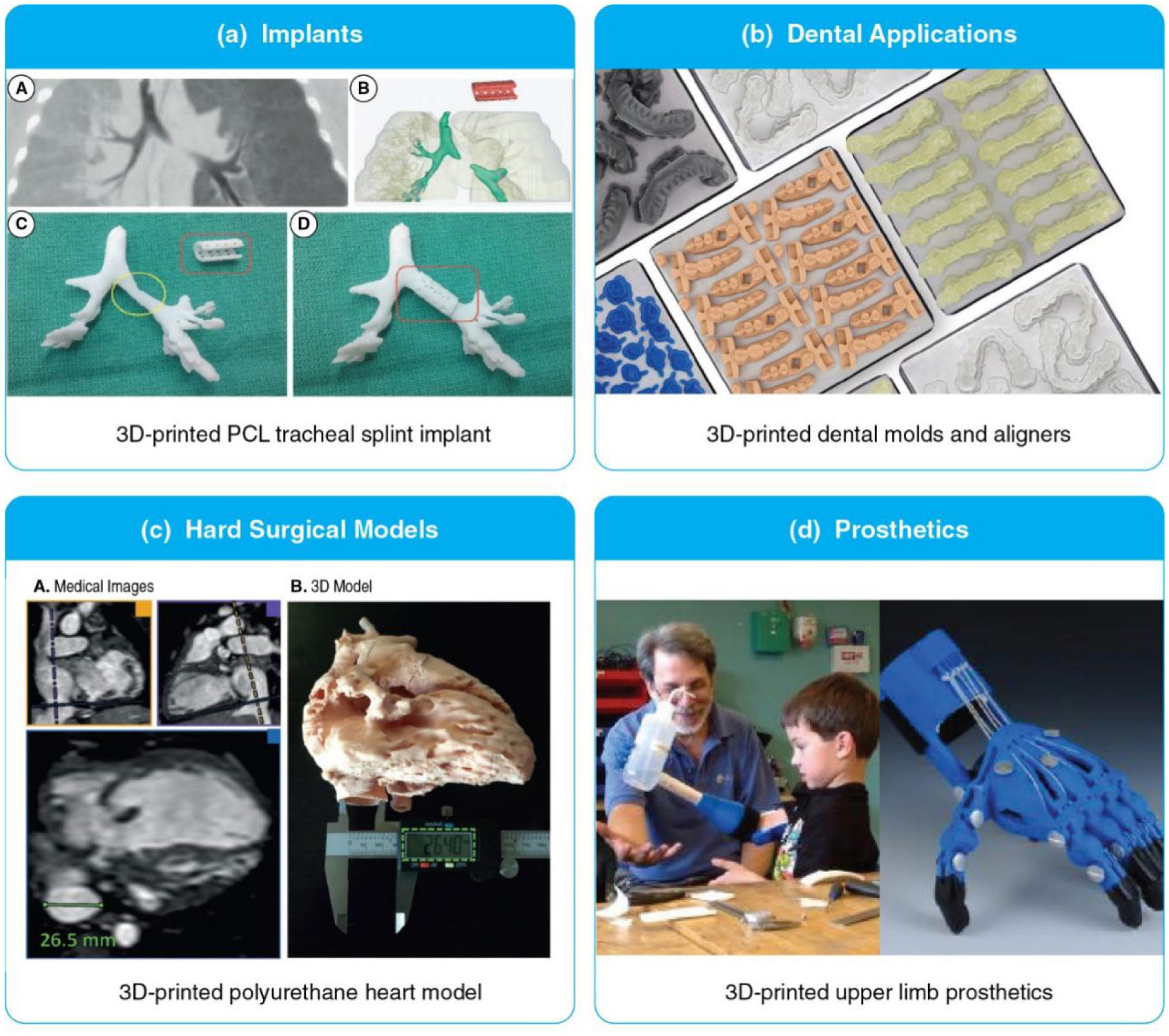
Zopf DA, Hollister SJ, Nelson ME, et al. (2013) Bioresorbable airway splint created with a three-dimensional printer. N Engl J Med 368: 2043–2045. doi: 10.1056/NEJMc1206319
![]() |
[53] | Form 2 dentistry resin (FormLabs) (2018) Available from: https://formlabs.com/industries/dental/ https://formlabs.com/materials/dental/. |
[54] |
Valverde I, Gomez-Ciriza G, Hussain T, et al. (2017) Three-dimensional printed models for surgical planning of complex congenital heart defects: An international multicentre study. Eur J Cardiothorac Surg 52: 1139–1148. doi: 10.1093/ejcts/ezx208
![]() |
[55] | e-NABLE 3D-printable prosethetic devices. Available from: https://3dprint.nih.gov/collections/prosthetics. |
[56] |
Hutmacher DW (2000) Scaffolds in tissue engineering bone and cartilage. Biomaterials 21: 2529–2543. doi: 10.1016/S0142-9612(00)00121-6
![]() |
[57] |
Park SH, Yun BG, Won JY, et al. (2017) New application of three-dimensional printing biomaterial in nasal reconstruction. Laryngoscope 127: 1036–1043. doi: 10.1002/lary.26400
![]() |
[58] |
Shim JH, Kim JY, Park M, et al. (2011) Development of a hybrid scaffold with synthetic biomaterials and hydrogel using solid freeform fabrication technology. Biofabrication 3: 034102. doi: 10.1088/1758-5082/3/3/034102
![]() |
[59] | Giordano RA, Wu BM, Borland SW, et al. (1996) Mechanical properties of dense polylactic acid structures fabricated by three dimensional printing J Biomater Sci, 8: 63–75. |
[60] |
Berretta S, Evans KE, Ghita O (2015) Processability of PEEK, a new polymer for high temperature laser sintering (HT-LS). Eur Polym J 68: 243–266. doi: 10.1016/j.eurpolymj.2015.04.003
![]() |
[61] |
Berretta S, Evans K, Ghita O (2018) Additive manufacture of PEEK cranial implants: Manufacturing considerations versus accuracy and mechanical performance. Mater Design 139: 141–152. doi: 10.1016/j.matdes.2017.10.078
![]() |
[62] |
Rengier F, Mehndiratta A, von Tengg-Kobligk H, et al. (2010) 3D printing based on imaging data: review of medical applications. Int J Comput Assist Radiol Surg 5: 335–341. doi: 10.1007/s11548-010-0476-x
![]() |
[63] | Sher D (2015) Materialise partners with University of Michigan for 3D printed tracheal splints, Available from: https://3dprintingindustry.com/news/materialise-partners-with-university-of-michigan-for-3d-printed-tracheal-splints-63111/. |
[64] |
Morrison RJ, Kashlan KN, Flanangan CL, et al. (2015) Regulatory considerations in the design and manufacturing of implantable 3D-printed medical devices. Clin Transl Sci 8: 594–600. doi: 10.1111/cts.12315
![]() |
[65] |
Mohseni M, Hutmacher D, Castro N (2018) Independent evaluation of medical-grade bioresorbable filaments for fused deposition modelling/fused filament fabrication of tissue engineered constructs. Polymers 10: 40. doi: 10.3390/polym10010040
![]() |
[66] |
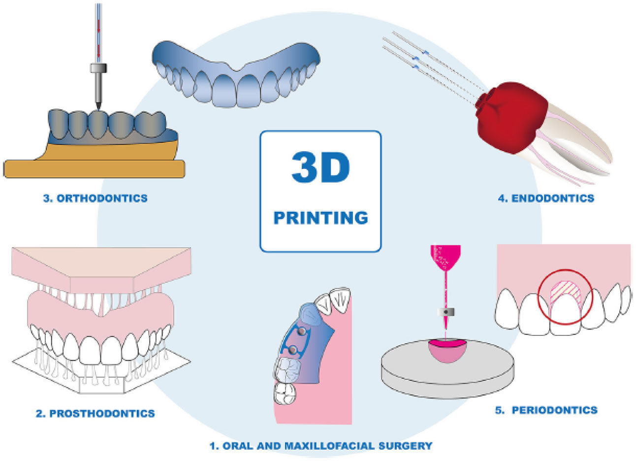
Dawood A, Marti Marti B, Sauret-Jackson V, et al. (2015) 3D printing in dentistry. Br Dent J 219: 521–529. doi: 10.1038/sj.bdj.2015.914
![]() |
[67] | Oberoi G, Nitsch S, Edelmayer M, et al. (2018) 3D printing-encompassing the facets of dentistry. Front Bioeng Biotechnol 6. |
[68] |
Rosen JE, Size A, Yang Y, et al. (2015) Artificial hand for minimally invasive surgery: design and testing of initial prototype. Surg Endosc 29: 61–67. doi: 10.1007/s00464-014-3657-9
![]() |
[69] |
Salmi M, Paloheimo KS, Tuomi J, et al. (2013) Accuracy of medical models made by additive manufacturing (rapid manufacturing). J Craniomaxillofac Surg 41: 603–609. doi: 10.1016/j.jcms.2012.11.041
![]() |
[70] | Lichtenberger JP, Tatum PS, Gada S, et al. (2018) Using 3D printing (additive manufacturing) to produce low-cost simulation models for medical training. Mil Med 183: 73–77. |
[71] |
Zhao L, Zhou S, Fan T, et al. (2018) Three-dimensional printing enhances preparation for repair of double outlet right ventricular surgery. J Card Surg 33: 24–27. doi: 10.1111/jocs.13523
![]() |
[72] | Meess KM, Izzo RL, Dryjski ML, et al. (2017) 3D printed abdominal aortic aneurysm phantom for image guided surgical planning with a patient specific fenestrated endovascular graft system. Proc SPIE Int Soc Opt Eng 10138. |
[73] |
Torres IO, De Luccia N (2017) A simulator for training in endovascular aneurysm repair: The use of three dimensional printers. Eur J Vasc Endovasc Surg 54: 247–253. doi: 10.1016/j.ejvs.2017.05.011
![]() |
[74] | Raos P, Klapan I, Galeta T (2015) Additive manufacturing of medical models--Applications in rhinology. Coll Antropol 39: 667–673. |
[75] |
Rose AS, Webster CE, Harrysson OL, et al. (2015) Pre-operative simulation of pediatric mastoid surgery with 3D-printed temporal bone models. Int J Pediatr Otorhinolaryngol 79: 740–744. doi: 10.1016/j.ijporl.2015.03.004
![]() |
[76] |
Vujaklija I, Farina D (2018) 3D printed upper limb prosthetics. Expert Rev Med Dev 15: 505–512. doi: 10.1080/17434440.2018.1494568
![]() |
[77] | Cuellar JS, Smit G, Zadpoor AA, et al. (2018) Ten guidelines for the design of non-assembly mechanisms: The case of 3D-printed prosthetic hands. P I Mech Eng H 232: 962–971. |
[78] |
Gopinathan J, Noh I (2018) Recent trends in bioinks for 3D printing. Biomater Res 22: 11. doi: 10.1186/s40824-018-0122-1
![]() |
[79] | Jang T-S, Jung H-D, Pan HM, et al. (2018) 3D printing of hydrogel composite systems: Recent advances in technology for tissue engineering. Int J Bioprint 4: 126–127. |
[80] |
Derakhshanfar S, Mbeleck R, Xu K, et al. (2018) 3D bioprinting for biomedical devices and tissue engineering: A review of recent trends and advances. Bioact Mater 3: 144–156. doi: 10.1016/j.bioactmat.2017.11.008
![]() |
[81] | OIS J&J's plans for smart & 3D printable contact lenses Available from: https://ois.net/jjs-plans-for-smart-3d-printable-contact-lenses/. |
[82] |
Homan KA, Kolesky DB, Skylar-Scott MA, et al. (2016) Bioprinting of 3D convoluted renal proximal tubules on perfusable chips. Sci Rep 6: 34845. doi: 10.1038/srep34845
![]() |
[83] |
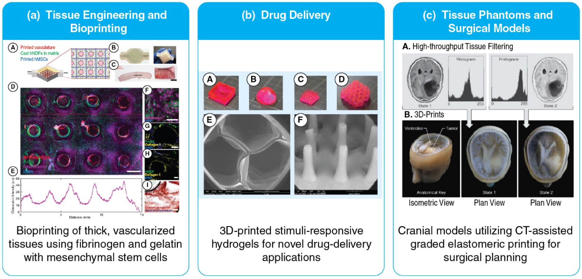
Kolesky DB, Homan KA, Skylar-Scott MA, et al. (2016) Three-dimensional bioprinting of thick vascularized tissues. Proc Nati Acad Sci USA 113: 3179–3184. doi: 10.1073/pnas.1521342113
![]() |
[84] |
Kolesky DB, Truby RL, Gladman AS, et al. (2014) 3D bioprinting of vascularized, heterogeneous cell-laden tissue constructs. Adv Mater 26: 3124–3130. doi: 10.1002/adma.201305506
![]() |
[85] |
He Y, Yang F, Zhao H, et al. (2016) Research on the printability of hydrogels in 3D bioprinting. Sci rep 6: 29977. doi: 10.1038/srep29977
![]() |
[86] |
Bode F, da Silva MA, Smith P, et al. (2013) Hybrid gelation processes in enzymatically gelled gelatin: impact on nanostructure, macroscopic properties and cellular response. Soft Matter 9: 6986–6999. doi: 10.1039/C3SM00125C
![]() |
[87] |
Burdick JA, Prestwich GD (2011) Hyaluronic acid hydrogels for biomedical applications. Adv Mater 23: H41–H56. doi: 10.1002/adma.201003963
![]() |
[88] |
Yue K, Trujillo-de Santiago G, Alvarez MM, et al. (2015) Synthesis, properties, and biomedical applications of gelatin methacryloyl (GelMA) hydrogels. Biomaterials 73: 254–271. doi: 10.1016/j.biomaterials.2015.08.045
![]() |
[89] | Ondeck MG, Engler AJ (2016) Mechanical characterization of a dynamic and tunable methacrylated hyaluronic acid hydrogel. J Biomech Eng 138: 0210031–0210036. |
[90] |
Poldervaart MT, Goversen B, de Ruijter M, et al. (2017) 3D bioprinting of methacrylated hyaluronic acid (MeHA) hydrogel with intrinsic osteogenicity. PLoS One 12: e0177628. doi: 10.1371/journal.pone.0177628
![]() |
[91] |
McBeth C, Lauer J, Ottersbach M, et al. (2017) 3D bioprinting of GelMA scaffolds triggers mineral deposition by primary human osteoblasts. Biofabrication 9: 015009. doi: 10.1088/1758-5090/aa53bd
![]() |
[92] |
Huettner N, Dargaville TR, Forget A (2018) Discovering cell-adhesion peptides in tissue engineering: Beyond RGD. Trends Biotechnol 36: 372–383. doi: 10.1016/j.tibtech.2018.01.008
![]() |
[93] | Estomba CMC, Fernández IG, Otero MAI (2017) 3D printing for biomedical applications: Where are we now? EMJ 2: 16–22. |
[94] |
Li J, Chen M, Fan X, et al. (2016) Recent advances in bioprinting techniques: Approaches, applications and future prospects. J Transl Med 14: 271. doi: 10.1186/s12967-016-1028-0
![]() |
[95] | Iwanaga S, Arai K, Nakamura M, (2015) Inkjet bioprinting. In: Atala A, Yoo JJ, Essentials of 3D Biofabrication and Translation, 4 Eds., Boston: Academic Press, 61–79. |
[96] | Liang H, He J, Chang J, et al. (2017) Coaxial nozzle-assisted electrohydrodynamic printing for microscale 3D cell-laden constructs. Int J Bioprint 4: 127. |
[97] |
Campbell J, McGuinness I, Wirz H, et al. (2015) Multimaterial and multiscale three-dimensional bioprinter. J Nanotechno Eng Medicine 6: 021005–021020. doi: 10.1115/1.4031230
![]() |
[98] | Hu N, Zhang YS, (2018) 3D bioprinting blood vessels. In: Thomas DJ, Jessop ZM, Whitaker IS, 3D Bioprinting for Reconstructive Surgery, 18 Eds Woodhead Publishing, 377–391. |
[99] | Lepowsky E, Tasoglu S (2018) 3D printing for drug manufacturing: A perspective on the future of pharmaceuticals. Int J Bioprint 4 |
[100] | FDA (2018) SPRITAM (levetiracetam) tablets, Available from: https://www.accessdata.fda.gov/drugsatfda_docs/nda/2015/207958Orig1s000TOC.cfm. |
[101] | Spritam What is SPRITAM? Available from: https://www.spritam.com/#/patient/about-spritam/what-is-spritam. |
[102] | Horst DJ (2018) 3D printing of pharmaceutical drug delivery systems. Arc Org Inorg Chem Sci 1: 65–69. |
[103] |
Larush L, Kaner I, Fluksman A, et al. (2017) 3D printing of responsive hydrogels for drug-delivery systems. J 3D Print Medicine 1: 219–229. doi: 10.2217/3dp-2017-0009
![]() |
[104] |
Gupta MK, Meng F, Johnson BN, et al. (2015) 3D printed programmable release capsules. Nano lett 15: 5321–5329. doi: 10.1021/acs.nanolett.5b01688
![]() |
[105] |
Buwalda SJ, Vermonden T, Hennink WE (2017) Hydrogels for therapeutic delivery: Current developments and future directions. Biomacromolecules 18: 316–330. doi: 10.1021/acs.biomac.6b01604
![]() |
[106] |
Lim SH, Kathuria H, Tan JJY, et al. (2018) 3D printed drug delivery and testing systems - a passing fad or the future? Adv Drug Deliver Rev 132: 139–168. doi: 10.1016/j.addr.2018.05.006
![]() |
[107] |
Cloonan AJ, Shahmirzadi D, Li RX, et al. (2014) 3D-printed tissue-mimicking phantoms for medical imaging and computational validation applications. 3D print addi manuf 1: 14–23. doi: 10.1089/3dp.2013.0010
![]() |
[108] |
Wang K, Ho C-C, Zhang C, et al. (2017) A review on the 3D printing of functional structures for medical phantoms and regenerated tissue and organ applications. Engineering 3: 653–662. doi: 10.1016/J.ENG.2017.05.013
![]() |
[109] |
Tappa K, Jammalamadaka U (2018) Novel biomaterials used in medical 3D printing techniques. J Funct Biomater 9: 17–33. doi: 10.3390/jfb9010017
![]() |
[110] |
Hosny A, Keating SJ, Dilley JD, et al. (2018) From improved diagnostics to presurgical planning: high-resolution functionally graded multimaterial 3D printing of biomedical tomographic data sets. 3D print addi manuf 5: 103–113. doi: 10.1089/3dp.2017.0140
![]() |
[111] |
Shusteff M, Browar AEM, Kelly BE, et al. (2017) One-step volumetric additive manufacturing of complex polymer structures. Sci Adv 3: eaao5496. doi: 10.1126/sciadv.aao5496
![]() |
[112] |
Gladman AS, Matsumoto EA, Nuzzo RG, et al. (2016) Biomimetic 4D printing. Nat Mater 15: 413–418. doi: 10.1038/nmat4544
![]() |
1. | Hatim Alqurashi, Ilida Ortega Asencio, Daniel W. Lambert, The Emerging Potential of Extracellular Vesicles in Cell-Free Tissue Engineering and Regenerative Medicine, 2020, 1937-3368, 10.1089/ten.teb.2020.0222 | |
2. | Matteo Gastaldi, Francesca Cardano, Marco Zanetti, Guido Viscardi, Claudia Barolo, Silvia Bordiga, Shlomo Magdassi, Andrea Fin, Ignazio Roppolo, Functional Dyes in Polymeric 3D Printing: Applications and Perspectives, 2021, 3, 2639-4979, 1, 10.1021/acsmaterialslett.0c00455 | |
3. | Saad Saleh Alghamdi, Sabu John, Namita Roy Choudhury, Naba K. Dutta, Additive Manufacturing of Polymer Materials: Progress, Promise and Challenges, 2021, 13, 2073-4360, 753, 10.3390/polym13050753 | |
4. | Anahita Rohani Shirvan, Alireza Nouri, Cuie Wen, 2021, 9780128188316, 395, 10.1016/B978-0-12-818831-6.00010-0 | |
5. | Amit Kumar, Chhotu Ram, Agave biomass: a potential resource for production of value-added products, 2021, 2523-8922, 10.1007/s42398-021-00172-y | |
6. | Jian Yang, Xin Gao, Xiaotao Xing, Haisen Huang, Qi Tang, Shixing Ma, Xun Xu, Cheng Liang, Maojiao Li, Li Liao, Weidong Tian, An Isolation System to Collect High Quality and Purity Extracellular Vesicles from Serum, 2021, Volume 16, 1178-2013, 6681, 10.2147/IJN.S328325 | |
7. | Adil Wazeer, Apurba Das, Arijit Sinha, Kazuaki Inaba, Su Ziyi, Amit Karmakar, Additive manufacturing in biomedical field: a critical review on fabrication method, materials used, applications, challenges, and future prospects, 2022, 2363-9512, 10.1007/s40964-022-00362-y | |
8. | Kseniya I. Baskakova, Alexander V. Okotrub, Lyubov G. Bulusheva, Olga V. Sedelnikova, Manufacturing of Carbon Nanotube-Polystyrene Filament for 3D Printing: Nanoparticle Dispersion and Electromagnetic Properties, 2022, 2, 2673-687X, 292, 10.3390/nanomanufacturing2040017 | |
9. | César Augusto Ramos dos Santos, Emerson Gonçalves Martins de Siqueira, Guilherme Thizen, UTILIZAÇÃO DA MANUFATURA ADITIVA (IMPRESSÃO EM 3D) NO TRATAMENTO DE OSTEOSSARCOMA VERTEBRAL EM CÃO SUBMETIDO A VERTEBRECTOMIA LOMBAR: RELATO DE CASO, 2022, 29, 2178-3764, 1, 10.35172/rvz.2022.v29.890 | |
10. | John D. Kechagias, Materials for Additive Manufacturing, 2022, 9, 2372-0484, 785, 10.3934/matersci.2022048 | |
11. | Luciana Paula Benício Arcas, Kusai Baroudi, Laís Regiane Silva-Concílio, Cristiane Aparecida de Assis Claro, Marina Amaral, Effect of different fabrication methods of occlusal devices on periradicular stress distribution: A photoelastic analysis, 2021, 00223913, 10.1016/j.prosdent.2021.06.037 | |
12. | Mohammad Amir Mishan, Sahar Balagholi, Tahereh Chamani, Sepehr Feizi, Zahra-Soheila Soheili, Mozhgan Rezaei Kanavi, Comparative culture of human corneal endothelial cells following treatment with human platelet lysate/fibrin hydrogel versus Y-27632 ROCK inhibitor: in vitro and ex vivo study, 2022, 42, 1573-2630, 1469, 10.1007/s10792-021-02136-x | |
13. | Smith Salifu, Olugbenga Ogunbiyi, Peter Apata Olubambi, Potentials and challenges of additive manufacturing techniques in the fabrication of polymer composites, 2022, 122, 0268-3768, 577, 10.1007/s00170-022-09976-6 | |
14. | Abhishek Kulkarni, Surendra Kumar Yadav, Virendra Kumar, Ashu Yadav, Shyam Sunder Sharma, A Critical Review on Materials in Additive Manufacturing Technologies, 2024, 2250-2122, 10.1007/s40033-024-00813-6 | |
15. | Marija Đurić, Václav Motyka, Petre I. Dobrev, Daniel Nedvěd, Eva Pokorná, Angelina Subotić, Milana Trifunović-Momčilov, Snežana Milošević, Changes in the Endogenous Phytohormone Status of Drought-Stressed Impatiens walleriana Hook. f. Plants Treated with Methyl Jasmonate, 2024, 0721-7595, 10.1007/s00344-024-11541-2 | |
16. | Mohammad Azadi, 2024, 9780323996907, 527, 10.1016/B978-0-323-99690-7.00018-2 | |
17. | Jungbin Yoon, Hohyeon Han, Jinah Jang, Nanomaterials-incorporated hydrogels for 3D bioprinting technology, 2023, 10, 2196-5404, 10.1186/s40580-023-00402-5 | |
18. | Md Aminul Islam, Md Hosne Mobarak, Md Israfil Hossain Rimon, Md Zobair Al Mahmud, Jubaraz Ghosh, Md Mir Shakib Ahmed, Nayem Hossain, Additive manufacturing in polymer research: Advances, synthesis, and applications, 2024, 132, 01429418, 108364, 10.1016/j.polymertesting.2024.108364 | |
19. | Asim Syed Sheeraz, Edoth Aiswarya, B. N. Kumara, J. Sonia, Relisha Viyona Rodrigues, Nazmin Sheikh, Sachin Vidyasagar, Rachana. A. Kunder, Selvakumar Elangovan, Priti Sundar Mohanty, K. Sudhakara Prasad, Additive-manufactured paper–PMMA hybrid microfluidic chip for simultaneous monitoring of creatinine and pH in artificial urine , 2024, 149, 0003-2654, 3882, 10.1039/D4AN00796D | |
20. | Rita Hani, Luna Khayat, Abir Abdel Rahman, Nada Alaaeddine, Effect of stem cell secretome in skin rejuvenation: a narrative review, 2023, 50, 0301-4851, 7745, 10.1007/s11033-023-08622-y | |
21. | Senthil Maharaj Kennedy, Amudhan K, Jerold John Britto J, Ezhilmaran V, Jeen Robert RB, Transformative applications of additive manufacturing in biomedical engineering: bioprinting to surgical innovations, 2024, 48, 0309-1902, 151, 10.1080/03091902.2024.2399017 | |
22. | Gianpaolo Serino, Fabio Distefano, Elisabetta M. Zanetti, Giulia Pascoletti, Gabriella Epasto, Multiscale Mechanical Characterization of Polyether-2-ketone (PEKK) for Biomedical Application, 2024, 11, 2306-5354, 244, 10.3390/bioengineering11030244 | |
23. | Maria D. Pissolato, Tamires S. Martins, Yutcelia C. G. Fajardo, Gustavo M. Souza, Eduardo C. Machado, Rafael V. Ribeiro, Stress memory in crops: what we have learned so far, 2024, 36, 2197-0025, 535, 10.1007/s40626-024-00315-6 | |
24. | Prakash Chander Thapliyal, 2025, 9781119791713, 177, 10.1002/9781119792314.ch8 | |
25. | Aleksei N. Shvetsov, Roman A. Vdovin, 2025, 3268, 0094-243X, 030011, 10.1063/5.0260904 | |
26. | Lorena Veliz, Cédric Lambin, Tyler T. Cooper, W. Michael McCarvell, Gilles A. Lajoie, Lynne-Marie Postovit, François Lagugné-Labarthet, Emerging SERS and TERS MoS2 platforms for the characterization of plasma-derived extracellular vesicles, 2025, 2040-3364, 10.1039/D4NR04926H | |
27. | Jyotsna G. Vitore, Amol D. Gholap, Diksha Nunse, Ranjitsinh Pawar, Sanjeevani Deshkar, Prabhanjan Giram, 2025, 9780443265167, 669, 10.1016/B978-0-443-26516-7.00021-3 |
Company Name | Device Name | 510(k) number | 510k Review Panel | Clearance Date | Material |
Additive Orthopaedics | Patient Specific 3D Printed Bone Segments | K180239 | Orthopedic | 5/16/2018 | Medical Grade Titanium Alloy (Ti-6Al-4V) |
Stryker | Intervertebral Fusion Device with Bone Graft | K1734776 | Orthopedic | 1/18/2018 | Titanium Alloy (Ti-6Al-4V) |
Clariance | Idys TLIF 3DTi Cages | K172465 | Orthopedic | 12/15/2017 | Titanium Alloy (Ti-6Al-4V) |
Medicrea International | IMPIX 3D Print Cages | K163595 | Orthopedic | 11/13/2017 | Titanium Alloy (Ti-6Al-4V) |
RTI Surgical | Fortilink Interbody Fusion with TETRAfuse 3D Technology | K172343 | Orthopedic | 10/23/2017 | Radiolucent Polyetherketoneketone (PEKK) |
Medtronic Sofamor Danek | ArTiC-L 3D Ti Spinal System with TiONIC Tech | K171689 | Orthopedic | 10/5/2017 | Titanium |
Medacta International | 3DMetal Tibial Cones | K170149 | Orthopedic | 8/18/2017 | Titanium Alloy |
Materialise | TruMatch CMF Titanium 3D Printed Implant System | K170272 | Dental | 8/8/2017 | Commercially Pure Titanium |
HT Medical | NeoFuse Ti3D PLIF//TLIF/Cervical Interbody | K170318 | Orthopedic | 7/12/2017 | Titanium Grade 23 |
Additive Orthopaedics | Bunion System | K163593 | Orthopedic | 6/20/2017 | Medical Grade Titanium Alloy (Ti-6Al-4V) |
MicroPort Orthopedics | BIOFOAM AM | K170288 | Orthopedic | 6/15/2017 | Commercially Pure Titanium |
RTI Surgical | Fortilink-C with TETRAfuse 3D Technology | K163673 | Orthopedic | 5/23/2017 | Radiolucent Polyetherketoneketone (PEKK) |
SI-Bone | iFuse Implant System - iFuse-3D implant | K162733 | Orthopedic | 3/10/2017 | Medical Grade Titanium Alloy (Ti-6Al-4V) |
Additive Orthopaedics | Hammertoe Correction System | K160264 | Orthopedic | 6/1/2016 | Medical Grade Titanium Alloy (Ti-6Al-4V) |
Renovis Surgical Technologies | Anterior Lumbar Interbody Fusion System | K142095 | Orthopedic | 10/15/2014 | Titanium |
Oxford Performance Materials | Osteofab Patient Specific Facial Device | K133809 | General & Plastic Surgery | 7/28/2014 | Polyetherketoneketone (PEKK) |
Renovis Surgical Technologies | Tesera Trabecular Technology (T3) Acetabular Shell System | K132312 | Orthopedic | 4/11/2014 | Titanium Alloy (Ti-6Al-4V) |
Stryker Orthopaedics | Triathlon Tritanium Metal-Backed Patella | K132624 | Orthopedic | 11/26/2013 | Titanium |
Tissue Regeneration Systems | TRS PCL Cranial Bone Void Filler | K123633 | Neurology | 8/16/2013 | Polycaprolactone (PCL) |
Polymer | Abbreviation | Type | Structure | Examples of use in FDA approved devices |
Polylactic acid | PLA | Biodegradable polyester | SmartNail bone fixation nail (ConMed) [32] | |
Polycaprolactone | PCL | Biodegradable polyester | Monocryl sutures (Ethicon) [25] | |
Poly(glycolic acid) | PGA | Biodegradable polyester | Monocryl sutures (Ethicon) [25] | |
Polydioxanone | PDO | Biodegradable polyester | Ventrio Hernia Patch (BD Bard) [30] | |
Polyetheretherketone | PEEK | Thermoplastic | Rampart O Interbody Fusion (Spineology) [33] | |
Polyetherketoneketone | PEKK | Thermoplastic | Fortilink™-C Interbody Fusion (RTI Surgical) [34] |
Class of Soft Polymer | Example polymers | Example structure |
Natural polymer | hyaluronic acid | |
alginate | ||
Synthetic polymer | Pluronic F127 | |
silicone | ||
Hybrid soft polymers | acrylate-PEG-RGD | |
GelMA |