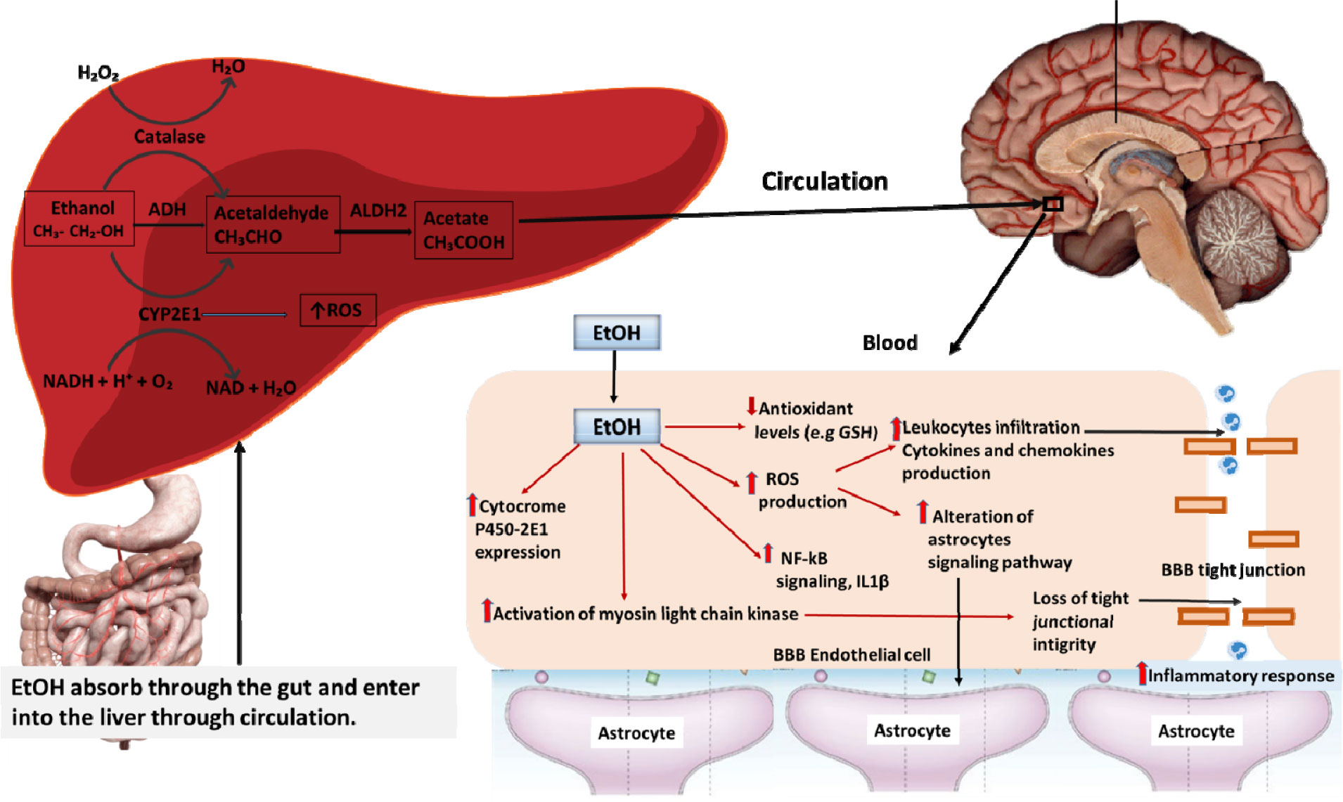
The central nervous system (CNS) is the major target for adverse effects of alcohol and extensively promotes the development of a significant number of neurological diseases such as stroke, brain tumor, multiple sclerosis (MS), Alzheimer's disease (AD), and amyotrophic lateral sclerosis (ALS). Excessive alcohol consumption causes severe neuro-immunological changes in the internal organs including irreversible brain injury and it also reacts with the defense mechanism of the blood-brain barrier (BBB) which in turn leads to changes in the configuration of the tight junction of endothelial cells and white matter thickness of the brain. Neuronal injury associated with malnutrition and oxidative stress-related BBB dysfunction may cause neuronal degeneration and demyelination in patients with alcohol use disorder (AUD); however, the underlying mechanism still remains unknown. To address this question, studies need to be performed on the contributing mechanisms of alcohol on pathological relationships of neurodegeneration that cause permanent neuronal damage. Moreover, alcohol-induced molecular changes of white matter with conduction disturbance in neurotransmission are a likely cause of myelin defect or axonal loss which correlates with cognitive dysfunctions in AUD. To extend our current knowledge in developing a neuroprotective environment, we need to explore the pathophysiology of ethanol (EtOH) metabolism and its effect on the CNS. Recent epidemiological studies and experimental animal research have revealed the association between excessive alcohol consumption and neurodegeneration. This review supports an interdisciplinary treatment protocol to protect the nervous system and to improve the cognitive outcomes of patients who suffer from alcohol-related neurodegeneration as well as clarify the pathological involvement of alcohol in causing other major neurological disorders.
Citation: Zinia Pervin, Julia M Stephen. Effect of alcohol on the central nervous system to develop neurological disorder: pathophysiological and lifestyle modulation can be potential therapeutic options for alcohol-induced neurotoxication[J]. AIMS Neuroscience, 2021, 8(3): 390-413. doi: 10.3934/Neuroscience.2021021
The central nervous system (CNS) is the major target for adverse effects of alcohol and extensively promotes the development of a significant number of neurological diseases such as stroke, brain tumor, multiple sclerosis (MS), Alzheimer's disease (AD), and amyotrophic lateral sclerosis (ALS). Excessive alcohol consumption causes severe neuro-immunological changes in the internal organs including irreversible brain injury and it also reacts with the defense mechanism of the blood-brain barrier (BBB) which in turn leads to changes in the configuration of the tight junction of endothelial cells and white matter thickness of the brain. Neuronal injury associated with malnutrition and oxidative stress-related BBB dysfunction may cause neuronal degeneration and demyelination in patients with alcohol use disorder (AUD); however, the underlying mechanism still remains unknown. To address this question, studies need to be performed on the contributing mechanisms of alcohol on pathological relationships of neurodegeneration that cause permanent neuronal damage. Moreover, alcohol-induced molecular changes of white matter with conduction disturbance in neurotransmission are a likely cause of myelin defect or axonal loss which correlates with cognitive dysfunctions in AUD. To extend our current knowledge in developing a neuroprotective environment, we need to explore the pathophysiology of ethanol (EtOH) metabolism and its effect on the CNS. Recent epidemiological studies and experimental animal research have revealed the association between excessive alcohol consumption and neurodegeneration. This review supports an interdisciplinary treatment protocol to protect the nervous system and to improve the cognitive outcomes of patients who suffer from alcohol-related neurodegeneration as well as clarify the pathological involvement of alcohol in causing other major neurological disorders.

| [1] |
Muneer PMA, Alikunju S, Szlachetka AM, et al. (2011) Inhibitory effects of alcohol on glucose transport across the blood-brain barrier leads to neurodegeneration: Preventive role of acetyl-L-carnitine. Psychopharmacology (Berl) 214: 707-718. doi: 10.1007/s00213-010-2076-4

|
| [2] | Alcohol Facts and Statistics, National Institute on Alcohol Abuse and Alcoholism (NIAAA) (2016) Natl Inst Alcohol Abus Alcohol 1-7. |
| [3] |
Ferrari AJ, Norman RE, Freedman G, et al. (2014) The burden attributable to mental and substance use disorders as risk factors for suicide: Findings from the Global Burden of Disease Study 2010. PLoS One 9: e91936. doi: 10.1371/journal.pone.0091936
|
| [4] |
Wyss-Coray T (2016) Ageing, neurodegeneration and brain rejuvenation. Nature 539: 180-186. doi: 10.1038/nature20411
|
| [5] | Lieber CS, Victor M (1992) The Effects of Alcohol on the Nervous System. Med Nutr Complicat Alcohol 413-457. |
| [6] |
Sutherland GT, Sheedy D, Kril JJ (2014) Neuropathology of alcoholism. Handbook of Clinical Neurology Elsevier BV, 603-615. doi: 10.1016/B978-0-444-62619-6.00035-5
|
| [7] |
Peng B, Yang Q, Joshi RB, et al. (2020) Role of alcohol drinking in Alzheimer's disease, Parkinson's disease, and amyotrophic lateral sclerosis. Int J Mol Sci 21: 2316. doi: 10.3390/ijms21072316
|
| [8] |
Harper C (2009) The Neuropathology of Alcohol-Related Brain Damage. Alcohol Alcohol 44: 136-140. doi: 10.1093/alcalc/agn102
|
| [9] |
Alcaide ML, Jayaweera D, Espinoza L, et al. (2003) Wernicke's encephalopathy in AIDS: A preventable cause of fatal neurological deficit. Int J STD AIDS 14: 712-713. doi: 10.1258/095646203322387992
|
| [10] | Kwok CL (2016) Central Nervous System Neurotoxicity of Chronic Alcohol Abuse. Asia Pac J Med Toxicol 2: 70-71. |
| [11] |
Crews FT, Nixon K (2009) Mechanisms of Neurodegeneration and Regeneration in Alcoholism. Alcohol Alcohol 44: 115-127. doi: 10.1093/alcalc/agn079
|
| [12] |
Crews FT (1999) Alcohol and neurodegeneration. CNS Drug Rev 5: 379-394. doi: 10.1111/j.1527-3458.1999.tb00112.x
|
| [13] | Diamond I, Francisco S (1993) Neurologic Effects of Alcoholism. West J Med 161: 279-287. |
| [14] |
Dam AM, Fuglsang-Frederiksen A, Svarre-Olsen U, et al. (1985) Late-Onset Epilepsy: Etiologies, Types of Seizure, and Value of Clinical Investigation, EEG, and Computerized Tomography Scan. Epilepsia 26: 227-231. doi: 10.1111/j.1528-1157.1985.tb05410.x
|
| [15] |
Freedland ES, McMicken DB (1993) Alcohol-related seizures, part I: Pathophysiology, differential diagnosis, and evaluation. J Emerg Med 11: 463-473. doi: 10.1016/0736-4679(93)90251-2
|
| [16] |
Przedborski S, Vila M, Jackson-Lewis V (2003) Series Introduction: Neurodegeneration: What is it and where are we? J Clin Invest 111: 3-10. doi: 10.1172/JCI200317522
|
| [17] |
Tabrizi S (2006) Neurodegenerative diseases neurobiology pathogenesis and therapeutics. J Neurol Neurosurg Psychiatry 77: 284. doi: 10.1136/jnnp.2005.072710
|
| [18] |
Collins MA, Corso TD, Neafsey EJ (1996) Neuronal degeneration in rat cerebrocortical and olfactory regions during subchronic “binge” intoxication with ethanol: Possible explanation for olfactory deficits in alcoholics. Alcohol Clin Exp Res 20: 284-292. doi: 10.1111/j.1530-0277.1996.tb01641.x
|
| [19] |
Przedborski S (2008) Neurodegeneration. Neuroimmune Pharmacology Springer US, 229-237. doi: 10.1007/978-0-387-72573-4_17
|
| [20] | Tyas SL (2002) Alcohol Use and the Risk of Developing Alzheimer's Disease. Alcohol Res Health 25: 299-306. |
| [21] |
Joseph J, Cole G, Head E, et al. (2009) Nutrition, brain aging, and neurodegeneration. J Neurosci 29: 12795-12801. doi: 10.1523/JNEUROSCI.3520-09.2009
|
| [22] |
Brust JCM (2010) Ethanol and cognition: Indirect effects, neurotoxicity and neuroprotection: A review. Int J Environ Res Public Health 7: 1540-1557. doi: 10.3390/ijerph7041540
|
| [23] |
Abrahao KP, Salinas AG, Lovinger DM (2017) Alcohol and the Brain: Neuronal Molecular Targets, Synapses, and Circuits. Neuron 96: 1223-1238. doi: 10.1016/j.neuron.2017.10.032
|
| [24] |
Kranzler HR, Zhou H, Kember RL, et al. (2019) Genome-wide association study of alcohol consumption and use disorder in 274,424 individuals from multiple populations. Nat Commun 10: 1499. doi: 10.1038/s41467-019-09480-8
|
| [25] |
Haorah J, Knipe B, Leibhart J, et al. (2005) Alcohol-induced oxidative stress in brain endothelial cells causes blood-brain barrier dysfunction. J Leukoc Biol 78: 1223-1232. doi: 10.1189/jlb.0605340
|
| [26] |
Walker I, Coleman MD (1995) The blood-brain barrier: In vitro methods and toxicological applications. Toxicol Vitr 9: 191-204. doi: 10.1016/0887-2333(94)00202-6
|
| [27] |
Daneman R, Prat A (2015) The Blood-Brain Barrier. Cold Spring Harb Perspect Biol 7: a020412. doi: 10.1101/cshperspect.a020412
|
| [28] |
Haorah J, Heilman D, Knipe B, et al. (2005) Ethanol-induced activation of myosin light chain kinase leads to dysfunction of tight junctions and blood-brain barrier compromise. Alcohol Clin Exp Res 29: 999-1009. doi: 10.1097/01.ALC.0000166944.79914.0A
|
| [29] |
Liu X, Sui B, Sun J (2017) Blood-brain barrier dysfunction induced by silica NPs in vitro and in vivo: Involvement of oxidative stress and Rho-kinase/JNK signaling pathways. Biomaterials 121: 64-82. doi: 10.1016/j.biomaterials.2017.01.006
|
| [30] | Zakhari S (2012) Alcohol metabolism and epigenetics changes. Alcohol Res Curr Rev 35: 6-16. |
| [31] |
Edenberg HJ, McClintick JN (2018) Alcohol Dehydrogenases, Aldehyde Dehydrogenases, and Alcohol Use Disorders: A Critical Review. Alcohol Clin Exp Res 42: 2281-2297. doi: 10.1111/acer.13904
|
| [32] | Wu DF, Cederbaum AI (2003) Alcohol, oxidative stress, and free radical damage. Alcohol Res Heal 27: 277-284. |
| [33] |
Hernández JA, López-Sánchez RC, Rendón-Ramírez A (2016) Lipids and Oxidative Stress Associated with Ethanol-Induced Neurological Damage. Oxid Med Cell Longev 2016: 1-15. doi: 10.1155/2016/1543809
|
| [34] |
Warner M, Gustafsson JÅ (1994) Effect of ethanol on cytochrome P450 in the rat brain. Proc Natl Acad Sci U S A 91: 1019-1023. doi: 10.1073/pnas.91.3.1019
|
| [35] | NIAAA PublicationsAvailable from: https://pubs.niaaa.nih.gov/publications/arh294/266-273.htm. |
| [36] |
Zehendner CM, Librizzi L, Hedrich J, et al. (2013) Moderate Hypoxia Followed by Reoxygenation Results in Blood-Brain Barrier Breakdown via Oxidative Stress-Dependent Tight-Junction Protein Disruption. PLoS One 8: e82823. doi: 10.1371/journal.pone.0082823
|
| [37] |
Haorah J, Ramirez SH, Floreani N, et al. (2008) Mechanism of alcohol-induced oxidative stress and neuronal injury. Free Radic Biol Med 45: 1542-1550. doi: 10.1016/j.freeradbiomed.2008.08.030
|
| [38] |
Maffi SK, Rathinam ML, Cherian PP, et al. (2008) Glutathione content as a potential mediator of the vulnerability of cultured fetal cortical neurons to ethanol-induced apoptosis. J Neurosci Res 86: 1064-1076. doi: 10.1002/jnr.21562
|
| [39] |
Adachi J, Mizoi Y, Fukunaga T, et al. (1989) Comparative Study on Ethanol Elimination and Blood Acetaldehyde between Alcoholics and Control Subjects. Alcohol Clin Exp Res 13: 601-604. doi: 10.1111/j.1530-0277.1989.tb00389.x
|
| [40] |
Anandatheerthavarada HK, Shankar SK, Bhamre S, et al. (1993) Induction of brain cytochrome P-450IIE1 by chronic ethanol treatmen. Brain Res 601: 279-285. doi: 10.1016/0006-8993(93)91721-4
|
| [41] |
Mandyam CD, Villalpando EG, Steiner NL, et al. (2017) Platelet endothelial cell adhesion molecule-1 and oligodendrogenesis: Significance in alcohol use disorders. Brain Sci 7: 131. doi: 10.3390/brainsci7100131
|
| [42] |
Collins MA, Neafsey EJ (2016) Alcohol, Excitotoxicity and Adult Brain Damage: An Experimentally Unproven Chain-of-Events. Front Mol Neurosci 9: 8. doi: 10.3389/fnmol.2016.00008
|
| [43] | Yu H, Wang CL, Wang XL, et al. (2017) Long-term exposure to ethanol downregulates tight junction proteins through the protein kinase Cα signaling pathway in human cerebral microvascular endothelial cells. Exp Ther Med 14: 4789-4796. |
| [44] |
Amano M, Nakayama M, Kaibuchi K (2010) Rho-kinase/ROCK: A key regulator of the cytoskeleton and cell polarity. Cytoskeleton (Hoboken) 67: 545-554. doi: 10.1002/cm.20472
|
| [45] |
Sweeney MD, Ayyadurai S, Zlokovic BV (2016) Pericytes of the neurovascular unit: Key functions and signaling pathways. Nat Neurosci 19: 771-783. doi: 10.1038/nn.4288
|
| [46] |
Thameem Dheen S, Kaur C, Ling EA (2007) Microglial Activation and its Implications in the Brain Diseases. Curr Med Chem 14: 1189-1197. doi: 10.2174/092986707780597961
|
| [47] |
Collins MA, Neafsey EJ (2012) Neuroinflammatory pathways in binge alcohol-induced neuronal degeneration: Oxidative stress cascade involving aquaporin, brain edema, and phospholipase A2 activation. Neurotox Res 21: 70-78. doi: 10.1007/s12640-011-9276-5
|
| [48] |
Deleo JA, Colburn RW, Rickman AJ (1997) Cytokine and growth factor immunohistochemical spinal profiles in two animal models of mononeuropathy. Brain Res 759: 50-57. doi: 10.1016/S0006-8993(97)00209-6
|
| [49] |
Noor S, Sanchez JJ, Sun MS, et al. (2020) The LFA-1 antagonist BIRT377 reverses neuropathic pain in prenatal alcohol-exposed female rats via actions on peripheral and central neuroimmune function in discrete pain-relevant tissue regions. Brain Behav Immun 87: 339-358. doi: 10.1016/j.bbi.2020.01.002
|
| [50] |
Tajuddin N, Moon KH, Marshall SA, et al. (2014) Neuroinflammation and Neurodegeneration in Adult Rat Brain from Binge Ethanol Exposure: Abrogation by Docosahexaenoic Acid. PLoS One 9: e101223. doi: 10.1371/journal.pone.0101223
|
| [51] |
Katada R, Nishitani Y, Honmou O, et al. (2012) Expression of aquaporin-4 augments cytotoxic brain edema after traumatic brain injury during acute ethanol exposure. Am J Pathol 180: 17-23. doi: 10.1016/j.ajpath.2011.09.011
|
| [52] |
Badaut J, Lasbennes F, Magistretti PJ, et al. (2002) Aquaporins in brain: Distribution, physiology, and pathophysiology. J Cereb Blood Flow Metab 22: 367-378. doi: 10.1097/00004647-200204000-00001
|
| [53] |
Dhir S, Tarasenko M, Napoli E, et al. (2019) Neurological, psychiatric, and biochemical aspects of thiamine deficiency in children and adults. Front Psychiatry 10: 207. doi: 10.3389/fpsyt.2019.00207
|
| [54] |
Niccoli T, Cabecinha M, Tillmann A, et al. (2016) Increased Glucose Transport into Neurons Rescues Ab Toxicity in Drosophila Highlights d Overexpression of glucose transporter Glut1 rescues a Drosophila Ab toxicity model d Glut1 overexpression reduces Grp78 protein levels and induces the UPR d A Grp78 dominant-negative mutant also rescues Ab toxicity in Drosophila d Metformin rescues Ab toxicity and leads to reduced Grp78 expression. Curr Biol 26: 2291-2300. doi: 10.1016/j.cub.2016.07.017
|
| [55] |
Shah K, DeSilva S, Abbruscato T (2012) The role of glucose transporters in brain disease: Diabetes and Alzheimer's disease. Int J Mol Sci 13: 12629-12655. doi: 10.3390/ijms131012629
|
| [56] |
Barros LF, San Martín A, Ruminot I, et al. (2017) Near-critical GLUT1 and Neurodegeneration. J Neurosci Res 95: 2267-2274. doi: 10.1002/jnr.23998
|
| [57] |
Hagen TM, Liu J, Lykkesfeldt J, et al. (2002) Feeding acetyl-L-carnitine and lipoic acid to old rats significantly improves metabolic function while decreasing oxidative stress. Proc Natl Acad Sci U S A 99: 1870-1875. doi: 10.1073/pnas.261708898
|
| [58] |
Choi DW, Koh JY, Peters S (1988) Pharmacology of glutamate neurotoxicity in cortical cell culture: Attenuation by NMDA antagonists. J Neurosci 8: 185-196. doi: 10.1523/JNEUROSCI.08-01-00185.1988
|
| [59] |
Zhou M, Baudry M (2006) Developmental changes in NMDA neurotoxicity reflect developmental changes in subunit composition of NMDA receptors. J Neurosci 26: 2956-2963. doi: 10.1523/JNEUROSCI.4299-05.2006
|
| [60] |
Chandrasekar R (2013) Alcohol and NMDA receptor: Current research and future direction. Front Mol Neurosci 6: 14. doi: 10.3389/fnmol.2013.00014
|
| [61] |
Bondy SC, Guo SX (1996) Effect of an NMDA receptor antagonist and a ganglioside GM1 derivative upon ethanol-induced modification of parameters of oxidative stress in several brain regions. Brain Res 716: 165-170. doi: 10.1016/0006-8993(96)00008-X
|
| [62] |
Bhave SV, Ghoda L, Hoffman PL (1999) Brain-derived neurotrophic factor mediates the anti-apoptotic effect of NMDA in cerebellar granule neurons: Signal transduction cascades and site of ethanol action. J Neurosci 19: 3277-3286. doi: 10.1523/JNEUROSCI.19-09-03277.1999
|
| [63] |
Miguel-Hidalgo JJ (2018) Molecular neuropathology of astrocytes and oligodendrocytes in alcohol use disorders. Front Mol Neurosci 11: 78. doi: 10.3389/fnmol.2018.00078
|
| [64] |
Alfonso-Loeches S, Pascual M, Gómez-Pinedo U, et al. (2012) Toll-like receptor 4 participates in the myelin disruptions associated with chronic alcohol abuse. Glia 60: 948-964. doi: 10.1002/glia.22327
|
| [65] |
Kengne FG, Nicaise C, Soupart A, et al. (2011) Astrocytes are an early target in osmotic demyelination syndrome. J Am Soc Nephrol 22: 1834-1845. doi: 10.1681/ASN.2010111127
|
| [66] |
Cullen CL, Young KM (2016) How does transcranial magnetic stimulation influence glial cells in the central nervous system? Front Neural Circuits 10: 26. doi: 10.3389/fncir.2016.00026
|
| [67] | Crews FT (2008) Alcohol-related neurodegeneration and recovery: mechanisms from animal models. Alcohol Res Health 31: 377-388. |
| [68] |
Melbourne JK, Thompson KR, Peng H, et al. (2019) Its complicated: The relationship between alcohol and microglia in the search for novel pharmacotherapeutic targets for alcohol use disorders. Prog Mol Bio Transl Sci 167: 179-221. doi: 10.1016/bs.pmbts.2019.06.011
|
| [69] |
Fede SJ, Grodin EN, Dean SF, et al. (2019) Resting state connectivity best predicts alcohol use severity in moderate to heavy alcohol users. NeuroImage Clin 22: 101782. doi: 10.1016/j.nicl.2019.101782
|
| [70] | Zahr NM, Pfefferbaum A (2017) Alcohol's Effects on the Brain: Neuroimaging Results in Humans and Animal Models. Alcohol Res 38: 183-206. |
| [71] |
Bühler M, Mann K (2011) Alcohol and the human brain: A systematic review of different neuroimaging methods. Alcohol Clin Exp Res 35: 1771-1793. doi: 10.1111/j.1530-0277.2011.01540.x
|
| [72] |
Zuccoli G, Siddiqui N, Cravo I, et al. (2010) Neuroimaging findings in alcohol-related encephalopathies. AJR Am J Roentgenol 195: 1378-1384. doi: 10.2214/AJR.09.4130
|
| [73] |
Charness ME, DeLaPaz RL (1987) Mamillary body atrophy in Wernicke's encephalopathy: Antemortem identification using magnetic resonance imaging. Ann Neurol 22: 595-600. doi: 10.1002/ana.410220506
|
| [74] |
Sullivan EV, Marsh L (2003) Hippocampal volume deficits in alcoholic Korsakoff's syndrome. Neurology 61: 1716-1719. doi: 10.1212/01.WNL.0000098940.31882.BB
|
| [75] |
Logan C, Asadi H, Kok HK, et al. (2017) Neuroimaging of chronic alcohol misuse. J Med Imaging Radiat Oncol 61: 435-440. doi: 10.1111/1754-9485.12572
|
| [76] |
Weyerer S, Schäufele M, Wiese B, et al. (2011) Current alcohol consumption and its relationship to incident dementia: Results from a 3-year follow-up study among primary care attenders aged 75 years and older. Age Ageing 40: 456-463. doi: 10.1093/ageing/afr007
|
| [77] |
Cao J, Hou JW, Ping J, et al. (2018) Advances in developing novel therapeutic strategies for Alzheimer's disease. Mol Neurodegener 13: 64. doi: 10.1186/s13024-018-0299-8
|
| [78] |
Liu R, Guo X, Park Y, et al. (2013) Alcohol Consumption, Types of Alcohol, and Parkinson's Disease. PLoS One 8: e66452. doi: 10.1371/journal.pone.0066452
|
| [79] |
Fukushima W, Miyake Y, Tanaka K, et al. (2010) Alcohol drinking and risk of Parkinson's disease: A case-control study in Japan. BMC Neurol 10: 111. doi: 10.1186/1471-2377-10-111
|
| [80] |
D'Ovidio F, Rooney JPK, Visser AE, et al. (2019) Association between alcohol exposure and the risk of amyotrophic lateral sclerosis in the Euro-MOTOR study. J Neurol Neurosurg Psychiatry 90: 11-19. doi: 10.1136/jnnp-2018-318559
|
| [81] |
Ji J, Sundquist J, Sundquist K (2016) Association of alcohol use disorders with amyotrophic lateral sclerosis: a Swedish national cohort study. Eur J Neurol 23: 270-275. doi: 10.1111/ene.12667
|
| [82] |
Koch M, Fitzpatrick AL, Rapp SR, et al. (2019) Alcohol Consumption and Risk of Dementia and Cognitive Decline Among Older Adults With or Without Mild Cognitive Impairment. JAMA Netw Open 2: e1910319. doi: 10.1001/jamanetworkopen.2019.10319
|
| [83] |
Moriyama Y, Mimura M, Kato M, et al. (2006) Primary alcoholic dementia and alcohol-related dementia. Psychogeriatrics 6: 114-118. doi: 10.1111/j.1479-8301.2006.00168.x
|
| [84] |
Ivashynka A, Copetti M, Naldi P, et al. (2019) The impact of lifetime alcohol and cigarette smoking loads on multiple sclerosis severity. Front Neurol 10: 866. doi: 10.3389/fneur.2019.00866
|
| [85] |
Banaie M, Sarbaz Y, Gharibzadeh S, et al. (2008) Huntington's disease: modeling the gait disorder and proposing novel treatments. J Theor Biol 254: 361-367. doi: 10.1016/j.jtbi.2008.05.023
|
| [86] |
Durazzo TC, Gazdzinski S, Yeh PH, et al. (2008) TREATMENT Combined Neuroimaging, Neurocognitive and Psychiatric Factors to Predict Alcohol Consumption Following Treatment for Alcohol Dependence. Alcohol Alcohol 43: 683-691. doi: 10.1093/alcalc/agn078
|
| [87] |
Niciu MJ, Mason GF (2014) Neuroimaging in Alcohol and Drug Dependence. Curr Behav Neurosci Reports 1: 45-54. doi: 10.1007/s40473-013-0005-7
|
| [88] |
Eriksson PS, Perfilieva E, Björk-Eriksson T, et al. (1998) Neurogenesis in the adult human hippocampus. Nat Med 4: 1313-1317. doi: 10.1038/3305
|
| [89] | Kumar A, Pareek V, Faiq MA, et al. (2019) ADULT NEUROGENESIS IN HUMANS: A Review of Basic Concepts, History, Current Research, and Clinical Implications. Innov Clin Neurosci 16: 30-37. |
| [90] |
Boldrini M, Fulmore CA, Tartt AN, et al. (2018) Human Hippocampal Neurogenesis Persists throughout Aging. Cell Stem Cell 22: 589-599. doi: 10.1016/j.stem.2018.03.015
|
| [91] |
Phillips LJ, Mcgorry PD, Garner B, et al. (2006) Stress, the Hippocampus and the Hypothalamic-Pituitary-Adrenal Axis: Implications for the Development of Psychotic Disorders. Aust New Zeal J Psychiatry 40: 725-741. doi: 10.1080/j.1440-1614.2006.01877.x
|
| [92] | Kang W, Balordi F, Su N, et al. (2014) Astrocyte activation is suppressed in both normal and injured brain by FGF signaling. Proc Natl Acad Sci U S A 111. |
| [93] |
Cservenka A, Casimo K, Fair DA, et al. (2014) Resting state functional connectivity of the nucleus accumbens in youth with a family history of alcoholism. Psychiatry Res 221: 210-219. doi: 10.1016/j.pscychresns.2013.12.004
|
| [94] |
Vergara VM, Weiland BJ, Hutchison KE, et al. (2018) The Impact of Combinations of Alcohol, Nicotine, and Cannabis on Dynamic Brain Connectivity. Neuropsychopharmacology 43: 877-890. doi: 10.1038/npp.2017.280
|
| [95] |
Claus ED, Hutchison KE (2012) Neural Mechanisms of Risk Taking and Relationships with Hazardous Drinking. Alcohol Clin Exp Res 36: 932-940. doi: 10.1111/j.1530-0277.2011.01694.x
|
| [96] |
Li CSR, Luo X, Yan P, et al. (2009) Altered impulse control in alcohol dependence: Neural measures of stop signal performance. Alcohol Clin Exp Res 33: 740-750. doi: 10.1111/j.1530-0277.2008.00891.x
|
| [97] |
Alexander AL, Lee JE, Lazar M, et al. (2007) Diffusion Tensor Imaging of the Brain. Neurotherapeutics 4: 316-329. doi: 10.1016/j.nurt.2007.05.011
|
| [98] |
Soares JM, Marques P, Alves V, et al. (2013) A hitchhiker's guide to diffusion tensor imaging. Front Neurosci 7: 31. doi: 10.3389/fnins.2013.00031
|
| [99] | Osier N, Dixon EC (2016) Injury Models of the Central Nervous System. Springer Protocols 1462: 177-192. |
| [100] |
Schulte T, Oberlin BG, Kareken DA, et al. (2012) How Acute and Chronic Alcohol Consumption Affects Brain Networks: Insights from Multimodal Neuroimaging. Alcohol Clin Exp Res 36: 2017-2027. doi: 10.1111/j.1530-0277.2012.01831.x
|
| [101] |
Durazzo TC, Mon A, Gazdzinski S, et al. (2017) Regional brain volume changes in alcohol-dependent individuals during early abstinence: associations with relapse following treatment. Addict Biol 22: 1416-1425. doi: 10.1111/adb.12420
|
| [102] |
Cunningham AS, Salvador R, Coles JP, et al. (2005) Physiological thresholds for irreversible tissue damage in contusional regions following traumatic brain injury. Brain 128: 1931-1942. doi: 10.1093/brain/awh536
|
| [103] | Zahr NM, Pfefferbaum A (2017) Alcohol's Effects on the Brain: Neuroimaging Results in Humans and Animal Models. Alcohol Res 38: 183-206. |
| [104] |
Jagannathan NR, Desai NG, Raghunathan P (1996) Brain metabolite changes in alcoholism: An in vivo proton magnetic resonance spectroscopy (MRS) study. Magn Reson Imaging 14: 553-557. doi: 10.1016/0730-725X(96)00048-3
|
| [105] |
Meyerhoff DJ, Durazzo TC (2008) Proton magnetic resonance spectroscopy in alcohol use disorders: A potential new endophenotype? Alcohol Clin Exp Res 32: 1146-1158. doi: 10.1111/j.1530-0277.2008.00695.x
|
| [106] |
Zahr NM, Alt C, Mayer D, et al. (2014) Associations between in vivo neuroimaging and postmortem brain cytokine markers in a rodent model of Wernicke's encephalopathy. Exp Neurol 261: 109-119. doi: 10.1016/j.expneurol.2014.06.015
|
| [107] |
Gass JT, Olive MF (2008) Glutamatergic substrates of drug addiction and alcoholism. Biochem Pharmacol 75: 218-265. doi: 10.1016/j.bcp.2007.06.039
|
| [108] |
Kähkönen S (2005) MEG and TMS combined with EEG for mapping alcohol effects. Alcohol 37: 129-133. doi: 10.1016/j.alcohol.2006.03.003
|
| [109] |
Nikulin VV, Nikulina AV, Yamashita H, et al. (2005) Effects of alcohol on spontaneous neuronal oscillations: A combined magnetoencephalography and electroencephalography study. Prog Neuro Psychopharmacol Biol Psychiatry 29: 687-693. doi: 10.1016/j.pnpbp.2005.04.014
|
| [110] |
Rowland JA, Stapleton-Kotloski JR, Alberto GE, et al. (2017) Changes in nonhuman primate brain function following chronic alcohol consumption in previously naïve animals. Drug Alcohol Depend 177: 244-248. doi: 10.1016/j.drugalcdep.2017.03.036
|
| [111] |
Büttner A (2014) Neuropathology of Chronic Alcohol Abuse. Acad Forensic Pathol 4: 180-187. doi: 10.23907/2014.029
|
| [112] | Venkataraman A, Kalk N, Sewell G, et al. (2017) Alcohol and Alzheimer's Disease-Does Alcohol Dependence Contribute to Beta-Amyloid Deposition, Neuroinflammation and Neurodegeneration in Alzheimer's Disease? Alcohol Alcohol 52: 151-158. |
| [113] |
Motaghinejad M, Safari S, Feizipour S, et al. (2019) Crocin may be useful to prevent or treatment of alcohol induced neurodegeneration and neurobehavioral sequels via modulation of CREB/BDNF and Akt/GSK signaling pathway. Med Hypotheses 124: 21-25. doi: 10.1016/j.mehy.2019.01.017
|
| [114] | Reddy SK, Husain K, Schlorff EC, et al. (1999) Dose response of ethanol ingestion on antioxidant defense system in rat brain subcellular fractions. Neurotoxicology 20: 977-987. |
| [115] | Poljsak B (2011) Strategies for reducing or preventing the generation of oxidative stress. Oxid Med Cell Longev 2011: 194586. |
| [116] |
Fujita K, Yamafuji M, Nakabeppu Y, et al. (2012) Therapeutic Approach to Neurodegenerative Diseases by Medical Gases: Focusing on Redox Signaling and Related Antioxidant Enzymes. Oxid Med Cell Longev 2012: 324256. doi: 10.1155/2012/324256
|
| [117] | Patel F, Mandal P (2019) Neurodegenerative Diseases and Their Therapeutic Approaches. Neurons Dendrites and Axons IntechOpen. |
| [118] |
Hussain R, Zubair H, Pursell S, et al. (2018) Neurodegenerative diseases: Regenerative mechanisms and novel therapeutic approaches. Brain Sci 8: 177. doi: 10.3390/brainsci8090177
|
| [119] |
Bulck M Van, Sierra-Magro A, Alarcon-Gil J, et al. (2019) Novel approaches for the treatment of alzheimer's and parkinson's disease. Int J Mol Sci 20: 719. doi: 10.3390/ijms20030719
|
| [120] |
Heilig M, Augier E, Pfarr S, et al. (2019) Developing neuroscience-based treatments for alcohol addiction: A matter of choice? Transl Psychiatry 9: 1-11. doi: 10.1038/s41398-019-0591-6
|
| [121] |
Mohammed HO, Starkey SR, Stipetic K, et al. (2008) The role of dietary antioxidant insufficiency on the permeability of the blood-brain barrier. J Neuropathol Exp Neurol 67: 1187-1193. doi: 10.1097/NEN.0b013e31818f8f51
|
| [122] |
González-Reimers E, Fernández-Rodríguez CM, Candelaria Martín-González M, et al. (2014) Antioxidant Vitamins and Brain Dysfunction in Alcoholics. Alcohol Alcohol 49: 45-50. doi: 10.1093/alcalc/agt150
|
| [123] |
Lubos E, Handy DE, Loscalzo J (2008) Role of oxidative stress and nitric oxide in atherothrombosis. Front Biosci 13: 5323-5344. doi: 10.2741/3084
|
| [124] |
Wiecek M, Szymura J, Maciejczyk M, et al. (2018) Anaerobic Exercise-Induced Activation of Antioxidant Enzymes in the Blood of Women and Men. Front Physiol 9: 1006. doi: 10.3389/fphys.2018.01006
|
| [125] | Zou J, Crews F (2006) CREB and NF-κB Transcription Factors Regulate Sensitivity to Excitotoxic and Oxidative Stress Induced Neuronal Cell Death. Cell Mol Neurobiol 26: 385-405. |
| [126] | Alcohol-Related Brain Damage—Alcohol Rehab Guide, 2019 Available from: https://www.alcoholrehabguide.org/resources/medical-conditions/alcohol-related-brain-damage/. |
| [127] |
Jung YC, Namkoong K (2006) Pharmacotheraphy for alcohol dependence: Anticraving medications for relapse prevention. Yonsei Med J 47: 167-178. doi: 10.3349/ymj.2006.47.2.167
|
| [128] |
Palpacuer C, Duprez R, Huneau A, et al. (2018) Pharmacologically controlled drinking in the treatment of alcohol dependence or alcohol use disorders: a systematic review with direct and network meta-analyses on nalmefene, naltrexone, acamprosate, baclofen and topiramate. Addiction 113: 220-237. doi: 10.1111/add.13974
|
| [129] |
Vellas B, Black R, Thal L, et al. (2009) Long-Term Follow-Up of Patients Immunized with AN1792: Reduced Functional Decline in Antibody Responders. Curr Alzheimer Res 6: 144-151. doi: 10.2174/156720509787602852
|
| [130] |
Plotkin SS, Cashman NR (2020) Passive immunotherapies targeting Aβ and tau in Alzheimer's disease. Neurobiol Dis 144: 105010. doi: 10.1016/j.nbd.2020.105010
|
| [131] |
Theunis C, Crespo-Biel N, Gafner V, et al. (2013) Efficacy and safety of a liposome-based vaccine against protein Tau, assessed in Tau.P301L mice that model tauopathy. PLoS One 8: e72301. doi: 10.1371/journal.pone.0072301
|
| [132] |
Hanger DP, Hughes K, Woodgett JR, et al. (1992) Glycogen synthase kinase-3 induces Alzheimer's disease-like phosphorylation of tau: Generation of paired helical filament epitopes and neuronal localisation of the kinase. Neurosci Lett 147: 58-62. doi: 10.1016/0304-3940(92)90774-2
|
| [133] |
Holla B, Karthik S, Biswal J, et al. (2018) Brain Functional Magnetic Resonance Imaging Cue-reactivity Can Predict Baclofen Response in Alcohol Use Disorders. Clin Psychopharmacol Neurosci 16: 290-301. doi: 10.9758/cpn.2018.16.3.290
|
| [134] |
Berman T, Bayati A (2018) What are Neurodegenerative Diseases and How Do They Affect the Brain? Front Young Minds 6: 70. doi: 10.3389/frym.2018.00070
|
| [135] |
Campanella S, Schroder E, Kajosch H, et al. (2019) Why cognitive event-related potentials (ERPs) should have a role in the management of alcohol disorders. Neurosci Biobehav Rev 106: 234-244. doi: 10.1016/j.neubiorev.2018.06.016
|
| [136] |
Campanella S, Petit G, Maurage P, et al. (2009) Chronic alcoholism: Insights from neurophysiology. Neurophysiol Clin 39: 191-207. doi: 10.1016/j.neucli.2009.08.002
|
| [137] |
Pashut T, Wolfus S, Friedman A, et al. (2011) Mechanisms of magnetic stimulation of central nervous system neurons. PLoS Comput Biol 7: e1002022. doi: 10.1371/journal.pcbi.1002022
|
| [138] | Pridmore S, Morey R, Pridmore S, et al. (2020) Anhedonia in Major Depressive Disorder (MDD) is Reduced by Transcranial Magnetic Stimulation (TMS). Neurol Neurobiol 2020: 1-3. |
| [139] |
Pini L, Manenti R, Cotelli M, et al. (2018) Non-invasive brain stimulation in dementia: A complex network story. Neurodegener Dis 18: 281-301. doi: 10.1159/000495945
|
| [140] |
Marron EM, Viejo-Sobera R, Quintana M, et al. (2018) Transcranial magnetic stimulation intervention in Alzheimer's disease: A research proposal for a randomized controlled trial NCT03121066 NCT. BMC Res Notes 11: 648. doi: 10.1186/s13104-018-3757-z
|
| [141] |
Murphy TJ, Pagano RR, Marlatt GA (1986) Lifestyle modification with heavy alcohol drinkers: Effects of aerobic exercise and meditation. Addict Behav 11: 175-186. doi: 10.1016/0306-4603(86)90043-2
|
| [142] |
Popa-Wagner A, Dumitrascu D, Capitanescu B, et al. (2020) Dietary habits, lifestyle factors and neurodegenerative diseases. Neural Regen Res 15: 394-400. doi: 10.4103/1673-5374.266045
|
| [143] |
Carratalà J, Mykietiuk A, Fernández-Sabé N, et al. (2007) Health care-associated pneumonia requiring hospital admission: Epidemiology, antibiotic therapy, and clinical outcomes. Arch Intern Med 167: 1393-1399. doi: 10.1001/archinte.167.13.1393
|
| [144] |
Corella D, Tucker K, Lahoz C, et al. (2001) Alcohol drinking determines the effect of the APOE locus on LDL-cholesterol concentrations in men: The Framingham Offspring Study. Am J Clin Nutr 73: 736-745. doi: 10.1093/ajcn/73.4.736
|
| [145] | Zhou SM, Zhou R, Zhong TT, et al. (2014) Association of Smoking and Alcohol Drinking with Dementia Risk Among Elderly Men in China. Curr Alzheimer Res 11: 899-907. |
| [146] |
Hagger-Johnson G, Sabia S, Brunner EJ, et al. (2013) Combined impact of smoking and heavy alcohol use on cognitive decline in early old age: Whitehall II prospective cohort study. Br J Psychiatry 203: 120-125. doi: 10.1192/bjp.bp.112.122960
|
| [147] |
Esang M, Ahmed S (2018) A Closer Look at Substance Use and Suicide. Am J Psychiatry Resid J 13: 6-8. doi: 10.1176/appi.ajp-rj.2018.130603
|
| [148] |
Bednarski SR, Zhang S, Hong KI, et al. (2011) Deficits in default mode network activity preceding error in cocaine dependent individuals. Drug Alcohol Depend 119: e51-e57. doi: 10.1016/j.drugalcdep.2011.05.026
|
| [149] |
Fratiglioni L, Qiu C (2009) Prevention of common neurodegenerative disorders in the elderly. Exp Gerontol 44: 46-50. doi: 10.1016/j.exger.2008.06.006
|
| [150] |
Karpyak VM, Biernacka JM, Geske JR, et al. (2016) Gender-specific effects of comorbid depression and anxiety on the propensity to drink in negative emotional states. Addiction 111: 1366-1375. doi: 10.1111/add.13386
|
| [151] |
Djoussé L, Myers RH, Province MA, et al. (2002) Influence of Apolipoprotein E, Smoking, and Alcohol Intake on Carotid Atherosclerosis National Heart, Lung, and Blood Institute Family Heart Study. Stroke 33: 1357-1361. doi: 10.1161/01.STR.0000014325.54063.1A
|
| [152] |
Bhatti GK, Reddy AP, Reddy PH, et al. (2020) Lifestyle Modifications and Nutritional Interventions in Aging-Associated Cognitive Decline and Alzheimer's Disease. Front Aging Neurosci 11: 369. doi: 10.3389/fnagi.2019.00369
|
| [153] |
Baghaiee B, Aliparasti MR, Almasi S, et al. (2016) Antioxidant expression response to free radicals in active men and women fallowing to a session incremental exercise; Numerical relationship between antioxidants and free radicals. Asian J Sports Med 7: e29901. doi: 10.5812/asjsm.29901
|
| [154] |
Topiwala H, Terrera GM, Stirland L, et al. (2018) Lifestyle and neurodegeneration in midlife as expressed on functional magnetic resonance imaging: A systematic review. Alzheimer Dement 4: 182-194. doi: 10.1016/j.trci.2018.04.001
|
Figures(2) / Tables(2)

Zinia Pervin, Julia M Stephen. Effect of alcohol on the central nervous system to develop neurological disorder: pathophysiological and lifestyle modulation can be potential therapeutic options for alcohol-induced neurotoxication[J]. AIMS Neuroscience, 2021, 8(3): 390-413. doi: 10.3934/Neuroscience.2021021


 DownLoad: