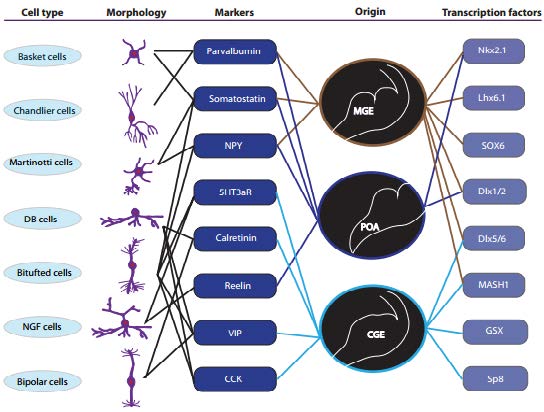
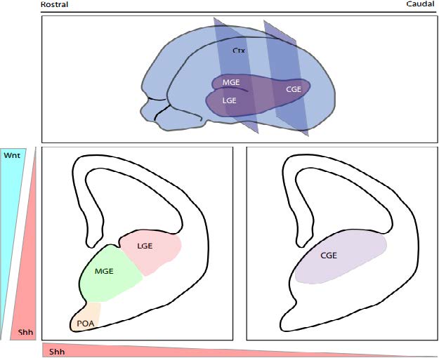
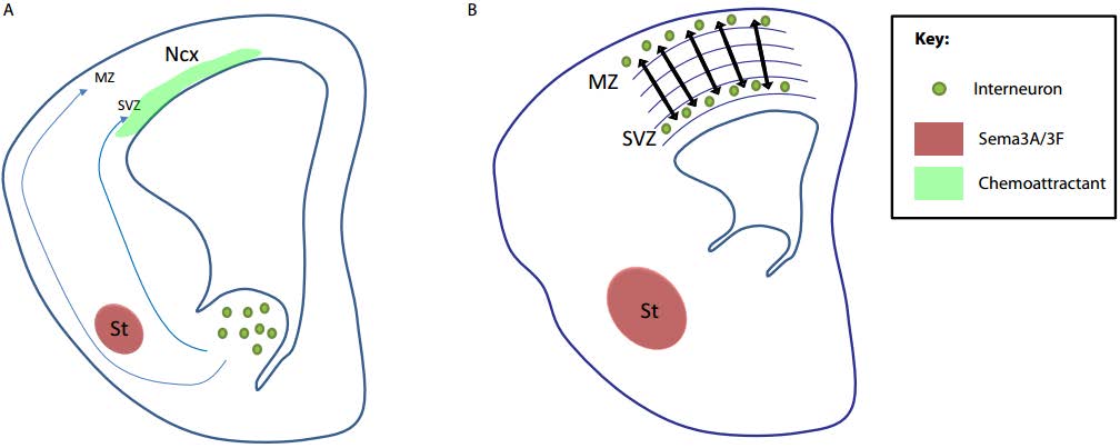
Schizophrenia is a devastating neuropsychiatric disorder widely believed to arise from defects during brain development. Indeed, dysfunction in the formation and function of GABAergic cortical interneurons has been implicated as a central pathogenic mechanism in this, and other, neurodevelopmental disorders. Understanding the coordination and timing of interneuron development including the complex processes of specification, proliferation, migration and their incorporation into finely tuned cortical networks is therefore essential in determining their role in neurodevelopmental disease. Studies using mouse models have highlighted the functional relevance of transcription factor networks and common signalling pathways in interneuron development but have faced challenges in identifying clear time windows where these factors are essential. Here we discuss recent developments highlighting critical time frames in the specification and migration of cortical interneurons and the impact of aberrant development to aetiology and treatments of schizophrenia.
Citation: Zarina Greenberg, Hayley Ramshaw, Quenten Schwarz. Time Windows of Interneuron Development: Implications to Our Understanding of the Aetiology and Treatment of Schizophrenia[J]. AIMS Neuroscience, 2015, 2(4): 294-321. doi: 10.3934/Neuroscience.2015.4.294
Schizophrenia is a devastating neuropsychiatric disorder widely believed to arise from defects during brain development. Indeed, dysfunction in the formation and function of GABAergic cortical interneurons has been implicated as a central pathogenic mechanism in this, and other, neurodevelopmental disorders. Understanding the coordination and timing of interneuron development including the complex processes of specification, proliferation, migration and their incorporation into finely tuned cortical networks is therefore essential in determining their role in neurodevelopmental disease. Studies using mouse models have highlighted the functional relevance of transcription factor networks and common signalling pathways in interneuron development but have faced challenges in identifying clear time windows where these factors are essential. Here we discuss recent developments highlighting critical time frames in the specification and migration of cortical interneurons and the impact of aberrant development to aetiology and treatments of schizophrenia.

| [1] | Hendry SH, Schwark HD, Jones EG, et al. (1987) Numbers and proportions of GABA-immunoreactive neurons in different areas of monkey cerebral cortex. J Neurosci 7: 1503-1519. |
| [2] |
Meinecke DL, Peters A (1987) GABA immunoreactive neurons in rat visual cortex. J Comp Neurol 261: 388-404. doi: 10.1002/cne.902610305

|
| [3] |
Levitt P, Eagleson KL, Powell EM (2004) Regulation of neocortical interneuron development and the implications for neurodevelopmental disorders. Trends Neurosci 27: 400-406. doi: 10.1016/j.tins.2004.05.008
|
| [4] |
Lewis DA (2000) GABAergic local circuit neurons and prefrontal cortical dysfunction in schizophrenia. Brain Res Brain Res Rev 31: 270-276. doi: 10.1016/S0165-0173(99)00042-9
|
| [5] |
Rubenstein JL, Merzenich MM (2003) Model of autism: increased ratio of excitation/inhibition in key neural systems. Genes Brain Behav 2: 255-267. doi: 10.1034/j.1601-183X.2003.00037.x
|
| [6] | Perry TL, Kish SJ, Buchanan J, et al. (1979) Gamma-aminobutyric-acid deficiency in brain of schizophrenic patients. Lancet 1: 237-239. |
| [7] |
Hashimoto T, Volk DW, Eggan SM, et al. (2003) Gene expression deficits in a subclass of GABA neurons in the prefrontal cortex of subjects with schizophrenia. J Neurosci 23: 6315-6326. |
| [8] |
Nakazawa K, Zsiros V, Jiang Z, et al. (2012) GABAergic interneuron origin of schizophrenia pathophysiology. Neuropharmacology 62: 1574-1583. doi: 10.1016/j.neuropharm.2011.01.022
|
| [9] |
Schmidt MJ, Mirnics K (2015) Neurodevelopment, GABA system dysfunction, and schizophrenia. Neuropsychopharmacology 40: 190-206. doi: 10.1038/npp.2014.95
|
| [10] |
Gupta S, Kulhara P (2010) What is schizophrenia: A neurodevelopmental or neurodegenerative disorder or a combination of both? A critical analysis. Indian J Psychiatry 52: 21-27. doi: 10.4103/0019-5545.58891
|
| [11] |
DeFelipe J (2002) Cortical interneurons: from Cajal to 2001. Prog Brain Res 136: 215-238. doi: 10.1016/S0079-6123(02)36019-9
|
| [12] |
Gonchar Y, Burkhalter A (1997) Three distinct families of GABAergic neurons in rat visual cortex. Cereb Cortex 7: 347-358. doi: 10.1093/cercor/7.4.347
|
| [13] |
Kawaguchi Y, Kubota Y (1997) GABAergic cell subtypes and their synaptic connections in rat frontal cortex. Cereb Cortex 7: 476-486. doi: 10.1093/cercor/7.6.476
|
| [14] |
DeFelipe J, López-Cruz PL, Benavides-Piccione R, et al. (2013) New insights into the classification and nomenclature of cortical GABAergic interneurons. Nat Rev Neurosci 14: 202-216. |
| [15] |
Petilla Interneuron Nomenclature G, Ascoli GA, Alonso-Nanclares L, et al. (2008) Petilla terminology: nomenclature of features of GABAergic interneurons of the cerebral cortex. Nat Rev Neurosci 9: 557-568. doi: 10.1038/nrn2402
|
| [16] |
Lee S, Hjerling-Leffler J, Zagha E, et al. (2010) The largest group of superficial neocortical GABAergic interneurons expresses ionotropic serotonin receptors. J Neurosci 30: 16796-16808. doi: 10.1523/JNEUROSCI.1869-10.2010
|
| [17] | Rudy B, Fishell G, Lee S, et al. (2011) Three groups of interneurons account for nearly 100% of neocortical GABAergic neurons. Dev Neurobiol 71: 45-61. |
| [18] |
Fanselow EE, Richardson KA, Connors BW (2008) Selective, state-dependent activation of somatostatin-expressing inhibitory interneurons in mouse neocortex. J Neurophysiol 100: 2640-2652. |
| [19] | Hu H, Gan J, Jonas P (2014) Interneurons. Fast-spiking, parvalbumin(+) GABAergic interneurons: from cellular design to microcircuit function. Science 345: 1255263. |
| [20] |
Xu H, Jeong HY, Tremblay R, et al. (2013) Neocortical somatostatin-expressing GABAergic interneurons disinhibit the thalamorecipient layer 4. Neuron 77: 155-167. doi: 10.1016/j.neuron.2012.11.004
|
| [21] |
Takada N, Pi HJ, Sousa VH, et al. (2014) A developmental cell-type switch in cortical interneurons leads to a selective defect in cortical oscillations. Nat Commun 5: 5333. doi: 10.1038/ncomms6333
|
| [22] |
Fung SJ, Webster MJ, Sivagnanasundaram S, et al. (2010) Expression of interneuron markers in the dorsolateral prefrontal cortex of the developing human and in schizophrenia. Am J Psychiatry 167: 1479-1488. doi: 10.1176/appi.ajp.2010.09060784
|
| [23] |
Volk DW, Austin MC, Pierri JN, et al. (2000) Decreased glutamic acid decarboxylase67 messenger RNA expression in a subset of prefrontal cortical gamma-aminobutyric acid neurons in subjects with schizophrenia. Arch Gen Psychiatry 57: 237-245. doi: 10.1001/archpsyc.57.3.237
|
| [24] |
Vitalis T, Rossier J (2011) New insights into cortical interneurons development and classification: contribution of developmental studies. Dev Neurobiol 71: 34-44. doi: 10.1002/dneu.20810
|
| [25] | Peyre E, Silva CG, Nguyen L (2015) Crosstalk between intracellular and extracellular signals regulating interneuron production, migration and integration into the cortex. Front Cell Neurosci 9: 129. |
| [26] |
Ericson J, Muhr J, Placzek M, et al. (1995) Sonic hedgehog induces the differentiation of ventral forebrain neurons: a common signal for ventral patterning within the neural tube. Cell 81: 747-756. doi: 10.1016/0092-8674(95)90536-7
|
| [27] | Flandin P, Zhao Y, Vogt D, et al. (2001) Lhx6 and Lhx8 coordinately induce neuronal expression of Shh that controls the generation of interneuron progenitors. Neuron 70: 939-950. |
| [28] | Shimamura K, Hartigan DJ, Martinez S, et al. (1995) Longitudinal organization of the anterior neural plate and neural tube. Development 121: 3923-3933. |
| [29] | Sussel L, Marin O, Kimura S, et al. (1999) Loss of Nkx2.1 homeobox gene function results in a ventral to dorsal molecular respecification within the basal telencephalon: evidence for a transformation of the pallidum into the striatum. Development 126: 3359-3370. |
| [30] |
Fuccillo M, Rallu M, McMahon AP, et al. (2004) Temporal requirement for hedgehog signaling in ventral telencephalic patterning. Development 131: 5031-5040. doi: 10.1242/dev.01349
|
| [31] |
Wonders CP, Taylor L, Welagen J, et al. (2008) A spatial bias for the origins of interneuron subgroups within the medial ganglionic eminence. Dev Biol 314: 127-136. doi: 10.1016/j.ydbio.2007.11.018
|
| [32] |
Xu Q, Cobos I, Cruz EDL, et al. (2004) Origins of cortical interneuron subtypes. J Neurosci 24: 2612-2622. doi: 10.1523/JNEUROSCI.5667-03.2004
|
| [33] |
Xu Q, Wonders CP, Anderson SA (2005) Sonic hedgehog maintains the identity of cortical interneuron progenitors in the ventral telencephalon. Development 132: 4987-4998. doi: 10.1242/dev.02090
|
| [34] |
Xu Q, Guo L, Moore H, et al. (2010) Sonic hedgehog signaling confers ventral telencephalic progenitors with distinct cortical interneuron fates. Neuron 65: 328-340. doi: 10.1016/j.neuron.2010.01.004
|
| [35] | Gulacsi A, Anderson SA (2006) Shh maintains Nkx2.1 in the MGE by a Gli3-independent mechanism. Cereb Cortex 16: i89-95. |
| [36] |
Batista-Brito R, Rossignol E, Hjerling-Leffler J, et al. (2009) The cell-intrinsic requirement of Sox6 for cortical interneuron development. Neuron 63: 466-481. doi: 10.1016/j.neuron.2009.08.005
|
| [37] |
Lei Q, Jeong Y, Misra K, et al. (2006) Wnt signaling inhibitors regulate the transcriptional response to morphogenetic Shh-Gli signaling in the neural tube. Dev Cel 11: 325-337. doi: 10.1016/j.devcel.2006.06.013
|
| [38] |
Aoto K, Nishimura T, Eto K, et al. (2002) Mouse GLI3 regulates Fgf8 expression and apoptosis in the developing neural tube, face, and limb bud. Dev Biol 251: 320-332. doi: 10.1006/dbio.2002.0811
|
| [39] |
Gulacsi A, Lillien L (2003) Sonic hedgehog and bone morphogenetic protein regulate interneuron development from dorsal telencephalic progenitors in vitro. J Neurosci 23: 9862-9872. |
| [40] | Anderson RM, Lawrence AR, Stottmann RW, et al. (2002) Chordin and noggin promote organizing centers of forebrain development in the mouse. Development 129: 4975-4987. |
| [41] |
Mukhopadhyay A, McGuire T, Peng CY, et al. (2009) Differential effects of BMP signaling on parvalbumin and somatostatin interneuron differentiation. Development 136: 2633-2642. doi: 10.1242/dev.034439
|
| [42] |
Samanta J, Burke GM, McGuire T, et al. (2007) BMPR1a signaling determines numbers of oligodendrocytes and calbindin-expressing interneurons in the cortex. J Neurosci 27: 7397-7407. doi: 10.1523/JNEUROSCI.1434-07.2007
|
| [43] |
Wine-Lee L, Ahn KJ, Richardson RD, et al. (2004) Signaling through BMP type 1 receptors is required for development of interneuron cell types in the dorsal spinal cord. Development 131: 5393-5403. doi: 10.1242/dev.01379
|
| [44] |
Gutin G, Marie F, Laura P, et al. (2006) FGF signalling generates ventral telencephalic cells independently of SHH. Development 133: 2937-2946. doi: 10.1242/dev.02465
|
| [45] |
Ohkubo Y, Chiang C, Rubenstein JL (2002) Coordinate regulation and synergistic actions of BMP4, SHH and FGF8 in the rostral prosencephalon regulate morphogenesis of the telencephalic and optic vesicles. Neuroscience 111: 1-17. doi: 10.1016/S0306-4522(01)00616-9
|
| [46] |
Machon O, van den Bout CJ, Backman M, et al. (2003) Role of beta-catenin in the developing cortical and hippocampal neuroepithelium. Neuroscience 122: 129-143. doi: 10.1016/S0306-4522(03)00519-0
|
| [47] |
Woodhead GJ, Mutch CA, Olson EC, et al. (2006) Cell-autonomous beta-catenin signaling regulates cortical precursor proliferation. J Neurosci 26: 12620-12630. doi: 10.1523/JNEUROSCI.3180-06.2006
|
| [48] | Zhou CJ, Borello U, Rubenstein JL, et al. (2006) Neuronal production and precursor proliferation defects in the neocortex of mice with loss of function in the canonical Wnt signaling pathway. Neuroscience 142: 1119-1131. |
| [49] |
Zhou CJ, Zhao C, Pleasure SJ (2004) Wnt signaling mutants have decreased dentate granule cell production and radial glial scaffolding abnormalities. J Neurosci 24: 121-126. doi: 10.1523/JNEUROSCI.4071-03.2004
|
| [50] |
Backman M, Machon O, Mygland L, et al. (2005) Effects of canonical Wnt signaling on dorso-ventral specification of the mouse telencephalon. Dev Biol 279: 155-168. doi: 10.1016/j.ydbio.2004.12.010
|
| [51] |
Gulacsi AA, Anderson SA (2008) Beta-catenin-mediated Wnt signaling regulates neurogenesis in the ventral telencephalon. Nat Neurosci 11: 1383-1391. doi: 10.1038/nn.2226
|
| [52] |
Martinez-Morales PL, Quiroga AC, Barbas JA, et al. (2010) SOX5 controls cell cycle progression in neural progenitors by interfering with the WNT-beta-catenin pathway. EMBO Rep 11: 466-472. doi: 10.1038/embor.2010.61
|
| [53] | Pino D, Choe Y, Pleasure SJ (2011) Wnt5a controls neurite development in olfactory bulb interneurons. ASN Neuro 3: e00059. |
| [54] |
Vidaki M, Tivodar S, Doulgeraki K, et al. (2012) Rac1-dependent cell cycle exit of MGE precursors and GABAergic interneuron migration to the cortex. Cereb Cortex 22: 680-692. doi: 10.1093/cercor/bhr145
|
| [55] |
Marin O, Yaron A, Bagri A, et al. (2001) Sorting of striatal and cortical interneurons regulated by semaphorin-neuropilin interactions. Science 293: 872-875. doi: 10.1126/science.1061891
|
| [56] | Marin O, Anderson SA, Rubenstein JL (2000) Origin and molecular specification of striatal interneurons. J Neurosci 20: 6063-6076. |
| [57] |
Lumb R, Wiszniak S, Kabbara S, et al. (2014) Neuropilins define distinct populations of neural crest cells. Neural Dev 9: 24. doi: 10.1186/1749-8104-9-24
|
| [58] |
Schwarz Q, Maden CH, Vieira JM, et al. (2009) Neuropilin 1 signaling guides neural crest cells to coordinate pathway choice with cell specification. Proc Natl Acad Sci U S A 106: 6164-6169. doi: 10.1073/pnas.0811521106
|
| [59] | Schwarz Q, Ruhrberg C (2010) Neuropilin, you gotta let me know: Should I stay or should I go? Cell Adh Migr 4. |
| [60] |
Hernandez-Miranda LR., Anna C, Clare F, et al. (2011) Robo1 regulates semaphorin signaling to guide the migration of cortical interneurons through the ventral forebrain. J Neurosci 31: 6174-6187. doi: 10.1523/JNEUROSCI.5464-10.2011
|
| [61] | Marin O, Baker J, Puelles L, et al. (2002) Patterning of the basal telencephalon and hypothalamus is essential for guidance of cortical projections. Development 129: 761-773. |
| [62] |
Zimmer G, Rudolph J, Landmann J, et al. (2011) Bidirectional ephrinB3/EphA4 signaling mediates the segregation of medial ganglionic eminence- and preoptic area-derived interneurons in the deep and superficial migratory stream. J Neurosci 31: 18364-18380. doi: 10.1523/JNEUROSCI.4690-11.2011
|
| [63] |
Zimmer G, Garcez P, Rudolph J, et al. (2008) Ephrin-A5 acts as a repulsive cue for migrating cortical interneurons. Eur J Neurosci 28: 62-73. doi: 10.1111/j.1460-9568.2008.06320.x
|
| [64] | Friocourt G, Parnavelas JG (2010) Mutations in ARX Result in Several Defects Involving GABAergic Neurons. Front Cell Neurosci 4: 4. |
| [65] |
Flames N, Long JE, Garratt AN, et al. (2004) Short- and long-range attraction of cortical GABAergic interneurons by neuregulin-1. Neuron 44: 251-261. doi: 10.1016/j.neuron.2004.09.028
|
| [66] |
Li H, Chou SJ, Hamasaki T, et al. (2012) Neuregulin repellent signaling via ErbB4 restricts GABAergic interneurons to migratory paths from ganglionic eminence to cortical destinations. Neural Dev 7: 10. doi: 10.1186/1749-8104-7-10
|
| [67] |
Balabanian K, Lagane B, Infantino S, et al. (2005) The chemokine SDF-1/CXCL12 binds to and signals through the orphan receptor RDC1 in T lymphocytes. J Biol Chem 280: 35760-35766. doi: 10.1074/jbc.M508234200
|
| [68] |
Wang Y, Li G, Stanco A, et al. (2011) CXCR4 and CXCR7 have distinct functions in regulating interneuron migration. Neuron 69: 61-76. doi: 10.1016/j.neuron.2010.12.005
|
| [69] | Hevner RF, Daza RA, Englund C, et al. (2004) Postnatal shifts of interneuron position in the neocortex of normal and reeler mice: evidence for inward radial migration. Neuroscience 124: 605-618. |
| [70] |
Nadarajah B, Alifragis P, Wong RO, et al. (2003) Neuronal migration in the developing cerebral cortex: observations based on real-time imaging. Cereb Cortex 13: 607-611. doi: 10.1093/cercor/13.6.607
|
| [71] |
Nadarajah B, Parnavelas JG (2002) Modes of neuronal migration in the developing cerebral cortex. Nat Rev Neurosci 3: 423-432. doi: 10.1038/nrn845
|
| [72] | Polleux F, Whitford KL, Dijkhuizen PA, et al. (2002) Control of cortical interneuron migration by neurotrophins and PI3-kinase signaling. Development 129: 3147-3160. |
| [73] |
Lysko DE, Putt M, Golden JA (2011) SDF1 regulates leading process branching and speed of migrating interneurons. J Neurosci 31: 1739-1745. doi: 10.1523/JNEUROSCI.3118-10.2011
|
| [74] |
Miyoshi G, Fishell G (2011) GABAergic interneuron lineages selectively sort into specific cortical layers during early postnatal development. Cereb Cortex 21: 845-852. doi: 10.1093/cercor/bhq155
|
| [75] |
Vogt D, Hunt RF, Mandal S, et al. (2014) Lhx6 directly regulates Arx and CXCR7 to determine cortical interneuron fate and laminar position. Neuron 82: 350-364. doi: 10.1016/j.neuron.2014.02.030
|
| [76] |
English JA, Dicker P, Föcking M, et al (2009) 2-D DIGE analysis implicates cytoskeletal abnormalities in psychiatric disease. Proteomics 9: 3368-3382. doi: 10.1002/pmic.200900015
|
| [77] |
Insel TR (2010) Rethinking schizophrenia. Nature 468: 187-193. doi: 10.1038/nature09552
|
| [78] |
Walsh T, McClellan JM, McCarthy SE, et al. (2008) Rare structural variants disrupt multiple genes in neurodevelopmental pathways in schizophrenia. Science 320: 539-543. doi: 10.1126/science.1155174
|
| [79] |
Lewis DA, Levitt P (2002) Schizophrenia as a disorder of neurodevelopment. Annu Rev Neurosci 25: 409-432. doi: 10.1146/annurev.neuro.25.112701.142754
|
| [80] | Benes FM, Vincent SL, Alsterberg G, et al. (1992) Increased GABAA receptor binding in superficial layers of cingulate cortex in schizophrenics. J Neurosci 12: 924-929. |
| [81] |
Konradi C, Yang CK, Zimmerman EI, et al. (2011) Hippocampal interneurons are abnormal in schizophrenia. Schizophr Res 131: 165-173. doi: 10.1016/j.schres.2011.06.007
|
| [82] |
Wang AY, Lohmann KM, Yang CK, et al. (2011) Bipolar disorder type 1 and schizophrenia are accompanied by decreased density of parvalbumin- and somatostatin-positive interneurons in the parahippocampal region. Acta Neuropathol 122: 615-626. doi: 10.1007/s00401-011-0881-4
|
| [83] |
Joshi D, Fung SJ, Rothwell A, et al. (2012) Higher gamma-aminobutyric acid neuron density in the white matter of orbital frontal cortex in schizophrenia. Biol Psychiatry 72: 725-733. doi: 10.1016/j.biopsych.2012.06.021
|
| [84] |
Daviss SR, Lewis DA (1995) Local circuit neurons of the prefrontal cortex in schizophrenia: selective increase in the density of calbindin-immunoreactive neurons. Psychiatry Res 59: 81-96. doi: 10.1016/0165-1781(95)02720-3
|
| [85] |
Ikeda K, Ikeda K, Iritani S, et al. (2004) Distribution of neuropeptide Y interneurons in the dorsal prefrontal cortex of schizophrenia. Prog Neuropsychopharmacol Biol Psychiatry 28: 379-383. doi: 10.1016/j.pnpbp.2003.11.008
|
| [86] |
Morris HM, Hashimoto T, Lewis DA (2008) Alterations in somatostatin mRNA expression in the dorsolateral prefrontal cortex of subjects with schizophrenia or schizoaffective disorder. Cereb Cortex 18: 1575-1587. doi: 10.1093/cercor/bhm186
|
| [87] |
Yang Y, Fung SJ, Rothwell A, et al. (2011) Increased interstitial white matter neuron density in the dorsolateral prefrontal cortex of people with schizophrenia. Biol Psychiatry 69: 63-70. doi: 10.1016/j.biopsych.2010.08.020
|
| [88] |
Di Rosa E, Crow TJ, Walker MA, et al. (2009) Reduced neuron density, enlarged minicolumn spacing and altered ageing effects in fusiform cortex in schizophrenia. Psychiatry Res 166: 102-115. |
| [89] |
Chance SA, Walker M, Crow TJ (2005) Reduced density of calbindin-immunoreactive interneurons in the planum temporale in schizophrenia. Brain Res 1046: 32-37. doi: 10.1016/j.brainres.2005.03.045
|
| [90] |
Akbarian S, Huang HS (2006) Molecular and cellular mechanisms of altered GAD1/GAD67 expression in schizophrenia and related disorders. Brain Res Rev 52: 293-304. doi: 10.1016/j.brainresrev.2006.04.001
|
| [91] |
Akbarian S, Kim JJ, Potkin SG, et al. (1995) Gene expression for glutamic acid decarboxylase is reduced without loss of neurons in prefrontal cortex of schizophrenics. Arch Gen Psychiatry 52: 258-266. doi: 10.1001/archpsyc.1995.03950160008002
|
| [92] | Costa E, Davis JM, Dong E, et al. (2004) A GABAergic cortical deficit dominates schizophrenia pathophysiology. Crit Rev Neurobiol 16: 1-23. |
| [93] |
Huang HS, Akbarian S (2007) GAD1 mRNA expression and DNA methylation in prefrontal cortex of subjects with schizophrenia. PLoS One 2: e809. doi: 10.1371/journal.pone.0000809
|
| [94] |
Impagnatiello F, Guidotti AR, Pesold C, et al. (1998) A decrease of reelin expression as a putative vulnerability factor in schizophrenia. Proc Natl Acad Sci U S A 95: 15718-15723. doi: 10.1073/pnas.95.26.15718
|
| [95] |
Kalkman HO, Loetscher E (2003) GAD(67): the link between the GABA-deficit hypothesis and the dopaminergic- and glutamatergic theories of psychosis. J Neural Transm (Vienna) 110: 803-812. |
| [96] |
Knable MB, Barci BM, Bartko JJ, et al. (2002) Molecular abnormalities in the major psychiatric illnesses: Classification and Regression Tree (CRT) analysis of post-mortem prefrontal markers. Mol Psychiatry 7: 392-404. doi: 10.1038/sj.mp.4001034
|
| [97] |
Lewis DA, Hashimoto T, Volk DW (2005) Cortical inhibitory neurons and schizophrenia. Nat Rev Neurosci 6: 312-324. doi: 10.1038/nrn1648
|
| [98] |
Mirnics K, Middleton FA, Marquez A, et al. (2000) Molecular characterization of schizophrenia viewed by microarray analysis of gene expression in prefrontal cortex. Neuron 28: 53-67. doi: 10.1016/S0896-6273(00)00085-4
|
| [99] |
Thompson RM, Weickert CS, Wyatt E, et al. (2011) Decreased BDNF, trkB-TK+ and GAD67 mRNA expression in the hippocampus of individuals with schizophrenia and mood disorders. J Psychiatry Neurosci 36: 195-203. doi: 10.1503/jpn.100048
|
| [100] |
Volk DW, Lewis DA (2002) Impaired prefrontal inhibition in schizophrenia: relevance for cognitive dysfunction. Physiol Behav 77: 501-505. doi: 10.1016/S0031-9384(02)00936-8
|
| [101] |
Hashimoto T, Arion D, Unger T, et al. (2008) Alterations in GABA-related transcriptome in the dorsolateral prefrontal cortex of subjects with schizophrenia. Mol Psychiatry 13: 147-161. doi: 10.1038/sj.mp.4002011
|
| [102] |
Hashimoto T, Bazmi HH, Mirnics K, et al. (2008) Conserved regional patterns of GABA-related transcript expression in the neocortex of subjects with schizophrenia. Am J Psychiatr 165: 479-489. doi: 10.1176/appi.ajp.2007.07081223
|
| [103] |
Hoftman GD, Volk DW, Bazmi HH, et al. (2015) Altered cortical expression of GABA-related genes in schizophrenia: illness progression vs developmental disturbance. Schizophr Bull 41: 180-191. doi: 10.1093/schbul/sbt178
|
| [104] |
Maldonado-Aviles JG, Curley AA, Hashimoto T, et al. (2009) Altered markers of tonic inhibition in the dorsolateral prefrontal cortex of subjects with schizophrenia. Am J Psychiatry 166: 450-459. doi: 10.1176/appi.ajp.2008.08101484
|
| [105] |
Chong VZ, Thompson M, Beltaifa S, et al. (2008) Elevated neuregulin-1 and ErbB4 protein in the prefrontal cortex of schizophrenic patients. Schizophr Res 100: 270-280. doi: 10.1016/j.schres.2007.12.474
|
| [106] |
Harrison PJ, Law AJ (2006) Neuregulin 1 and schizophrenia: genetics, gene expression, and neurobiology. Biol Psychiatry 60: 132-140. doi: 10.1016/j.biopsych.2005.11.002
|
| [107] |
Hashimoto R, Straub RE, Weickert CS, et al. (2004) Expression analysis of neuregulin-1 in the dorsolateral prefrontal cortex in schizophrenia. Mol Psychiatry 9: 299-307. doi: 10.1038/sj.mp.4001434
|
| [108] |
Law AJ, Lipska BK, Weickert CS, et al. (2006) Neuregulin 1 transcripts are differentially expressed in schizophrenia and regulated by 5' SNPs associated with the disease. Proc Natl Acad Sci U S A 103: 6747-6752. doi: 10.1073/pnas.0602002103
|
| [109] |
Iritani S, Kuroki N, Niizato K, et al. (2000) Morphological changes in neuropeptide Y-positive fiber in the hippocampal formation of schizophrenics. Prog Neuropsychopharmacol Biol Psychiatry 24: 241-249. doi: 10.1016/S0278-5846(99)00102-5
|
| [110] |
Kuromitsu J, Yokoi A, Kawai T, et al. (2001) Reduced neuropeptide Y mRNA levels in the frontal cortex of people with schizophrenia and bipolar disorder. Brain Res Gene Expr Patterns 1: 17-21. doi: 10.1016/S1567-133X(01)00003-5
|
| [111] |
Mellios N, Huang HS, Baker SP, et al. (2009) Molecular determinants of dysregulated GABAergic gene expression in the prefrontal cortex of subjects with schizophrenia. Biol Psychiatry 65: 1006-1014. doi: 10.1016/j.biopsych.2008.11.019
|
| [112] |
Volk DW, Matsubara T, Li S, et al. (2012) Deficits in transcriptional regulators of cortical parvalbumin neurons in schizophrenia. Am J Psychiatry 169: 1082-1091. doi: 10.1176/appi.ajp.2012.12030305
|
| [113] |
Straub RE, Lipska BK, Egan MF, et al. (2007) Allelic variation in GAD1 (GAD67) is associated with schizophrenia and influences cortical function and gene expression. Mol Psychiatry 12: 854-869. doi: 10.1038/sj.mp.4001988
|
| [114] |
Costa E, Grayson DR, Mitchell CP, et al. (2003) GABAergic cortical neuron chromatin as a putative target to treat schizophrenia vulnerability. Crit Rev Neurobiol 15: 121-142. doi: 10.1615/CritRevNeurobiol.v15.i2.20
|
| [115] |
Grayson DR, Chen Y, Costa E, et al. (2006) The human reelin gene: transcription factors (+), repressors (-) and the methylation switch (+/-) in schizophrenia. Pharmacol Ther 111: 272-286. doi: 10.1016/j.pharmthera.2005.01.007
|
| [116] |
Grayson DR, Jia X, Chen Y, et al. (2005) Reelin promoter hypermethylation in schizophrenia. Proc Natl Acad Sci U S A 102: 9341-9346. doi: 10.1073/pnas.0503736102
|
| [117] |
Tochigi M, Iwamoto K, Bundo M, et al. (2008) Methylation status of the reelin promoter region in the brain of schizophrenic patients. Biol Psychiatry 63: 530-533. doi: 10.1016/j.biopsych.2007.07.003
|
| [118] |
Veldic M, Caruncho HJ, Liu WS, et al. (2004) DNA-methyltransferase 1 mRNA is selectively overexpressed in telencephalic GABAergic interneurons of schizophrenia brains. Proc Natl Acad Sci U S A 101: 348-353. doi: 10.1073/pnas.2637013100
|
| [119] |
Veldic M, Kadriu B, Maloku E, et al. (2007) Epigenetic mechanisms expressed in basal ganglia GABAergic neurons differentiate schizophrenia from bipolar disorder. Schizophr Res 91: 51-61. doi: 10.1016/j.schres.2006.11.029
|
| [120] | Gorski JA, Caruncho HJ, Liu WS, et al. (2007) Brain-derived neurotrophic factor is required for the maintenance of cortical dendrites. J Neurosci 23: 6856-6865. |
| [121] | Knusel B, Rabin SJ, Hefti F, et al. (1994) Regulated neurotrophin receptor responsiveness during neuronal migrationand early differentiation. J Neurosci 14: 1542-1554. |
| [122] |
Egan MF, Kojima M, Callicott JH, et al. (2003) The BDNF val66met polymorphism affects activity-dependent secretion of BDNF and human memory and hippocampal function. Cell 112: 257-269. doi: 10.1016/S0092-8674(03)00035-7
|
| [123] |
Gratacos M, González JR, Mercader JM, et al. (2007) Brain-derived neurotrophic factor Val66Met and psychiatric disorders: meta-analysis of case-control studies confirm association to substance-related disorders, eating disorders, and schizophrenia. Biol Psychiatry 61: 911-922. doi: 10.1016/j.biopsych.2006.08.025
|
| [124] |
Fromer M, Pocklington AJ, Kavanagh DH, et al. (2014) De novo mutations in schizophrenia implicate synaptic networks. Nature 506: 179-184. doi: 10.1038/nature12929
|
| [125] |
Hosak L (2013) New findings in the genetics of schizophrenia. World J Psychiatry 3: 57-61. doi: 10.5498/wjp.v3.i3.57
|
| [126] |
Silberberg G, Darvasi A, Pinkas-Kramarski R, et al. (2006) The involvement of ErbB4 with schizophrenia: association and expression studies. Am J Med Genet B Neuropsychiatr Genet 141B: 142-148. doi: 10.1002/ajmg.b.30275
|
| [127] |
Belforte JE, Zsiros V, Sklar ER, et al. (2010) Postnatal NMDA receptor ablation in corticolimbic interneurons confers schizophrenia-like phenotypes. Nat Neurosci 13: 76-83. doi: 10.1038/nn.2447
|
| [128] |
Del Arco A, Segovia G, Mora F (2008) Blockade of NMDA receptors in the prefrontal cortex increases dopamine and acetylcholine release in the nucleus accumbens and motor activity. Psychopharmacology. 201: 325-338. doi: 10.1007/s00213-008-1288-3
|
| [129] |
Inan M, Petros TJ, Anderson SA (2013) Losing your inhibition: linking cortical GABAergic interneurons to schizophrenia. Neurobiol Dis 53: 36-48. doi: 10.1016/j.nbd.2012.11.013
|
| [130] |
Chattopadhyaya B, Di Cristo G, Wu CZ, et al. (2007) GAD67-mediated GABA synthesis and signaling regulate inhibitory synaptic innervation in the visual cortex. Neuron 54: 889-903. doi: 10.1016/j.neuron.2007.05.015
|
| [131] |
Schmidt MJ, Horvath S, Ebert P, et al. (2014) Modulation of behavioral networks by selective interneuronal inactivation. Mol Psychiatry 19: 580-587. doi: 10.1038/mp.2013.167
|
| [132] |
Kvitsiani D, Ranade S, Hangya B, et al. (2013) Distinct behavioural and network correlates of two interneuron types in prefrontal cortex. Nature 498: 363-366. doi: 10.1038/nature12176
|
| [133] |
Fisahn A, Neddens J, Yan L, et al. (2009) Neuregulin-1 modulates hippocampal gamma oscillations: implications for schizophrenia. Cereb Cortex 19: 612-618. doi: 10.1093/cercor/bhn107
|
| [134] |
Del Pino I, García-Frigola C, Dehorter N, et al. (2013) Erbb4 deletion from fast-spiking interneurons causes schizophrenia-like phenotypes. Neuron 79: 1152-1168. doi: 10.1016/j.neuron.2013.07.010
|
| [135] |
Millar JK, Christie S, Semple CA, et al. (2000) Chromosomal location and genomic structure of the human translin-associated factor X gene (TRAX; TSNAX) revealed by intergenic splicing to DISC1, a gene disrupted by a translocation segregating with schizophrenia. Genomics 67: 69-77. doi: 10.1006/geno.2000.6239
|
| [136] |
Steinecke A, Gampe C, Valkova C, et al. (2012) Disrupted-in-Schizophrenia 1 (DISC1) is necessary for the correct migration of cortical interneurons. J Neurosci 32: 738-745. doi: 10.1523/JNEUROSCI.5036-11.2012
|
| [137] |
Matsumoto H, Higa HH (1966) Studies on methylazoxymethanol, the aglycone of cycasin: methylation of nucleic acids in vitro. Biochem J 98: 20C-22C. doi: 10.1042/bj0980020C
|
| [138] |
Lodge DJ, Behrens MM, Grace AA (2009) A loss of parvalbumin-containing interneurons is associated with diminished oscillatory activity in an animal model of schizophrenia. J Neurosci 29: 2344-2354. doi: 10.1523/JNEUROSCI.5419-08.2009
|
| [139] |
Penschuck S, Flagstad P, Didriksen M, et al. (2006) Decrease in parvalbumin-expressing neurons in the hippocampus and increased phencyclidine-induced locomotor activity in the rat methylazoxymethanol (MAM) model of schizophrenia. Eur J Neurosci 23: 279-284. doi: 10.1111/j.1460-9568.2005.04536.x
|
| [140] | Canetta SE, Brown AS (2012) Prenatal Infection, Maternal Immune Activation, and Risk for Schizophrenia. Transl Neurosci 3: 320-327. |
| [141] |
Meyer U (2014) Prenatal poly(i:C) exposure and other developmental immune activation models in rodent systems. Biol Psychiatry 75: 307-315. doi: 10.1016/j.biopsych.2013.07.011
|
| [142] |
Bitanihirwe BK, Peleg-Raibstein D, Mouttet F, et al. (2010) Late prenatal immune activation in mice leads to behavioral and neurochemical abnormalities relevant to the negative symptoms of schizophrenia. Neuropsychopharmacology 35: 2462-2478. doi: 10.1038/npp.2010.129
|
| [143] |
Deslauriers J, Larouche A, Sarret P, et al. (2013) Combination of prenatal immune challenge and restraint stress affects prepulse inhibition and dopaminergic/GABAergic markers. Prog Neuropsychopharmacol Biol Psychiatry 45: 156-164. doi: 10.1016/j.pnpbp.2013.05.006
|
| [144] |
Nyffeler M, Meyer U, Yee BK, et al. (2006) Maternal immune activation during pregnancy increases limbic GABAA receptor immunoreactivity in the adult offspring: implications for schizophrenia. Neuroscience 143: 51-62. doi: 10.1016/j.neuroscience.2006.07.029
|
| [145] |
Richetto J, Calabrese F, Meyer U, et al. (2013) Prenatal versus postnatal maternal factors in the development of infection-induced working memory impairments in mice. Brain Behav Immun 33: 190-200. doi: 10.1016/j.bbi.2013.07.006
|
| [146] |
Abazyan B, Nomura J, Kannan G, et al. (2010) Prenatal interaction of mutant DISC1 and immune activation produces adult psychopathology. Biol Psychiatry 68: 1172-1181. doi: 10.1016/j.biopsych.2010.09.022
|
| [147] |
Lipina TV, Niwa M, Jaaro-Peled H, et al. (2010) Enhanced dopamine function in DISC1-L100P mutant mice: implications for schizophrenia. Genes Brain Behav 9: 777-789. doi: 10.1111/j.1601-183X.2010.00615.x
|
| [148] |
Jones CA, Watson DJ, Fone KC (2011) Animal models of schizophrenia. Br J Pharmacol 164: 1162-1194. doi: 10.1111/j.1476-5381.2011.01386.x
|
| [149] |
Bloomfield C, French SJ, Jones DN, et al. (2008) Chandelier cartridges in the prefrontal cortex are reduced in isolation reared rats. Synapse 62: 628-631. doi: 10.1002/syn.20521
|
| [150] |
Harte MK, Powell SB, Swerdlow NR, et al. (2007) Deficits in parvalbumin and calbindin immunoreactive cells in the hippocampus of isolation reared rats. J Neural Transm (Vienna) 114: 893-898. doi: 10.1007/s00702-007-0627-6
|
| [151] |
Cassidy AW, Mulvany SK, Pangalos MN, et al. (2010) Developmental emergence of reelin deficits in the prefrontal cortex of Wistar rats reared in social isolation. Neuroscience 166: 377-385. doi: 10.1016/j.neuroscience.2009.12.045
|
| [152] |
Giovanoli S, Engler H, Engler A, et al. (2013) Stress in puberty unmasks latent neuropathological consequences of prenatal immune activation in mice. Science 339: 1095-1099. doi: 10.1126/science.1228261
|
| [153] |
Agim ZS, Esendal M, Briollais L, et al. (2013) Discovery, validation and characterization of Erbb4 and Nrg1 haplotypes using data from three genome-wide association studies of schizophrenia. PLoS One 8: e53042. doi: 10.1371/journal.pone.0053042
|
| [154] |
Lu CL, Wang YC, Chen JY, et al. (2010) Support for the involvement of the ERBB4 gene in schizophrenia: a genetic association analysis. Neurosci Lett 481: 120-125. doi: 10.1016/j.neulet.2010.06.067
|
| [155] | Shi J, Levinson DF, Duan J, et al. (2009) Common variants on chromosome 6p22.1 are associated with schizophrenia. Nature 460: 753-757. |
| [156] |
Kimura S, Hara Y, Pineau T, et al. (1996) The T/ebp null mouse: thyroid-specific enhancer-binding protein is essential for the organogenesis of the thyroid, lung, ventral forebrain, and pituitary. Genes Dev 10: 60-69. doi: 10.1101/gad.10.1.60
|
| [157] |
Chiang C, Litingtung Y, Lee E, et al. (1996) Cyclopia and defective axial patterning in mice lacking Sonic hedgehog gene function. Nature 383: 407-413. doi: 10.1038/383407a0
|
| [158] | Hafner H, Maurer K, Löffler W, et al. (1994) The epidemiology of early schizophrenia. Influence of age and gender on onset and early course. Br J Psychiatry Suppl: 29-38. |
| [159] |
Crews F, He J, Hodge C (2007) Adolescent cortical development: a critical period of vulnerability for addiction. Pharmacol Biochem Behav 86: 189-199. doi: 10.1016/j.pbb.2006.12.001
|
| [160] |
Spear LP (2000) The adolescent brain and age-related behavioral manifestations. Neurosci Biobehav Rev 24: 417-463. doi: 10.1016/S0149-7634(00)00014-2
|
| [161] |
Andersen SL, Teicher MH (2008) Stress, sensitive periods and maturational events in adolescent depression. Trends Neurosci 31: 183-191. doi: 10.1016/j.tins.2008.01.004
|
| [162] | Andersen SL, Thompson AT, Rutstein M, et al. (2000) Dopamine receptor pruning in prefrontal cortex during the periadolescent period in rats. Synapse 37: 167-169. |
| [163] |
Fiorentini A, Rosi MC, Grossi C, et al. (2010) Lithium improves hippocampal neurogenesis, neuropathology and cognitive functions in APP mutant mice. PLoS One 5: e14382. doi: 10.1371/journal.pone.0014382
|
| [164] |
Wang JM, Singh C, Liu L, et al. (2010) Allopregnanolone reverses neurogenic and cognitive deficits in mouse model of Alzheimer's disease. Proc Natl Acad Sci U S A 107: 6498-6503. doi: 10.1073/pnas.1001422107
|
| [165] |
Guidotti A, Auta J, Davis JM, et al. (2014) Toward the identification of peripheral epigenetic biomarkers of schizophrenia. J Neurogenet 28: 41-52. doi: 10.3109/01677063.2014.892485
|
| [166] |
Wichterle H, Garcia-Verdugo JM, Herrera DG, et al. (1999) Young neurons from medial ganglionic eminence disperse in adult and embryonic brain. Nat Neurosci 2: 461-466. doi: 10.1038/8131
|
| [167] |
Perez SM, Lodge DJ (2013) Hippocampal interneuron transplants reverse aberrant dopamine system function and behavior in a rodent model of schizophrenia. Mol Psychiatry 18: 1193-1198. doi: 10.1038/mp.2013.111
|
| [168] |
Tyson JA, Anderson SA (2014) GABAergic interneuron transplants to study development and treat disease. Trends Neurosci 37: 169-177. doi: 10.1016/j.tins.2014.01.003
|
Figures(3)

Zarina Greenberg, Hayley Ramshaw, Quenten Schwarz. Time Windows of Interneuron Development: Implications to Our Understanding of the Aetiology and Treatment of Schizophrenia[J]. AIMS Neuroscience, 2015, 2(4): 294-321. doi: 10.3934/Neuroscience.2015.4.294


 DownLoad: