Processing math: 100%
Research article Special Issues

CMEIAS bioimage informatics that define the landscape ecology of immature microbial biofilms developed on plant rhizoplane surfaces

  • Colonization of the rhizoplane habitat is an important activity that enables certain microorganisms to promote plant growth. Here we describe various types of computer-assisted microscopy that reveal important ecological insights of early microbial colonization behavior within biofilms on plant root surfaces grown in soil. Examples of the primary data are obtained by analysis of processed images of rhizoplane biofilm landscapes analyzed at single-cell resolution using the emerging technology of CMEIAS bioimage informatics software. Included are various quantitative analyses of the in situ biofilm landscape ecology of microbes during their pioneer colonization of white clover roots, and of a rhizobial biofertilizer strain colonized on rice roots where it significantly enhances the productivity of this important crop plant. The results show that spatial patterns of immature biofilms developed on rhizoplanes that interface rhizosphere soil are highly structured (rather than distributed randomly) when analyzed at the appropriate spatial scale, indicating that regionalized microbial cell-cell interactions and the local environment can significantly affect their cooperative and competitive colonization behaviors.

    Citation: Frank B Dazzo, Youssef G Yanni, Ashley Jones, Abdelgawad Y Elsadany. CMEIAS bioimage informatics that define the landscape ecology of immature microbial biofilms developed on plant rhizoplane surfaces[J]. AIMS Bioengineering, 2015, 2(4): 469-486. doi: 10.3934/bioeng.2015.4.469

    Related Papers:

    [1] Huafei Di, Yadong Shang, Jiali Yu . Existence and uniform decay estimates for the fourth order wave equation with nonlinear boundary damping and interior source. Electronic Research Archive, 2020, 28(1): 221-261. doi: 10.3934/era.2020015
    [2] Yi Cheng, Ying Chu . A class of fourth-order hyperbolic equations with strongly damped and nonlinear logarithmic terms. Electronic Research Archive, 2021, 29(6): 3867-3887. doi: 10.3934/era.2021066
    [3] Ibtissam Issa, Zayd Hajjej . Stabilization for a degenerate wave equation with drift and potential term with boundary fractional derivative control. Electronic Research Archive, 2024, 32(8): 4926-4953. doi: 10.3934/era.2024227
    [4] Xu Liu, Jun Zhou . Initial-boundary value problem for a fourth-order plate equation with Hardy-Hénon potential and polynomial nonlinearity. Electronic Research Archive, 2020, 28(2): 599-625. doi: 10.3934/era.2020032
    [5] Shuting Chang, Yaojun Ye . Upper and lower bounds for the blow-up time of a fourth-order parabolic equation with exponential nonlinearity. Electronic Research Archive, 2024, 32(11): 6225-6234. doi: 10.3934/era.2024289
    [6] Gongwei Liu . The existence, general decay and blow-up for a plate equation with nonlinear damping and a logarithmic source term. Electronic Research Archive, 2020, 28(1): 263-289. doi: 10.3934/era.2020016
    [7] Xiaoqiang Dai, Chao Yang, Shaobin Huang, Tao Yu, Yuanran Zhu . Finite time blow-up for a wave equation with dynamic boundary condition at critical and high energy levels in control systems. Electronic Research Archive, 2020, 28(1): 91-102. doi: 10.3934/era.2020006
    [8] Yang Liu, Wenke Li . A family of potential wells for a wave equation. Electronic Research Archive, 2020, 28(2): 807-820. doi: 10.3934/era.2020041
    [9] Hami Gündoğdu . RETRACTED ARTICLE: Impact of damping coefficients on nonlinear wave dynamics in shallow water with dual damping mechanisms. Electronic Research Archive, 2025, 33(4): 2567-2576. doi: 10.3934/era.2025114
    [10] Liangliang Li . Chaotic oscillations of 1D wave equation with or without energy-injections. Electronic Research Archive, 2022, 30(7): 2600-2617. doi: 10.3934/era.2022133
  • Colonization of the rhizoplane habitat is an important activity that enables certain microorganisms to promote plant growth. Here we describe various types of computer-assisted microscopy that reveal important ecological insights of early microbial colonization behavior within biofilms on plant root surfaces grown in soil. Examples of the primary data are obtained by analysis of processed images of rhizoplane biofilm landscapes analyzed at single-cell resolution using the emerging technology of CMEIAS bioimage informatics software. Included are various quantitative analyses of the in situ biofilm landscape ecology of microbes during their pioneer colonization of white clover roots, and of a rhizobial biofertilizer strain colonized on rice roots where it significantly enhances the productivity of this important crop plant. The results show that spatial patterns of immature biofilms developed on rhizoplanes that interface rhizosphere soil are highly structured (rather than distributed randomly) when analyzed at the appropriate spatial scale, indicating that regionalized microbial cell-cell interactions and the local environment can significantly affect their cooperative and competitive colonization behaviors.


    It has been almost two years since the outbreak of COVID-19. A large number of international research results have given us a certain understanding of the virus. This is a severe respiratory illness caused by the coronavirus [1,2]. The International Committee on Taxonomy of viruses named the virus SARS-CoV-2 is mainly transmitted through respiratory tract. The clinical symptoms of infected people are mostly fever, dry cough and fatigue, and some patients are asymptomatic infected people. When the disease is serious, it will also cause organ failure and even death [3,4].

    Currently, the COVID-19 pandemic is still ongoing. According to the latest outbreak of the World Health Organization, as of October 2021, there were more than 70 million newly confirmed cases in Europe [5]. The United Kingdom is one of the worst-affected countries in Europe, with more than 8 million cumulative cases, of which more than 7 million were diagnosed in England alone [6], accounting for 85% of the total cumulative cases reported in the United Kingdom.

    Since more than 80% of confirmed cases in Britain are concentrated in England, we intend to do some theoretical research based on the data of England for the reference of government decision-making. Figure 1 shows the daily new cases data of COVID-19 epidemic in England during 2020 [6]. In early March, due to the obvious increase of new cases, the British government began to implement the first large-scale social blockade at the end of March and gradually unsealed at the beginning of June. This phase corresponds to the phase in which the first small peak appears in Figure 1. In the three months after the first unsealing, the epidemic situation in the region had been relatively gentle. However, since September, the number of new cases per day had increased significantly. By the end of October, the number of new cases per day had reached about 20000.At this time, the British government once again announced that it would implement the second large-scale social blockade on November 5, which lasted until December 2nd. Soon after the second unsealing, the third wave of more large-scale epidemic began, and there were reports that a mutant virus with stronger transmission ability was detected, and the epidemic reached a stage that was difficult to control. On December 20th, Britain ushered in the third social blockade, which lasted until February 2021.

    Figure 1.  Daily new cases data of COVID-19 in England in 2020.

    H. Ward et al. based on antibody levels in 365,000 people, showed that at the start of the second wave of infections in England, only 4.4% of adults had IgG antibodies detected using text for lateral flow immunoassay (LFIA) [7]. A study on the antibody positive rate of asymptomatic and symptomatic infected people in Wanzhou District of Chongqing by Quanxin Long et al showed that after 8 weeks of infection, the antibody concentration of more than 90% of the participants decreased by more than 70% [8]. A study on the neutralizing antibody level of SARS-CoV-2 patients within three months after infection found that after analyzing the serum samples of 65 SARS-CoV-2 infected patients, 95% of the cases had seroconversion [9]. Some researchers found that the antibody titers of the two virus strains decreased significantly, and the delta variant virus strain decreased more significantly than the wild strain [10,11,12]. The infectious disease dynamics model is a key tool for studying the spread of infectious diseases. It is generally divided into four compartments, susceptible (S), exposed (E), infected (I) and recovered (R) compartments. It can clearly describe the dynamic relationship between various warehouses in the process of virus transmission, and give relatively accurate predictions on the development trend of infectious diseases. P. Wintachai [13] et al. predicted the change trend of COVID-19 cases over time by establishing SEIR infectious disease dynamics model considering different vaccination rates. A. Fuady [14] et al. established an infectious disease dynamics model considering different vaccination times to explore the inhibitory effect of changes in vaccination time on the spread of covid-19 epidemics. M. Angeli et. al. [15] et al introduced SAIVR mathematical model to forecast the covid-19 epidemic evolution during the vaccination campaign by employing a semi-supervised machine learning process. S. Zhai [16] et al. studied an SEIR-type epidemic model with time delay and vaccination control, the vaccination strategy was expressed as a state delayed feedback which was related to the current and previous state of the epidemic model and investigated the non-negativity and boundedness of the model. J. Medina [17] et al. modeled a trustable and reliable management system based on block chain for vaccine distribution by extending the SEIR model, which included prevention measures such as mask-wearing, social distancing, vaccination rate, and vaccination efficiency. A. Karabay [18] et al. presented a particle-based SEIR epidemic simulator as a tool to assess the impact of different vaccination strategies on viral propagation and to model sterilizing and effective immunization outcomes and the simulator included modules to support contact tracing of the interactions amongst individuals and epidemiological testing of the general population. J. Wieland [19] et al. developed a model for the simulation of the SARS Cov-2 spread in Germany in the context of a complementary course at the Leuphana University. It tried to simulate different scenarios of the number of cases with the help of ODE. Chen [20] et al. used compartmental models to model and analyze the COVID-19 dynamics of different considered populations as susceptible, exposed, infected and rcovered compartments (SEIR). They derived control-oriented compartmental models of the pandemic, together with constructive control laws based on the Lyapunov theory. Antibody levels decrease over time after a person recovers from COVID-19, there will be a chance of re-infection with the virus. In the first model, we explore the impact of declining antibody levels on the spread of the COVID-19 epidemic in the UK, taking into account that a certain proportion of recoveries would become susceptible again. In the optimized model, we add the vaccinated compartment and study the impact of the vaccine's effect on the outbreak.

    This study selects the daily new data of the COVID-19 epidemic in England from September 1 to October 31, 2020 as the data for model fitting, that is, the data in the middle and early stages of the second wave of the epidemic in England. Based on these possible factors: 1) The first wave of the epidemic has ended, and the level of antibodies in the infected people began to decline; 2) The British government did not have any intervention policy during this period; 3) There was no vaccination during this period; Therefore we used data for this time period when considering the impact of declining antibody levels on the spread of COVID-19 in this model.

    Because the spread of COVID-19 has a long incubation period and the incubation period is contagious, in our model, we consider the incubation period and adopt the SEIR model. We divide the total population N into four categories: susceptible (S), exposed (E), infected (I), and recovered (R). $ \beta $ is the rate of disease transmission, $ \sigma $ is the conversion rate from exposed people to infected people, $ \gamma $ is the disease recovery rate, $ \mu $ is the mortality due to disease, and $ P $ is the conversion rate from recovered group to susceptible group. The SEIR infectious disease dynamics model considering the decline of antibody level is shown in Figure 2.

    Figure 2.  Diagram of COVID-19 transmission considering declining antibody levels.

    According to the above COVID-19 spread diagram, we establish the infectious disease dynamics model as follows:

    $ {dS/dt=βS(E+I)+PR,dE/dt=βS(E+I)σE,dI/dt=σE(μ+γ)I,dR/dt=γIPR.
    $
    (2.1)

    We obtain, when $ I = 0 $, the disease-free equilibrium point of model (2.1) is $ E_{01} = (S_0, 0, 0, 0) $.When $ I\neq0 $, the endemic equilibrium point of model (2.1) is

    $ {E_1}^* = \left( {\frac{{{S_0}}}{{{R_0}}},\frac{{\gamma + \mu }}{\sigma }{I^*},{I^*},\frac{\gamma }{P}{I^*}} \right),{I^*} \ne 0. $

    The basic reproduction number refers to the average number of secondary cases after an infected individual enters the susceptible population [21], the next generation matrix is the most commonly used method to calculate the basic reproduction number.

    In order to calculate the basic reproduction number of model (2.1), we take

    $ \mathscr{F} = \left( βS(E+I)0
    \right), \mathscr{V} = \left( σEσE+(γ+μ)I
    \right). $

    The Jacobian matrices of F and V are

    $ F = \left( {βSβS00
    } \right), V = \left( {σ0σγ+μ
    } \right). $

    At disease-free equilibrium, we obtain

    $ {\left. {FV^{-1}} \right|_{{E_{01}}}} = \left( {βS0σ+βS0γ+μβS0γ+μ00
    } \right). $

    The basic reproduction number $ \mathscr{R}_0 $ is the spectral radius of $ {\left. {F{V^{ - 1}}} \right|_{{E_{01}}}} $, so the expression is

    $ {\mathscr{R}_0} = \frac{{\beta {S_0}\left( {\sigma+\gamma+\mu} \right)}}{{\sigma \left( {\gamma + \mu } \right)}}. $

    Theorem 1. When $ \mathscr{R}_0 < 0 $, model (2.1) is stable at the disease-free equilibrium point, otherwise unstable.

    Proof. The Jacobian matrix of model (2.1) at $ E_{01} $ is

    $ {\left. J \right|_{{E_{01}}}} = \left( {0βS0βS000βS0σβS000σ(γ+μ)000γP
    } \right). $

    The matrix eigenvalue satisfies the following formula:

    $ |λEJ|E01|=|λβS0βS000λβS0+σβS000σλ+γ+μ000γλ+P|=λ(λ+P)[λ2+(σ+γ+μβS0)λ+σ(γ+μ)βS0(σ+γ+μ)].
    $

    Obviously $ {\lambda _1} = 0, {\lambda _2} = - P < 0 $, the other two eigenvalues $ {\lambda _3}, \; {\lambda _4} $ satisfy the equation

    $ P\left( \lambda \right) = {\lambda ^2} + {a_1}\lambda + {a_2} = 0, $

    where

    $ {a_1} = \sigma + \gamma + \mu - \beta {S_0},\;{a_2} = \sigma \left( {\gamma + \mu } \right) - \beta {S_0}\left( {\sigma + \gamma + \mu } \right). $

    Easy to know

    $ \frac{{\beta {S_0}}}{{\sigma + \gamma + \mu }} = {\mathscr{R}_0}\frac{{\sigma \left( {\gamma + \mu } \right)}}{{{{\left( {\sigma + \gamma + \mu } \right)}^2}}} < 1, $

    therefore,

    $ a1=σ+γ+μβS0>0,a2=σ(γ+μ)βS0(σ+γ+μ)=σ(γ+μ)(1R0).
    $

    So, when $ \mathscr{R}_0 < 1 $, we obrain $ {a_1} > 0 $ and $ {a_2} > 0 $. According to Hurwitz discriminant method, the characteristic roots $ {\lambda _3}, \; {\lambda _4} $ of $ P(\lambda) $ have negative real parts.

    To sum up, among the eigenvalues of the Jacobian matrix of model (2.1), the real part of four eigenvalues is negative and one is zero. So, when $ \mathscr{R}_0 < 1 $, model (2.1) is stable at the disease-free equilibrium point, otherwise unstable.

    Antibody levels decline over time, and herd immunity approaches that rely on natural immunity become unfeasible. Therefore, on the basis of the above model (2.1), we add the vaccination compartment (people who were successfully vaccinated) to establish the SEIRV model. In this model, we consider daily vaccination of susceptible populations at a fixed vaccination rate and explore the impact of varying injection and failure rates on the outbreak in England. The propagation mechanism of the model is shown in the Figure 3. Here $ \alpha $ refers to the daily proportion of vaccination, $ \omega $ is the vaccine failure rate and refers to the reciprocal of the duration of vaccine effectiveness. The other parameters are the same of model (2.1).

    Figure 3.  Diagram of COVID-19 transmission considering declining antibody levels and vaccination.

    According to the above COVID-19 diagram, the infectious disease dynamics model we established is

    $ {dS/dt=αS(1α)βS(E+I)+PR+ωV,dE/dt=(1α)βS(E+I)σE,dI/dt=σE(γ+μ)I,dR/dt=γIPR,dV/dt=αSωV.
    $
    (3.1)

    Let

    $ \left\{ αS(1α)βS(E+I)+PR+ωV=0,(1α)βS(E+I)σE=0,σE(γ+μ)I=0,γIPR=0,αSωV=0.
    \right. $

    When $ I = 0 $, there are

    $ S = {S_0},E = 0,R = 0,V = \frac{{\alpha {S_0}}}{\omega }. $

    The disease-free equilibrium point of model (3.1) is

    $ {E_{02}} = ({S_0},0,0,0,\frac{{\alpha {S_0}}}{\omega }). $

    When $ I\neq0 $, we obtain

    $ {S^*} = \frac{{{S_0}}}{{{\mathscr{R}_0}}},{E^*} = \frac{{\gamma + \mu }}{\sigma }{I^*},{R^*} = \frac{\gamma }{P}{I^*},{V^*} = \frac{{\alpha {S_0}}}{{\omega {\mathscr{R}_C}}}. $

    Therefore, the endemic equilibrium point of model (3.1) is

    $ {E_2}^* = \left( {\frac{{{S_0}}}{{{\mathscr{R}_C}}},\frac{{\gamma + \mu }}{\sigma }{I^*},{I^*},\frac{\gamma }{P}{I^*},\frac{{\alpha {S_0}}}{{\omega {\mathscr{R}_C}}}} \right),{I^*} \ne 0. $

    The number of controlled reproduction refers to the number of people who can be infected by an infected person during the infection period under certain prevention and control measures [22]. It is also calculated by the method of next generation matrix.

    In order to calculate the control reproduction number of model (3.1), we take

    $ \mathscr{F} = \left( (1α)βS(E+I)0
    \right), \mathscr{V} = \left( σEσE+(γ+μ)I
    \right). $

    The Jacobian matrices of F and V are

    $ F = \left( {(1α)βS(1α)βS00
    } \right),V = \left( {σ0σγ+μ
    } \right). $

    At disease-free equilibrium, there is

    $ {\left. {F{V^{ - 1}}} \right|_{{E_{02}}}} = \left( {(1α)βS0σ+(1α)βS0γ+μ(1α)βS0γ+μ00
    } \right). $

    The control reproduction number is the spectral radius of $ {\left. {F{V^{ - 1}}} \right|_{{E_{02}}}} $, and the expression is

    $ {\mathscr{R}_C} = \frac{{\left( {1 - \alpha } \right)\beta {S_0}\left( {\sigma + \gamma + \mu } \right)}}{{\sigma \left( {\gamma + \mu } \right)}}. $

    Theorem 2. When $ {\mathscr{R}_C} < 1 $, the model (3.1) is locally asymptotically stable at the disease-free equilibrium point, otherwise it is unstable.

    Proof. The Jacobian matrix of model (3.1) at $ {E_{02}} $ is

    $ {\left. J \right|_{{E_{02}}}} = \left( {α(1α)βS0(1α)βS0Pω0(1α)βS0σ(1α)βS0000σ(γ+μ)0000γP0α000ω
    } \right). $

    The matrix eigenvalue satisfies the following formula:

    $ \begin{array}{*{20}{l}}  {\left| {\lambda E - J\left| {_{{E_{02}}}} \right.} \right|}& = &{\left| {\begin{array}{*{20}{c}}  {\lambda  + \alpha }&{\left( {1 - \alpha } \right)\beta {S_0}}&{\left( {1 - \alpha } \right)\beta {S_0}}&{ - P}&{ - \omega }\\  0&{\lambda  - \left( {1 - \alpha } \right)\beta {S_0} + \sigma }&{ - \left( {1 - \alpha } \right)\beta {S_0}}&0&0\\  0&{ - \sigma }&{\lambda  + \gamma  + \mu }&0&0\\  0&0&{ - \gamma }&{\lambda  + P}&0\\  { - \alpha }&0&0&0&{\lambda  + \omega }  \end{array}} \right|}\\  {}& = &\begin{array}{l}  \left( {\lambda  + P} \right)\left[ {{\lambda ^2} + \left( {\alpha  + \omega } \right)\lambda  + 2\alpha \omega } \right]\\  \left\{ {{\lambda ^2} + \left[ {\sigma  + \gamma  + \mu  - \left( {1 - \alpha } \right)\beta {S_0}} \right]\lambda  + \sigma \left( {\gamma  + \mu } \right) - \left( {1 - \alpha } \right)\beta {S_0}\left( {\sigma  + \gamma  + \mu } \right)} \right\}.   \end{array}
    \end{array} $

    Obviously $ {\lambda _1} = - P < 0 $, the other eigenvalues $ {\lambda _2}, \; {\lambda _3} $ satisfy the equation

    $ {P_1}\left( \lambda \right) = {\lambda ^2} + {A_1}\lambda + {A_2}, $

    where

    $ {A_1} = \omega + \alpha ,{A_2} = 2\omega \alpha . $

    We obtain

    $ {\Delta _1} = 1 > 0,{\Delta _2} = \left| {A110A2
    } \right| = \left| {ω+α102ωα
    } \right| > 0, $

    which means $ {\lambda _2} $ and $ {\lambda _3} $ have negative real parts. The last two eigenvalues $ {\lambda _4}, \; {\lambda _5} $ satisfy the equation

    $ {P_2}\left( \lambda \right) = {\lambda ^2} + {B_1}\lambda + {B_2}, $

    where

    $ {B_1} = \sigma + \gamma + \mu - \left( {1 - \alpha } \right)\beta {S_0},{B_2} = \sigma \left( {\gamma + \mu } \right) - \left( {1 - \alpha } \right)\beta {S_0}\left( {\sigma + \gamma + \mu } \right). $

    So, when $ \mathscr{R}_C < 1 $, we obrain $ {B_1} > 0 $ and $ {B_2} > 0 $. According to Hurwitz discriminant method, the characteristic roots $ {\lambda _4} $ and $ {\lambda _5} $ have negative real parts.

    To sum up, all eigenvalues of the Jacobian matrix of model (3.1) have negative real parts, so model (3.1) is locally asymptotically stable at the disease-free equilibrium point, otherwise it is unstable.

    According to model (2.1), the number of new cases per day in England from September 1 to October 31 is fitted by the nonlinear least squares method, and the fitting result is shown in Figure 4. Within the allowable range of error, we set the initial value of the susceptible as $ {S_0} = 55699400 $, the initial value of exposed people as $ {E_0} = 6500 $, the initial value of infected people as $ {I_0} = 1041 $, meaning the number of new cases in England on September 1, and the initial value of recovered people as 200. The conversion rate from exposed people to infected people is $ \sigma = 1/12\; (day^{-1}) $ [23], the recovery rate of disease is $ \gamma = 1/14 \; (day^{-1}) $ [24] and the mortality rate is $ \mu = 0.003 $ [25], which are selected according to the actual transmission of infectious diseases. And we obtain the transmission rate is $ \beta = 1.1295 \times {10^{ - 9}} $ and the conversion rate from the recovered group to susceptible group is $ P = 1.8888 \times 10{}^{ - 6} $ by fitting.

    Figure 4.  Fitting results for the number of daily new infected cases in England.

    The fitting results of the improved SEIR model are in good agreement with the England data, and can better reflect the growth trend of the COVID-19 epidemic in England.

    In the SEIRV model (3.1), $ \alpha $ refers to the daily vaccination rate, which is further explained here. The overall immunization rates corresponding to different daily vaccination rates (60 days after vaccination) is shown in Figure 5. The total population of England is about 50 million, $ \alpha = 0.005 $ means that 250,000 people are vaccinated on the first day. After 60 days of continuous vaccination, the overall immunization rate is 25.9%.

    Figure 5.  Population overall immunization rates under different vaccination rates (after 60 consecutive days of vaccination).

    In SEIRV model, the meaning of vaccine failure rate $ \omega $ refers to the reciprocal of the duration of vaccine effectiveness, that is, each corresponds to a duration of vaccine effectiveness. The data details are shown in Table 1.

    Table 1.  Vaccine effective time period corresponding to different vaccine failure rates.
    Vaccine failure rates Vaccine effective time period (days)
    0 FOREVER
    0.001 1000
    0.002 500
    0.003 333
    0.004 250
    0.005 200
    0.006 167
    0.007 143
    0.008 125
    0.009 111
    0.01 100
    0.02 50
    0.03 33
    0.04 25
    0.05 20

     | Show Table
    DownLoad: CSV

    In order to evaluate the role of vaccines in epidemic control, we investigate the impact of different vaccination rates and failure rates on the spread of COVID-19 epidemic in England. We change the vaccine failure rate when the fixed vaccination rate is 0.005, and simulate the impact of different failure rates on the spread of COVID-19 epidemic in England. The results are shown in Figures 68. In Figure 6, the failure rates are taken from 0 to 0.01 in steps of 0.001; in Figure 7, the failure rate is taken from 0.01 to 0.05 in steps of 0.01; in Figure 8, the failure rate is taken from 0.01 to 0.02 in steps of 0.001. As can be seen from Figures 68, with the decrease of vaccine failure rate, the peak of epidemic situation will gradually decrease. When the failure rate is less than 0.01, the peak time will advance with the decrease of failure rate; when the failure rate is greater than 0.01, the peak time will be delayed with the decrease of failure rate. If only Figures 6 and 7 are used to illustrate that 0.01 is the boundary value of peak time trend change, it may be coincidental because the failure rate less than 0.01 is taken as the step of 0.001, and the failure rate greater than 0.01 is taken as the step of 0.01. Therefore, we further take the failure rate from 0.01 to 0.02 in steps of 0.001 for simulation, as shown in Figure 8. It can be seen from Figure 8 that the time of peak value is delayed with the decrease of failure efficiency. In general, when the step of failure rate change is 0.001, the simulation shows that when the fixed vaccination rate is 0.005, the peak of epidemic situation will decrease with the decrease of failure efficiency. When the failure rate is less than 0.01, the peak time will advance with the decrease of failure efficiency; when the failure rate is greater than 0.01, the peak time will be delayed with the decrease of failure efficiency. When the vaccination rate is 0.005, the line chart of the epidemic peak under different vaccine failure rates is shown in Figure 9. When the value of failure rate is large (greater than 0.02), the peak will increase significantly. When the failure rate is 0.001 the peak value is 81.4% lower than the peak value when the failure rate is 0.01, and when the failure rate is 0.01 the peak value is 89.5% lower than the peak value when the failure rate is 0.02. Therefore, the lower the vaccine failure rate, the lower the peak of the epidemic. The peak values under different failure rates are shown in Table 2.

    Figure 6.  The number of infected cases under different vaccine failure rates when the vaccination rate is 0.005 (failure rates are taken from 0 to 0.01 in steps of 0.001).
    Figure 7.  The number of infected cases under different vaccine failure rates when the vaccination rate is 0.005 (failure rates are taken from 0.01 to 0.05 in steps of 0.01).
    Figure 8.  The number of infected cases under different vaccine failure rates when the vaccination rate is 0.005 (failure rates are taken from 0.01 to 0.02 in steps of 0.001).
    Figure 9.  The peak value of infected cases under different vaccine failure rates when the vaccination rate is 0.005.
    Table 2.  Peak value and peak time of infected cases and vaccine effective time period under different vaccine failure rates when the vaccination rate is 0.005.
    Vaccine failure rates Vaccine effective time period (days) Peak value Peak time (days)
    0 FOREVER 17126 101
    0.001 1000 17934 106
    0.002 500 18887 113
    0.003 333 20032 121
    0.004 250 21443 130
    0.005 200 23244 143
    0.006 167 25658 160
    0.007 143 29159 187
    0.008 125 35046 239
    0.009 111 49967 435
    0.01 100 96669 634
    0.02 50 924,980 445
    0.03 33 1,481,000 383
    0.04 25 1,823,700 355
    0.05 20 2,050,800 339

     | Show Table
    DownLoad: CSV

    When the vaccination rate is 0.005, the line chart of the peak time of epidemic situation under different vaccine failure rates is shown in Figure 10. It can be seen when the failure rate is less than 0.01, the peak time will advance with the decrease of failure rate; when the failure rate is greater than 0.01, the peak time will be delayed with the decrease of failure rate. When the failure rate is 0.01, the peak time is 528 days later than that when the failure rate is 0.001 and 295 days later than that when the failure rate is 0.05. The peak time under different failure rates is shown in Table 2.

    Figure 10.  The peak time of infected cases under different vaccine failure rates when the vaccination rate is 0.005.

    We also discuss the number of new cases per day at 30, 40, 50, 60 and 70 days in England with different failure rates when the vaccination rate is 0.005. The results are shown in Figure 11. It can be seen from the figure that at the same time, the smaller the failure rate, the fewer the number of cases. On the 60th day of vaccination, the vaccine with failure rate of 0.002 is 5.8% lower than the vaccine with failure rate of 0.01; on the 70th day of vaccination, the vaccine with failure rate of 0.002 is 9.1% lower than the vaccine with failure rate of 0.01. Therefore, with the extension of time, the vaccine with low failure rate is more effective in reducing the number of cases than the vaccine with high failure rate. The number of cases is shown in Table 3.

    Figure 11.  Infection cases under different vaccine failure rates at 30 days, 40 days, 50 days, 60 days and 70 days when the vaccination rate is 0.005 (The blue histogram means the vaccine failure rate is 0.002; the orange histogram means the vaccine failure rate is 0.006; the grey histogram means the failure rate is 0.01).
    Table 3.  Infection cases under different vaccine failure rates at 30 days, 40 days, 50 days, 60 days and 70 days when the vaccination rate is 0.005.
    30 days 40 days 50 days 60 days 70 days
    $ \omega=0.002 $ 8337 10214 12079 13862 15476
    $ \omega=0.006 $ 8366 10305 12298 14307 16276
    $ \omega=0.010 $ 8393 10390 12502 14720 17020

     | Show Table
    DownLoad: CSV

    When the vaccination rate is 0.01, we discuss the impact of different vaccine failure rates on the spread of the epidemic in England, the results are shown in Figures 1215. The simulation results show that the smaller the failure rate is, the lower the peak of epidemic situation is, and 0.019 is the boundary value of failure rate. When the failure rate is less than 0.019, the peak time will advance with the decrease of failure rate; when the failure rate is greater than 0.019, the peak time will be delayed with the decrease of failure rate. Some data are shown in Table 4.

    Figure 12.  The number of infected cases under different vaccine failure rates when the vaccination rate is 0.01 (failure rates are taken from 0 to 0.01 in steps of 0.001).
    Figure 13.  The number of infected cases under different vaccine failure rates when the vaccination rate is 0.01 (failure rates are taken from 0.01 to 0.05 in steps of 0.01).
    Figure 14.  The number of infected cases under different vaccine failure rates when the vaccination rate is 0.01 (failure rates are taken from 0.015 to 0.019 in steps of 0.001).
    Figure 15.  The number of infected cases under different vaccine failure rates when the vaccination rate is 0.01 (failure rates are taken from 0.019 to 0.023 in steps of 0.001).
    Table 4.  Peak value and peak time of infected cases under different vaccine failure rates when the vaccination rate is 0.01.
    failure rates 0 0.005 0.01 0.015 0.019 0.02 0.021
    Peak value 8856 9452 10354 12125 38455 66507 101150
    Peak time (days) 54 62 75 110 979 956 892

     | Show Table
    DownLoad: CSV

    When the vaccine failure rate is 0.005, the impact of different vaccination rates on the spread of the epidemic in England is shown in Figure 16. The vaccination rates change from 0.005 to 0.025 in steps of 0.005. According to the simulation, the peak of epidemic situation decreases with the increase of vaccination rate, and the peak time advances with the increase of vaccination rate. When the vaccination rate is 0.025, the peak value decrease by 74.8% and the peak time is 114 days earlier than that when the vaccination rate is 0.005. The result is given in Table 5.

    Figure 16.  The number of infected cases under different vaccination rates when the vaccine failure rate is 0.005 (the vaccination rates are taken from 0.005 to 0.025 in steps of 0.005).
    Table 5.  Peak value and peak time of infected cases under different vaccination rates when the vaccine failure rate is 0.005.
    Vaccination rate 0.005 0.01 0.015 0.02 0.025
    Peak value 23244 9452 7269 6370 5857
    Peak time (days) 143 62 42 34 29

     | Show Table
    DownLoad: CSV

    In order to comprehensively evaluate the effects of vaccination rate and failure rate on the peak infection cases and peak time, we add heat-maps for peak infection cases and peak times, the results are shown in Figures 17 and 18. The vaccination rates change from 0.002 to 0.005 in steps of 0.001 and the vaccine failure rates change from 0.001 to 0.01 in steps of 0.001.

    Figure 17.  The peak infection cases under different vaccination rates and vaccine failure rates.
    Figure 18.  The peak time of infected cases under different vaccination rates and vaccine failure rates.

    The first part of this study is to develop an improved SEIR model of COVID-19 transmission in England taking into account the decline in antibody levels in recovers. In the second part, a SEIRV vaccination model is established on the basis of the decline in antibody levels, taking into account that susceptible populations are vaccinated at a fixed vaccination rate every day. We calculate the basic reproduction number of model (2.1) and the control reproduction number of model (3.1), calculate the disease-free equilibrium point and endemic equilibrium point of the two models, and analyze the stability of the model.

    In the numerical simulation part, we further explain the vaccination rate and failure rate: $ \alpha = 0.005 $ means that 250,000 people are vaccinated every day on the basis of 50 million people in England, on the basis of this vaccination rate, the overall immunization rate is 25.9% after 60 days of continuous vaccination. In addition, the meaning of vaccine failure rate $ \omega $ refers to the reciprocal of the duration of vaccine effectiveness, that is, each corresponds to a duration of vaccine effectiveness.

    In model (3.1), we investigate the impact of different failure rates on the spread of the epidemic in England when the vaccination rates are 0.005 and 0.01. The results shows that when the fixed vaccination rate is 0.005, when the failure rate is 0.001, the peak value is 81.4% lower than that when the failure rate is 0.01, and when the failure rate is 0.01the peak value is 89.5% lower than that when the failure rate is 0.02. In addition, when the failure rate is 0.01, the peak time is 528 days later than when the failure rate is 0.001 and 295 days later than when the failure rate is 0.05. Therefore, the peak of epidemic situation will decrease with the decrease of failure efficiency. The peak time of the epidemic has two situations. When the failure rate is less than 0.01, the peak time will advance with the decrease of failure efficiency; when the failure rate is greater than 0.01, the peak time will be delayed with the decrease of failure efficiency. On the 60th day of vaccination, the vaccine with failure rate of 0.002 is 5.8% lower than the vaccine with failure rate of 0.01, on the 70th day of vaccination, the vaccine with failure rate of 0.002 is 9.1% lower than the vaccine with failure rate of 0.01. Therefore, with the extension of time, the vaccine with low failure rate is more effective in reducing the number of cases than the vaccine with high failure rate. When the fixed vaccination rate is 0.01, the peak of epidemic situation will decrease with the decrease of failure efficiency. When the failure rate is less than 0.019, the peak time will advance with the decrease of failure efficiency; when the failure rate is greater than 0.019, the peak time will be delayed with the decrease of failure efficiency. We also investigate the impact of different vaccination rates on the spread of the epidemic in England when the fixed vaccine failure rate is 0.005. The peak of epidemic situation decreases with the increase of vaccination rate, and the peak time is advanced with the increase of vaccination rate. When the vaccination rate is 0.025, the peak value decreases by 74.8% and the peak time is 114 days earlier than that when the vaccination rate was 0.005.

    The rationality of the fitting value of $ P = 1.8888 \times 10{}^{ - 6} $. This fitting value refers to the level of antibody decline in the actual recovered patients, including not only the number of hospitalizations officially counted, but also the number of uncounted patients with moderate and mild symptoms, and the number of confirmed asymptomatic infections. This value is somewhat underestimated. If only the number of recovered patients in hospital is counted according to official data from the UK government, this fitted value may have practical significance. Antibody levels take longer to drop due to higher antibody levels in severely recovered patients [26].

    In model (3.1), the meaning of the vaccine failure rate refers to the inverse of the effective duration of the vaccine, that is, the failure rate corresponds to the effective duration of an effective vaccine. The data details are shown in Table 1. Similarly, the vaccination rate corresponds to a time period in which vaccinations are completed, for example $ \alpha = 0.005 $ indicating that it takes 200 days to complete all vaccinations.

    In the numerical simulation part, the peak always decreases as the vaccine failure rate decreases. Peak arrival times may be related to boundary values. When the failure rate is less than the boundary value, the peak time will advance as the failure rate decreases; when the failure rate is greater than the boundary value, the peak time will be delayed as the failure rate decreases. In this study, when the vaccination rate was 0.005, the boundary value for the failure rate was 0.01; when the vaccination rate was assumed to be 0.01, the boundary value for the failure rate was 0.019. The boundary values in this paper are based on the failure rate with a step size of 0.001, when we take a smaller step size for simulation, we may get more accurate failure rate boundary values.

    Israel has one of the highest vaccination rates in the world [27]. On December 20, 2020, the country took the lead in launching the world's first universal vaccination program for COVID-19. According to the official website of the Israeli Ministry of Health [28], as of March 2, 2020, the proportion of Israel receiving one dose of the vaccine was 51.67%, and the proportion of receiving two doses of the new crown vaccine was 37.53%. By early August 2021, about 62% of Israel's 9.3 million population had received at least one dose of the vaccine, and about 58% had completed two doses. Israel, despite reaching such a high vaccination rate, saw a new peak of cases in early September 2021. According to the World Health Organization, on September 1, 2021, Israel recorded more than 10,000 new cases per day. By the end of August 2021, the vaccination rate in Singapore had reached 80%, and since September 2021, the number of new cases in Singapore has increased significantly, and by the end of October, there were more than 5,000 new cases in a single day.

    This article explores the impact of different vaccine failure rates on the spread of the COVID-19 outbreak in the UK, given a certain vaccination rates. Although many countries have achieved high vaccination rates, and even theoretically achieved rates of herd immunity, from the data alone, vaccination does not seem to have a significant inhibitory effect on the epidemic in these countries. The reason may be that the mutated virus is difficult to deal with, or the vaccine is not as effective as expected. In the next article, we will use data from severe cases to assess vaccine efficacy and protection. Vaccination rates can be increased through government policies, but if the protection rate of the vaccine itself is not high, increasing the vaccination rate will not help to fundamentally control the epidemic.

    This study was funded by Natural Science Foundation of China (NSFC 11901027, 11871093), the China Postdoctoral Science Foundation(2021M703426), Postgraduate Teaching Research and Quality Improvement Project of BUCEA (J2021010), BUCEA Post Graduate Innovation Project (2021098, 2021099). We thank all the individuals who generously shared their time and materials for this study.

    The authors declare there is no conflict of interest.

    [1] Dazzo FB, Gantner S (2012) Rhizosphere. In: Schmidt T, Schaechter M, Topics in Ecological and Environmental Microbiology, Massachusetts, USA: Elsevier Press, 467-479.
    [2] Dazzo FB, McGarrell D (2015) CMEIAS Center for Microbial Ecology Image Analysis System. Available from: http://cme.msu.edu/cmeias/.
    [3] Dazzo FB (2006) COST 631: Visualization of the rhizoplane microflora by computer-assisted microscopy and spatial analysis by CMEIAS image analysis. In: Luster J., Finlay R., Handbook of Methods Used in Rhizosphere Research, Section 4.1, Birmensdorf, Swiss Federal Research Institute.
    [4] Dazzo FB, Mateos P, Orgambide G, et al. (1993) The infection process in the Rhizobium-legume symbiosis and visualization of rhizoplane microorganisms by laser scanning confocal microscopy. In: Guerrero R., Pedros-Alio C., Trends in Microbial Ecology, Barcelona, Spain: Spanish Society for Microbiology, 259-262.
    [5] Folland I, Trione D, Dazzo FB (2014) Accuracy of biovolume formulas for CMEIAS computer-assisted microscopy and body size analysis of morphologically diverse microbial populations and communities. Microbial Ecology 68: 596-610. doi: 10.1007/s00248-014-0410-9
    [6] Loferer-Krobacher M, Klima J, Psenner R (1998) Determination of bacterial cell dry mass by transmission electron microscopy and densitometric image analysis. Appl Environ Microbiol 64: 688-694.
    [7] Dazzo FB, Joseph AR, Gomaa A, et al. (2003) Quantitative indices for the autecological biogeography of a Rhizobium endophyte of rice at macro and micro spatial scales. Symbiosis 35: 147-158.
    [8] Dazzo FB, Schmid M, Hartmann A (2007) Immunofluorescence microscopy and fluorescence in situ hybridization combined with CMEIAS and other image analysis tools for soil- and plant-associated microbial autecology. In: Garland J, Hurst C, Lipson D, et al., Manual of Environmental Microbiology, Vol 59, 3rd Ed, Washington DC, USA: American Society for Microbiology Press, 712-733.
    [9] Yanni YG, Rizk RY, Corish V, et al. (1997) Natural endophytic association between Rhizobium leguminosarum bv. trifolii and rice roots and assessment of its potential to promote rice growth. Plant and Soil 194: 99-114.
    [10] Yanni YG, Rizk RY, Abd El-Fattah FK, et al. (2001) The beneficial plant growth-promoting association of Rhizobium leguminosarum bv. trifolii with rice roots. Austr J Plant Physiol 28: 845-870.
    [11] Yanni YG, Dazzo FB (2010) Enhancement of rice production using endophytic strains of Rhizobium leguminosarum bv. trifolii in extensive field inoculation trials within the Egypt Nile delta. Plant and Soil 336: 129-142.
    [12] Liu J, Dazzo FB, Glagoleva O, et al. (2001) CMEIAS: a computer-aided system for the image analysis of bacterial morphotypes in microbial communities. Microbial Ecology 41: 173-194. doi: 10.1007/s002480000004
    [13] Gross, CA, Reddy C, Dazzo FB (2010) CMEIAS color segmentation: an improved computing technology to process color images for quantitative microbial ecology studies at single-cell resolution. Microbial Ecology 59: 400-414.
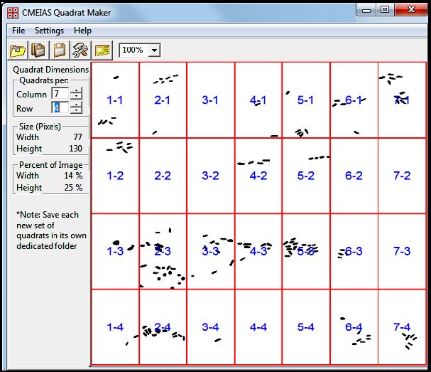
    [14] Dazzo F, Gross C (2012) CMEIAS Quadrat Maker: a digital software tool to optimize grid dimensions and produce quadrat images for landscape ecology spatial analysis. J Ecosystem and Ecography 3: 4 (136).
    [15] Ji Z, Card K, Dazzo FB (2015) CMEIAS JFrad: a new computing toolkit to discriminate the fractal geometry of landscape architecture and spatial patterns of individual cells in microbial biofilms. Microbial Ecology 69: 710-720.
    [16] Dazzo FB, Liu J, Gross C, et al. CMEIAS v3.10: Advanced Computational Tools of Image Analysis Software Designed to Strengthen Microscopy-Based Approaches for Understanding Microbial Ecology. All-Investigator Meeting Kellogg Biological Station Long-Term Ecological Research Mtg, 2015. Available from: http://lter.kbs.msu.edu/abstracts/493.
    [17] Towner H EcoStat: an Ecological Analysis Program, version 1.03. Exeter Software, Setauket, New York. 1999 available from: http://www.exetersoftware.com/cat/Trinity/ecostat.html.
    [18] Krebs C Ecological Methodology, version 7.2, Exeter Software, Setauket, NY, 2011, available from:http://www.exetersoftware.com/cat/ecometh/ecomethodology.html.
    [19] Hammer O, Harper DAT, Ryan PD (2001) PAST: Paleontological statistics software package for education and data analysis. Palaeontol Electron 4: 1-9.
    [20] Roberts A, Withers P. StatistiXL, Version 1.10. Broadway-Nedlands, Kalamunda, Australia, 2012, available from: http://www.statistixl.com/default.aspx.
    [21] Robertson GP GS+ Geostatistics, Version 9.0 Gamma Design Software, Plainwell, MI USA. 2004, Available from: http://www.gammadesign.com/default.aspx.
    [22] Paul EA, Harris D, Klug MJ, et al. (1999) The determination of microbial biomass. In: Robertson GP, Coleman DC, Bledsoe CS, et al., Standard Soil Methods for Long-Term Ecological Research. New York, USA: Oxford University Press, 291-317.
    [23] Dazzo FB, Klemmer KJ, Chandler R, et al. (2013) In situ ecophysiology of microbial biofilm communities analyzed by CMEIAS computer-assisted microscopy at single-cell resolution. Diversity 5: 426-460. doi: 10.3390/d5030426
    [24] Petchey OI, Long ZT, Morin PJ (2007) The consequences of body size in model microbial ecosystems. In: Hildrew A, Raffelli D, Edmonds-Brown R, Body Size: the Structure and Function of Aquatic Ecosystems. Cambridge UK: Cambridge University Press, 245-265.
    [25] Magurran AE (2004) Measuring Biological Diversity. Malden Massachusetts, USA: Blackwell.
    [26] Gonzalez JM, Sherr EB, Sherr BF (1990) Size-selective grazing on bacteria by natural assemblages of estuarine flagellates and ciliates. Appl Environ Microbiol 56: 583-589.
    [27] Fukuda M, Matsuyama J, Katano T et al. (2006) Assessing primary and bacterial production rates in biofilms on pebbles in Ishite Stream, Japan. Microbial Ecology 52: 1-9. doi: 10.1007/s00248-006-9114-0
    [28] Legendre P, Legendre L (2012) Numerical Ecology, 3rd Ed., Amsterdam The Netherlands: Elsevier.
    [29] Dazzo FB, Ganesan I, Yanni YG (2014) Spatial ecology of rhizobacterial colonization on roots analyzed by CMEIAS computer-assisted microscopy at single-cell resolution. In: Singh DP, Singh H, Trends in Soil Microbial Ecology, Chapter. 4, Houston USA: Studium Press, 67-92.
    [30] Gantner S, Schmid M, Dürr C, et al. (2006). In situ spatial scale of calling distances and population density-independent N-Acylhomoserine lactone mediated communication by rhizobacteria colonized on plant roots. FEMS Microbiol Ecol 56: 188-194. doi: 10.1111/j.1574-6941.2005.00037.x
    [31] Dazzo FB (2012) CMEIAS-aided microscopy of the spatial ecology of individual bacterial interactions involving cell-to-cell communication within biofilms. Sensors 12: 7047-7062. doi: 10.3390/s120607047
    [32] Perry J (1998) Measures of spatial patterns for counts. Ecology 79: 1008-1017.
    [33] Ripley BD (1979) Tests of “randomness” for spatial point patterns. J Roy Statist Soc B 41: 368-374.
    [34] Hammer Ø (2009) New methods for the statistical detection of point alignments. Computers and Geosciences 35: 659-666.
    [35] Robertson GP (1987) Geostatistics in ecology: interpolating with known variance. Ecology 68: 744-748. doi: 10.2307/1938482
    [36] Ettema CH, Wardle DH (2002) Spatial soil ecology. Trends in Ecology and Evolution 17: 177-183.
    [37] Zahid W, Ganczarczyk J (1994) A technique for a characterization of RBC biofilm surface. Water Res 28: 2229-2231. doi: 10.1016/0043-1354(94)90036-1
    [38] Hermanowicz S, Schindler U, Wilderer P (1995) Fractal structure of biofilms: new tools for investigation of morphology. Water Sci Technol 32: 99-105.
    [39] Yang X, Beyenal H, Harkin G, et al. (2000) Quantifying biofilm structure using image analysis. J Microbiol Methods 39: 109-119. doi: 10.1016/S0167-7012(99)00097-4
    [40] Ritchie ME (2010) Scale, Heterogeneity, and the Structure and Diversity of Ecological Communities. Princeton, NJ USA: Princeton University Press.
    [41] Sugihara G, May RM (1990) Applications of fractals in ecology. TREE 5: 79-86.
    [42] West G, Brown J, Enquist B (1999) The fourth dimension of life: fractal geometry and allometric scaling of organisms. Science 284: 1677-1679.
    [43] Dale MRT, Fortin MJ (2014) Spatial Analysis: a Guide for Ecologists. 2nd Ed. Cambridge UK: Cambridge University Press.
    [44] Hill MO (1973) The intensity of spatial pattern in plant communities. J Ecol 61: 225-235. doi: 10.2307/2258930
  • This article has been cited by:

    1. Zefang Song, Huafei Di, Instability analysis and regularization approximation to the forward/backward problems for fractional damped wave equations with random noise, 2023, 01689274, 10.1016/j.apnum.2022.12.017
    2. Yue Pang, Xingchang Wang, Furong Wu, Global existence and blowup in infinite time for a fourth order wave equation with damping and logarithmic strain terms, 2021, 14, 1937-1632, 4439, 10.3934/dcdss.2021115
    3. Quang-Minh Tran, Hong-Danh Pham, Global existence and blow-up results for a nonlinear model for a dynamic suspension bridge, 2021, 14, 1937-1632, 4521, 10.3934/dcdss.2021135
    4. Huafei Di, Zefang Song, F. Rabiei, Initial and Boundary Value Problems for a Class of Nonlinear Metaparabolic Equations, 2021, 2021, 1687-9139, 1, 10.1155/2021/6668355
    5. Nouri Boumaza, Billel Gheraibia, Global existence, nonexistence, and decay of solutions for a wave equation of p-Laplacian type with weak and p-Laplacian damping, nonlinear boundary delay and source terms, 2022, 129, 18758576, 577, 10.3233/ASY-211742
    6. Chao Yang, Yanbing Yang, Long-time behavior for fourth-order wave equations with strain term and nonlinear weak damping term, 2021, 14, 1937-1632, 4643, 10.3934/dcdss.2021110
    7. Yi Cheng, Ying Chu, A class of fourth-order hyperbolic equations with strongly damped and nonlinear logarithmic terms, 2021, 29, 2688-1594, 3867, 10.3934/era.2021066
    8. Le Thi Mai Thanh, Le Thi Phuong Ngoc, Nguyen Huu Nhan, Nguyen Thanh Long, FINITE-TIME BLOW UP OF SOLUTIONS FOR A FOURTH-ORDER VISCOELASTIC WAVE EQUATION WITH DAMPING TERMS, 2023, 13, 2156-907X, 3558, 10.11948/20230162
    9. Wen-jun Liu, Zhi-yu Tu, Equivalence between the internal observability and exponential decay for the Moore-Gibson-Thompson equation, 2024, 39, 1005-1031, 89, 10.1007/s11766-024-4133-5
  • Reader Comments
  • © 2015 the Author(s), licensee AIMS Press. This is an open access article distributed under the terms of the Creative Commons Attribution License (http://creativecommons.org/licenses/by/4.0)
通讯作者: 陈斌, bchen63@163.com
  • 1. 

    沈阳化工大学材料科学与工程学院 沈阳 110142

  1. 本站搜索
  2. 百度学术搜索
  3. 万方数据库搜索
  4. CNKI搜索

Metrics

Article views(7791) PDF downloads(1368) Cited by(8)

Figures and Tables

Figures(13)  /  Tables(2)

/

DownLoad:  Full-Size Img  PowerPoint
Return
Return

Catalog