Citation: Kamila Tomoko Yuyama, Thaís Souto Paula da Costa Neves, Marina Torquato Memória, Iago Toledo Tartuci, Wolf-Rainer Abraham. Aurantiogliocladin inhibits biofilm formation at subtoxic concentrations[J]. AIMS Microbiology, 2017, 3(1): 50-60. doi: 10.3934/microbiol.2017.1.50
[1] | Aynur Aybey, Elif Demirkan . Inhibition of Pseudomonas aeruginosa biofilm formation and motilities by human serum paraoxonase (hPON1). AIMS Microbiology, 2016, 2(4): 388-401. doi: 10.3934/microbiol.2016.4.388 |
[2] | Tatyana V. Polyudova, Daria V. Eroshenko, Vladimir P. Korobov . Plasma, serum, albumin, and divalent metal ions inhibit the adhesion and the biofilm formation of Cutibacterium (Propionibacterium) acnes. AIMS Microbiology, 2018, 4(1): 165-172. doi: 10.3934/microbiol.2018.1.165 |
[3] | Alexandra Soares, Ana Azevedo, Luciana C. Gomes, Filipe J. Mergulhão . Recombinant protein expression in biofilms. AIMS Microbiology, 2019, 5(3): 232-250. doi: 10.3934/microbiol.2019.3.232 |
[4] | Yunfeng Xu, Attila Nagy, Gary R. Bauchan, Xiaodong Xia, Xiangwu Nou . Enhanced biofilm formation in dual-species culture of Listeria monocytogenes and Ralstonia insidiosa. AIMS Microbiology, 2017, 3(4): 774-783. doi: 10.3934/microbiol.2017.4.774 |
[5] | Afraa Said Al-Adawi, Christine C. Gaylarde, Jan Sunner, Iwona B. Beech . Transfer of bacteria between stainless steel and chicken meat: A CLSM and DGGE study of biofilms. AIMS Microbiology, 2016, 2(3): 340-358. doi: 10.3934/microbiol.2016.3.340 |
[6] | Stephen H. Kasper, Ryan Hart, Magnus Bergkvist, Rabi A. Musah, Nathaniel C. Cady . Zein nanocapsules as a tool for surface passivation, drug delivery and biofilm prevention. AIMS Microbiology, 2016, 2(4): 422-433. doi: 10.3934/microbiol.2016.4.422 |
[7] | Bindu Subhadra, Man Hwan Oh, Chul Hee Choi . Quorum sensing in Acinetobacter: with special emphasis on antibiotic resistance, biofilm formation and quorum quenching. AIMS Microbiology, 2016, 2(1): 27-41. doi: 10.3934/microbiol.2016.1.27 |
[8] | Luciana C. Gomes, Joana M. R. Moreira, José D. P. Araújo, Filipe J. Mergulhão . Surface conditioning with Escherichia coli cell wall components can reduce biofilm formation by decreasing initial adhesion. AIMS Microbiology, 2017, 3(3): 613-628. doi: 10.3934/microbiol.2017.3.613 |
[9] | Luciana C. Gomes, Filipe J. Mergulhão . Effect of heterologous protein expression on Escherichia coli biofilm formation and biocide susceptibility. AIMS Microbiology, 2016, 2(4): 434-446. doi: 10.3934/microbiol.2016.4.434 |
[10] | Zoya Samoilova, Alexey Tyulenev, Nadezhda Muzyka, Galina Smirnova, Oleg Oktyabrsky . Tannic and gallic acids alter redox-parameters of the medium and modulate biofilm formation. AIMS Microbiology, 2019, 5(4): 379-392. doi: 10.3934/microbiol.2019.4.379 |
In many infections, pathogens are embedded in a complex matrix of macromolecules that adhere to various surfaces, a lifestyle called biofilm. Biofilm infections cause severe complications and 12% to 25% of bloodstream infections are attributable to patient mortality [1]. Biofilms form on any implant but also on mucosa, e. g. in the cystic fibrosis lung [2], the middle ear [3], the urinary tract [4] or gastric mucosa [5]. Biofilm formation protects the embedded microbial cells against hostile environments, including antibiotics or the immune system of the host. Biofilms are dynamic, adapting to changes in the environment and biofilm formation is reversible, enabling cells to disperse and to colonize new habitats if the conditions become unfavorable [6].
To form a biofilm, microbes have to coordinate their activities. This communication is achieved by small molecules acting as autoinducers to start genetic programs [7]. If the autoinducer concentration reaches a certain threshold, biofilm formation starts followed by the production of virulence factors. Because the process depends on cell density, this is called quorum sensing and used by many microbes [8]. To date more than 100 different autoinducers are known for bacteria, archaea and fungi. The best studied autoinducers are N-acyl-L-homoserine lactones, only known from Gram-negative bacteria [9]. Autoinducer-2, having a rather unusual cyclic boronic ester, is known both from Gram-positive and -negative bacteria [10]. In many Gram-positive bacteria small peptides act as autoinducers which are derived from larger precursor peptides [11].
The eradication of biofilm infections is a difficult task because the embedded pathogens are well protected against macrophages and antibiotics [12], e.g. 220 times higher antibiotic concentrations were needed to kill Escherichia coli in a biofilm compared to cells of the same strain living in planktonic form in the serum [13]. To overcome these difficulties together with increasing antibiotic resistances [14] novel antibiotics are needed to control biofilm infections [15]. One of these approaches aims at the communication between the cells in a biofilm by blocking quorum sensing [16,17]. On the one hand compounds from large chemical libraries can be screened but on the other hand natural compounds from various organisms can be tested for their ability to prevent biofilm formation or to disperse existing ones. The rationale behind this approach is that all organisms have to deal with the formation of biofilms on their bodies and probably have developed strategies to fight pathogenic biofilms. One potential resource for novel biofilm-modulating compounds are sporocarps of fungi which are exposed to a wet climate, favorable conditions for biofilm formation. As a proof of this concept it has been demonstrated for sporocarps of Basidiomycotina that ectomycorhizal fungi had a higher diversity of bacterial biofilms on their fruiting bodies than saprophytic ones which showed a higher percentage of biofilm-inhibiting extracts than ectomycorrhizal fungi [18].
Acetonitrile, ethyl acetate, chloroform and methanol were purchased from J. T. Baker (USA, Germany and Netherlands respectively), d-chloroform, formic acid 98%, potato dextrose (PD) and Luria-Bertani broth (LB) from Carl Roth GmbH (Karlsruhe, Germany). Bacto malt extract, Bacto peptone and agar were from BD (La Point de Claix, France), D-glucose from Merck (Darmstadt, Germany). N-hexanoyl-L-homoserine lactone was purchased from Cayman Chemical Company (USA), crystal violet from Fluka (Steinheim, Germany), tetracycline and RPMI 1640 medium from Sigma Aldrich (Germany, United Kingdom), respectively.
Bacillus cereus DSM 626, Staphylococcus epidermidis ATCC 35984, Streptococcus mutans UA59, Staphylococcus aureus DSM 1104, Candida albicans DSM 11225, Candida guilliermondii DSM 70052, Candida krusei DSM 6128, Candida parapsilosis (DSM 5784, Rhodotorula glutinis DSM 70398 and Yarrowia lipolytica DSM 70151 were purchased from the German collection of microorganisms and cell cultures (DSMZ). Fungi were used in antifungal assays. The bacteria and the mutant Chromobacterium violaceum CV026; Pseudomonas aeruginosa PA14 and Escherichia coli MT102 were applied in antimicrobial and antibiofilm assays. Bacterial strains were maintained on LB agar, and yeast strains on YM agar (0.3% yeast extract, 0.3% malt extract, 0.5% peptone, 1% glucose, 1.5% agar) at 4 °C.
Fungi were collected in Harz mountains (latitude 51°45'21”, longitude 10°32'17”, 724 m), Germany, and isolated on Potato Dextrose Agar (PDA). The identification of the isolates were made by sequencing of the internal spacer regions (ITS). For DNA extraction, the NucleoSpin Plant Ⅱ kit (Macherey, Nagel) was used. Fungal primers ITS1F and ITS4 were applied in PCR amplification following Bruns et al. [19]; the amplicons were sequenced by Eurofins, analyzed using Sequencher 4.10.1 and the phylogenetic tree was constructed with the MEGA 6 program. The sequence (LT635436) was deposited in European Nucleotide Archive (ENA).
Pellets (5 × 5 mm) of mycelia of the isolates grown on Malt extract agar (3% malt extract, 0.5% bacto peptone and 1.5% agar) were transferred to 2L Erlenmeyer flasks containing 1L of malt extract (ME) broth (3% malt extract, 0.5% bacto peptone). After 65 days of static incubation at 20 °C or 25 °C in the dark, the broth was filtrated, extracted with ethyl acetate and tested for antimicrobial and antibiofilm activities.
Aurantiogliocladin was purified in a gradient with an Agilent HPLC (C18 column Varian, Agilent Technologies 250 × 10 mm). Solvent A: acetonitrile, solvent B: MilliQ® water with 1% formic acid, gradient from 10% A at 0 min to 100% A at 25 min; UV absorbance was detected at 210, 230, 254, 260 and 280 nm; flow rate: 4 mL min-1. Molecular mass was identified in Agilent LC-MS/MS 1200 series system (C18 analytical column Nucleosil® 125 mm × 20 mm), with the same conditions described above. The structure was elucidated by 1H and 13C NMR recorded on a Bruker 600 MHz spectrometer, in 5 mm tube of d-chloroform. The chemical shifts were compared with the literature.
After overnight incubation at 30 °C in LB medium, C. violaceum CV026 was adjusted to OD 0.5 according to McFarland scale, mixed with 3.5 nmol mL-1 N-hexanoyl-L-homoserinelactone, and inoculated in 96 well no tissue microtiter plates (Falcon®Micro TestTM, USA) containing LB broth with serial diluted aurantiogliocladin (180 to 3 μg mL-1) dissolved in methanol. Plates were covered with a sterile adhesive porous paper (Kisker Biotech GmbH, Steinfurt, Germany) and incubated at 30 °C for 48 h. Methanol and LB broth were used as negative and tetracycline (100 μg mL-1) was used as positive control. Absence of violacein indicated quorum-quenching activity.
To determine the minimal inhibitory concentration (MIC) of the bioactive compound, bacteria and yeast were incubated in LB and in RPMI 1640 media respectively, C. violaceum, B. cereus, S. epidermidis and all yeasts at 30 °C and the other strains at 37 °C, containing serial diluted aurantiogliocladin (256 to 3 μg mL-1) dissolved in methanol. Bioscreen-C automated growth curve analysis system (Oy Growth Curves AB Ltd, Helsinki, Finland) was used to determine the bacterial/fungal growth over 24 h, employing one OD600 measurement each 15 min. Experiments were performed in triplicate. To evaluate bactericide/fungicide or bacteriostatic/fungistatic effects, aliquots from different concentrations in the well, after OD measurements, were inoculated in LB agar for bacterial assays and YM agar for fungal assays and incubated for 24 h. Methanol and LB/RMPI 1640 media were used as negative, tetracycline (100 μg mL-1) as positive control for bacteria.
For biofim inhibition bacteria were incubated as above. After 24h, biofilm formation in the microtiter wells was measured by the staining with crystal violet following a published protocol (O'Toole, 2011) [20].
To assess the potential of fungi collected at the Harz Mountains, Germany, several isolates were screened for their potential to infer with bacterial biofilm formation. Among the fungi, showing activities, isolate 1 was selected for further investigation of compounds against biofilms of Gram positive and negative bacteria. The isolate was identified by comparison of its ITS region with that of related type species and the sequence was deposited (LT635436). According to the sequence of the ITS region, isolate 1 belongs to the genus Clonostachys and is closely related to Clonostachys candelabrum (Figure 1).
From the culture broth of isolate 1 [LT635436] a compound was isolated which formed yellow crystals. It revealed in the ESI mass spectrum a [M + H]+ ion of 197.0822 in the positive mode which led to a molecular formula of C10H12O4. Such a molecular composition requires five double bond equivalents. The 1H NMR spectra had only two singlets at 2.05 and 3.98 (CDCL3) while the 13C NMR spectra displayed the resonances of two carbonyls and two double bonds demanding one ring to fulfill the molecular formula. The spectral data and comparison with literature data [21] led to the identification of the active compound as aurantiogliocladin 1 (Figure 2). Aurantiogliocladin has been discovered in the culture broth of a Gliocladium species [22] and its structure was determined in 1953 by Vischer [23]. Biological tests showed that it is a weak antibiotic but no activities against biofilms have been reported.
The antibiotic activity of aurantiogliocladin against several bacteria was low to moderate. No clear tendency could be seen but Gram-negative bacteria seemed to be more resistant against aurantiogliocladin than Gram-positive. S. epidermidis was found to be more sensitive against the antibiotic than the closely related S. aureus (Table 1). These differences were more pronounced in the biofilm inhibition assay. While for S. epidermidis only 51% biofilm inhibition was observed at MIC concentration (64 μg mL-1) and half of the MIC prevented only 19% biofilm formation, B. cereus revealed 44% biofilm inhibition at MIC but still 37% at one quarter of MIC. Both, S. epidermidis and B. cereus, still formed almost 50% of the biofilm when twice or even higher concentrations of MIC were applied. The most sensitive strain in the test was C. violaceum CV026 where 90% of the biofilm could be prevented at MIC and still 29 % at one eighth of MIC. These results indicate that biofilm inhibitions were almost independent from the minimal inhibition concentrations for planktonic cells, and also were associated with quorum-quenching activity (180-22.5 μg mL-1) due to the absence of violacein (Figure 3). Interesting is the finding that some inhibition of biofilm formation occurred even at subtoxic concentrations of aurantiogliocladin. To shed more light on these results bacteria we treated with aurantiogliocladin at MIC and were then plated on fresh agar plates. All bacteria grew again under these conditions indicating that aurantiogliocladin is not bactericidal at this concentration but bacteriostatic.
Strain | MIC [μg mL-1] | % biofilm inhibition |
Bacillus cereus DSM 626 | 128 | 54% (256 μg mL-1) 44% (128 μg mL-1) 37% (32 μg mL-1) |
Chromobacterium violaceum CV026 | 180 | 90% (180 μg mL-1) 62% (45 μg mL-1) 29% (22.5 μg mL-1) |
Escherichia coli MT102 | 300 | - |
Pseudomonas aeruginosa PA14 | >300 | - |
Staphylococcus aureus DSM 1104 | 300 | - |
Staphylococcus epidermidis ATCC 35984 | 64 | 55% (256 μg mL-1) 51% (64 μg mL-1) 19% (32 μg mL-1) 10% (16 μg mL-1) |
Streptococcus mutans UA59 | 300 | 34% (300 μg mL-1) 15% (150 μg mL-1) |
Aurantiogliocladin concentrations for the specific biofilm inhibitions are given in brackets. For easier visualization biofilm inhibitions at MIC are set in red. |
Because in the first publications of aurantiogliocladin activities against fungi have been reported we also tested the antifungal activity of this compounds against several yeasts (Table 2). Aurantiogliocladin was moderately active against all yeasts tested with the highest activities against C. guilliermondii and R. glutinis and the lowest activity against C. albicans. As already found for the tested bacteria at MIC the substance was fungistatic but not fungicidal.
Strain | MIC [μg mL-1] |
Candida albicans DSM 11225 | 128 |
Candida guilliermondii DSM 70052 | 32 |
Candida krusei DSM 6128 | 64 |
Candida parapsilosis DSM 5784 | 64 |
Rhodotorula glutinis DSM 70398 | 32 |
Yarrowia lipolytica DSM 70151 | 64 |
Aurantiogliocladin 1 is known for several decades and has been found in a number of strains. Various studies were directed towards the biosynthesis of aurantiogliocladin and it was found that it starts from acetate and runs via orsellic acid to the final metabolite [24]. 1,3-dihydroxy-4,5- dimethylbenzene, also reported from Gliocladium roseum maybe a precursor for the intermediate orsellic acid [25]. Aurantiogliocladin was discovered as a weak antibiotic but later found to be also weakly anthelmintic [26] and toxic for termites (Coptotermes formosanus) [27] and also for the second instar larvae of Aedes aegypti mosquito [28]. Furthermore it serves as attractant for the plant-parasitic nematode Neotylenchus linfordi which is associated with Gliocladium roseum [29].
Secondary metabolites with a quinone skeleton were described from several fungi. One of the first reports of a compound of this class was spinulosin 2 from Penicillium spinulosum [30]; from Aspergillus fumigatus several toluquinones have been reported, e. g. 4-hydroxy-3-methoxy-2,5- toluquinone 3, which are closely related to aurantiogliocladin [31]. 4-Methoxy-2,5-toluquinone 4, isolated from Lentinus adhaerens, has been shown to block the thromboxane A2 receptor [32]. Not only in fungi but also in animals this type of secondary metabolites can be found. The millipede Floridobolus penneri excretes for defense a number of benzoquinone which are closely related to aurantiogliocladin [33].
Aurantiogliocladin is a weak antibiotic acting both against a number of bacteria but also against some fungi. The selectivity of the antibiotic is remarkable. While S. aureus was not inhibited at all, S. epidermidis was more sensitive. The compound had not influence on the growth of P. aeruginosa or E. coli. In all these experiments it was found that plating on fresh medium after the inhibition with aurantiogliocladin resulted in the growth of the treated cells, demonstrating that the antibiotic stalls growth but does not kill the cells. This phenomenon has already been reported for less substituted 1,4-benzoquinone and S. aureus [34]. The action on biofilm formation is more complex. For some bacteria there are only little effects on the biofilm (e. g. S mutans or S. epidermidis), while for other strains biofilm inhibition is considerable large (e. g. C. violaceum) and lasting well below the MIC threshold (e. g. B. cereus).
Some insights into the structure-activity-relations of this class of compounds are gained by comparing literature data of related compounds. Both 4-methoxy-2,5-toluquinone 4 [35] and 3-methoxy-2,5-toluquinone 5 [36] are more active, especially against S. aureus and B. subtilis than aurantiogliocladin. A study assessing a large number of 1,4-benzoquinones and 2,5-toluquinones demonstrated a low sensitivity of E. coli against almost all compounds while the sensitivity of S. aureus varied across the different compounds. As tendencies, it was noted that the introduction of methoxy groups at the nucleus increased activity while hydroxyl had the opposite effect. The most active compounds were found to have a methoxy group at C-4 of 2,5-toluquinone and the author recommended 4,6-dimethoxy-2,5-toluquinone derivatives as the most active ones [37]. From these results one can speculate that the additional methyl group and the adjacent methoxy functions cause the decline in activity against S. aureus. Interestingly, another study could not find any activities (>800 mg mL-1) of 1,4-benzoquinone 6,2-methoxy-1,4-benzoquinone 7 and 3,5-dimethoxy-1,4- benzoquinone 8, confirming the very different action of this class of compounds on fungi compared to bacteria [38]. None of these studies included biofilms and as the results of the presented study shows MIC data are here a very poor indicator for the prediction of biofilm inhibition.
The rational that natural products from organisms living in a wet environment may be active against biofilms could be confirmed and fungi proved to be a rich source for biofilm-interfering compounds. In a screen of fungal isolates obtained from the Harz mountains in Germany several strains have been found producing compounds for the inhibition of biofilms. One of these strains has been identified as C. candelabrum producing aurantiogliocladin. Biological tests showed aurantiogliocladin as a weak antibiotic which was active against S. epidermidis but not S. aureus. Aurantiogliocladin could also inhibit biofilm formation of several tested bacterial strains. This inhibition, however, was never complete and biofilm inhibition activities were also found at concentrations below the minimal inhibitory concentrations. In agreement with this finding was the observation that aurantiogliocladin was bacteriostatic for the tested bacteria but not bactericidal. Many quorum-quenching compounds do not kill bacteria and it has been suggested to combine them with antibiotics for improved clearance of biofilm infections [39]. Because several closely related toluquinones with different antibiotic activities have been reported from various fungi, screening of a chemical library of toluquinones is suggested for the improvement of biofilm inhibition activities. These investigations should not be conducted only on pure strains because in nature microbes seldom live alone but in polymicrobial communities with complex and multifold interactions [40]. Any applicable drug for the control of biofilm infections has to address this issue in order not to suppress one pathogen but foster the development of another one in the same biofilm community.
K.T.Y., T. S. P. da C. N., M. T. M., and I. T. T. acknowledge the support by a stipend of the "Science without border (Ciências sem Fronteiras)" program of Coordenação de Aperfeiçoamento de Pessoal de Nível Superior (CAPES) and the Conselho Nacional de Desenvolvimento Científico e Tecnológico (CNPq) from Brazil and the German Academic Exchange Service (DAAD).
All authors declare no conflicts of interest in this paper.
[1] | Leone M,Dillon LR (2008) Catheter outcomes in home infusion. J Infus Nurs 31: 84–91. |
[2] | Singh PK, Schaefer AL,Parsek MR, et al. (2000) Quorum-sensing signals indicate that cystic fibrosis lungs are infected with bacterial biofilms. Nature 407: 762–764. |
[3] |
Hall-Stoodley L,Hu FZ, Gieseke A, et al. (2006) Direct detection of bacterial biofilms on the middle-ear mucosa of children with chronic otitis media. JAMA 296:202–211. doi: 10.1001/jama.296.2.202
![]() |
[4] |
Rosen Da HT, Stamm WE, Humphrey PA, et al. (2007) Detection of intracellular bacterial communities in human urinary tract infection. PLoS Med 4: e329. doi: 10.1371/journal.pmed.0040329
![]() |
[5] |
Carron MA, Tran VR, Sugawa C, et al. (2006) Identification of Helicobacter pylori biofilms in human gastric mucosa. J Gastrointest Surg 10:712–717. doi: 10.1016/j.gassur.2005.10.019
![]() |
[6] |
Hall-Stoodley L, Costerton JW, Stoodley P (2004) Bacterial biofilms: From the natural environment to infectious diseases. Nat Rev Microbiol 2: 95–108. doi: 10.1038/nrmicro821
![]() |
[7] |
Camilli A, Bassler BL (2006) Bacterial small-molecule signaling pathways. Science 311: 1113–1116. doi: 10.1126/science.1121357
![]() |
[8] |
Schauder S, Bassler BL (2001) The languages of bacteria. Genes Dev 15:1468–1480. doi: 10.1101/gad.899601
![]() |
[9] |
Abraham WR (2005) Controlling Gram-negative pathogenic bacteria by interfering with their biofilm formation. Drug Design Rev Online 2: 13–33. doi: 10.2174/1567269053390257
![]() |
[10] |
Chen X, Schauder S, Potier N, et al. (2002) Structural identification of a bacterial quorum-sensing signal containing boron. Nature 415:545–549. doi: 10.1038/415545a
![]() |
[11] |
Lyon GJ, Novick RP (2004) Peptide signaling in Staphylococcus aureus and other Gram-positive bacteria. Peptides 25: 1389–1403. doi: 10.1016/j.peptides.2003.11.026
![]() |
[12] |
Stewart PS, Costerton JW (2001) Antibiotic resistance of bacteria in biofilms. Lancet 358: 135–138. doi: 10.1016/S0140-6736(01)05321-1
![]() |
[13] | Joly V, Pangon B,Vallois JM, et al. (1987) Value of antibiotic levels in serum and cardiac vegetations for predicting antibacterial effect of ceftriaxone in experimental Escherichia coli endocarditis. Antimicrob Agents Chemother 31:1632–1639. |
[14] |
Austin DJ, Kristinsson KG, Anderson RM (1999) The relationship between the volume of antimicrobial consumption in human communities and the frequency of resistance. Proc Natl Acad Sci USA 96: 1152–1156. doi: 10.1073/pnas.96.3.1152
![]() |
[15] | Payne DJ (2008) Microbiology. Desperately seeking new antibiotics. Science 321: 1644–1645. |
[16] |
Worthington RJ, Richards JJ, Melander C (2014) Non-microbicidal control of bacterial biofilms with small molecules. Anti-Inf Agents 12: 120–138. doi: 10.2174/22113525113119990107
![]() |
[17] |
Zhu J, Kaufmann GF (2013) Quo vadis quorum quenching? Curr Opin Pharmacol 13: 688–898. doi: 10.1016/j.coph.2013.07.003
![]() |
[18] |
de Carvalho MP, Türck P, Abraham WR (2015) Secondary metabolites control the associated bacterial communities of saprophytic Basidiomycotina fungi. Microbes Environ 30: 196–198. doi: 10.1264/jsme2.ME14139
![]() |
[19] |
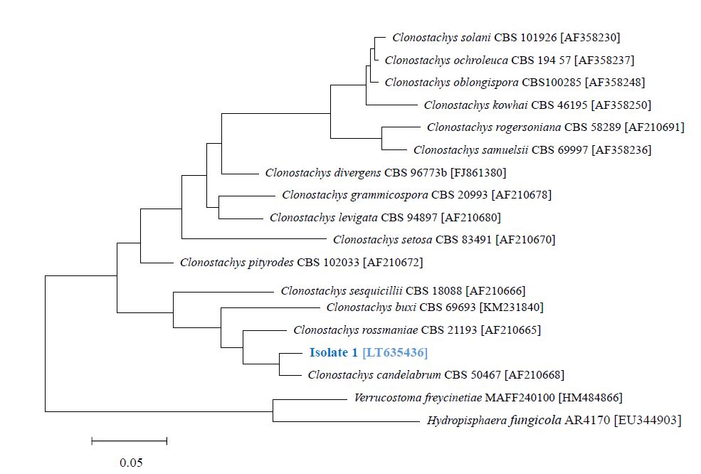
Bruns TD, Fogel R, White TJ, et al. (1990) Amplification and sequencing of DNA from fungal herbarium specimens. Mycologia 82: 175–184. doi: 10.2307/3759846
![]() |
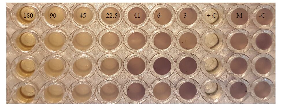
[20] | O'Toole GA (2011) Microtiter dish biofilm formation assay. J Vis Exp 47: e2437–e2437. |
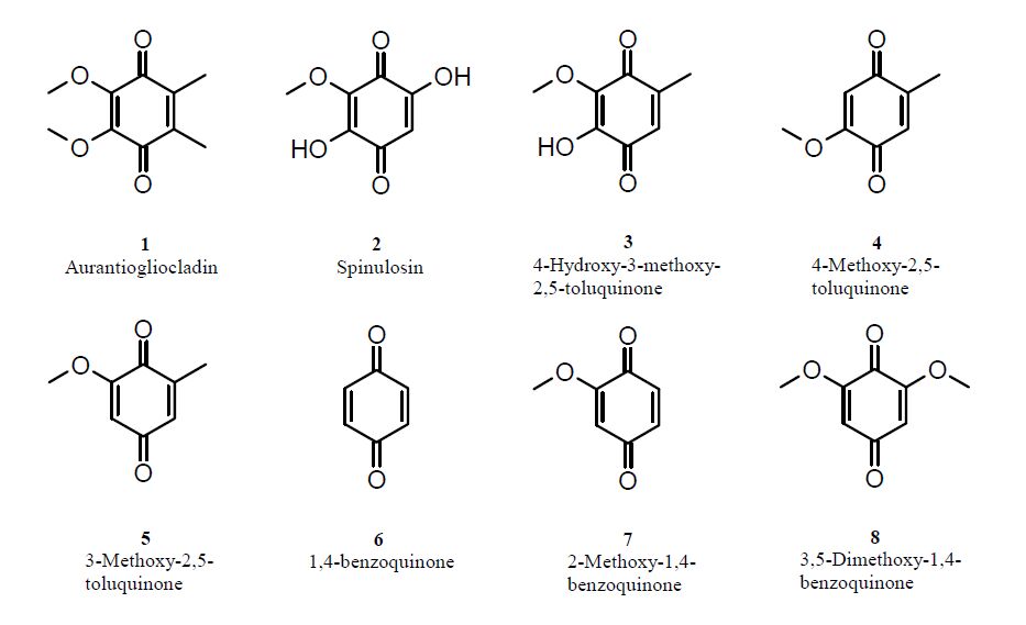
[21] | Franke C, Krohn K (2008) Aurantiogliocladin, In: Roth L, Rupp G, Roth collection of natural products data: concise descriptions and spectra , Weinheim, John Wiley & Sons. |
[22] |
Brian PW, Curtis PJ, Howland SR, et al. (1951) Three new antibiotics from a species of Gliocladium. Experientia 7: 266–267. doi: 10.1007/BF02154546
![]() |
[23] | Vischer EB (1953) The structures of aurantio-and rubro-gliocladin and gliorosein. J Chem Soc (Resumed) , 815–820. |
[24] | Bentley R, Lavate WV (1965) Studies on Coenzyme Q. The biosynthesis of aurantiogliocladin and coenzyme Q in molds. J Biol Chem 240: 532–540. |
[25] |
Pettersson G (1965) New phenolic metabolites from Gliocladium roseum. Acta Chem Scand 19:414–420. doi: 10.3891/acta.chem.scand.19-0414
![]() |
[26] |
Ayers S, Zink DL, Mohn K, et al. (2010). Anthelmintic constituents of Clonostachys candelabrum. J Antibiotics 63:119–122. doi: 10.1038/ja.2009.131
![]() |
[27] |
Mozaina K, Cantrell CL, Mims AB, et al. (2008) Activity of 1,4-benzoquinones against Formosan subterranean termites (Coptotermes formosanus ).J Agric Food Chem 56:4021–4026. doi: 10.1021/jf800331r
![]() |
[28] | Likhovidov VE, Isangalin FS, Naumov AN, et al. (2010) Agents against mosquitos. Russ patent , RU 2391389 C2 20100610. |
[29] | Klink JW, Dropkin VH, Mitchell JE (1970) Studies on the host-finding mechanisms of Neotylenchus linfordi. J Nematol 2:106–117. |
[30] |
Birkinshaw JH, Raistrick H (1931) On a new methoxy-dihydroxy-toluquinone produced from glucose by species of Penicillium of the P. spinulosum series.Phil Trans Roy Soc London. B 220: 245–254. doi: 10.1098/rstb.1931.0023
![]() |
[31] |
Petterson G (1963) Toluquinones from Aspergillus fumigatus. Acta Chem Scand 17:1771–1776. doi: 10.3891/acta.chem.scand.17-1771
![]() |
[32] | Lauer U, Anke T, Hansske F (1991) Antibiotics from basidiomycetes. XXXVIII. 2-Methoxy-5- methyl-1,4-benzoquinone, a thromboxane A2 receptor antagonist from Lentinus adhaerens. J Antibiot 44:59–65. |
[33] |
Attygalle AB, Xu SC, Meinwald J, et al. (1993). Defensive secretion of the millipede Floridobolus penneri. J Nat Prod 56:1700–1706. doi: 10.1021/np50100a007
![]() |
[34] | Nishina A, Uchibori T (1991) Antimicrobial activity of 2, 6-dimethoxy-p-benzoquinone, isolated from thick-stemmed bamboo, its analogs. Agric Biol Chem 55:2395–2398. |
[35] |
Anchel M, Hervey A, Kavanagh F, et al. (1948) Antibiotic substances from Basidiomycetes III. Coprinus similis and Lentinus degener. Proc Nat Acad Sci 34:498–502. doi: 10.1073/pnas.34.11.498
![]() |
[36] | Sood RS, Roy K, Reddy GCS, et al. (1982) 3-Methoxy-2, 5-toluquinone from Aspergillus sp. HPL Y-30,212. Fermentation, isolation, characterization and biological properties. J Antibiot 35:985–987. |
[37] | Oxford AE (1942) Bacteriostatic powers of the methyl ethers of fumigatin and spinulosin and other hydroxy-, methoxy- and hydroxymethoxy-derivatives of toluquinone and benzoquinone. Chem Ind 61:189–192. |
[38] |
Lana EJ, Carazza F, Takahashi JA (2006) Antibacterial evaluation of 1, 4-benzoquinone derivatives. J Agric Food Chem 54:2053–2056. doi: 10.1021/jf052407z
![]() |
[39] |
Estrela AB, Abraham W-R (2010) Combining biofilm-controlling compounds and antibiotics as a promising new way to control biofilm infections. Pharmaceuticals 3:1374–1393. doi: 10.3390/ph3051374
![]() |
[40] |
Wargo MJ, Hogan DA (2006) Fungal-bacterial interactions: a mixed bag of mingling microbes. Curr Opin Microbiol 9: 359–364. doi: 10.1016/j.mib.2006.06.001
![]() |
1. | Abolfazl Narmani, Rémy Bertrand Teponno, Soleiman E. Helaly, Mahdi Arzanlou, Marc Stadler, Cytotoxic, anti-biofilm and antimicrobial polyketides from the plant associated fungus Chaetosphaeronema achilleae, 2019, 139, 0367326X, 104390, 10.1016/j.fitote.2019.104390 | |
2. | Kamila Tomoko Yuyama, Clara Chepkirui, Lucile Wendt, Diana Fortkamp, Marc Stadler, Wolf-Rainer Abraham, Bioactive Compounds Produced by Hypoxylon fragiforme against Staphylococcus aureus Biofilms, 2017, 5, 2076-2607, 80, 10.3390/microorganisms5040080 | |
3. | Clara Chepkirui, Kamila T. Yuyama, Lucy A. Wanga, Cony Decock, Josphat C. Matasyoh, Wolf-Rainer Abraham, Marc Stadler, Microporenic Acids A–G, Biofilm Inhibitors, and Antimicrobial Agents from the BasidiomyceteMicroporusSpecies, 2018, 81, 0163-3864, 778, 10.1021/acs.jnatprod.7b00764 | |
4. | Kamila Tomoko Yuyama, Manfred Rohde, Gabriella Molinari, Marc Stadler, Wolf-Rainer Abraham, Unsaturated Fatty Acids Control Biofilm Formation of Staphylococcus aureus and Other Gram-Positive Bacteria, 2020, 9, 2079-6382, 788, 10.3390/antibiotics9110788 | |
5. | Fazlurrahman Khan, Dung T.N. Pham, Sandra F. Oloketuyi, Young-Mog Kim, Antibiotics Application Strategies to Control Biofilm Formation in Pathogenic Bacteria, 2020, 21, 13892010, 270, 10.2174/1389201020666191112155905 | |
6. | Denis Swolana, Małgorzata Kępa, Agata Kabała-Dzik, Radosław Dzik, Robert D. Wojtyczka, Sensitivity of Staphylococcal Biofilm to Selected Compounds of Plant Origin, 2021, 10, 2079-6382, 607, 10.3390/antibiotics10050607 | |
7. | Kamila Tomoko Yuyama, Lucile Wendt, Frank Surup, Robin Kretz, Clara Chepkirui, Kathrin Wittstein, Chollaratt Boonlarppradab, Sarunyou Wongkanoun, Jennifer Luangsa-ard, Marc Stadler, Wolf-Rainer Abraham, Cytochalasans Act as Inhibitors of Biofilm Formation of Staphylococcus aureus, 2018, 8, 2218-273X, 129, 10.3390/biom8040129 |
Strain | MIC [μg mL-1] | % biofilm inhibition |
Bacillus cereus DSM 626 | 128 | 54% (256 μg mL-1) 44% (128 μg mL-1) 37% (32 μg mL-1) |
Chromobacterium violaceum CV026 | 180 | 90% (180 μg mL-1) 62% (45 μg mL-1) 29% (22.5 μg mL-1) |
Escherichia coli MT102 | 300 | - |
Pseudomonas aeruginosa PA14 | >300 | - |
Staphylococcus aureus DSM 1104 | 300 | - |
Staphylococcus epidermidis ATCC 35984 | 64 | 55% (256 μg mL-1) 51% (64 μg mL-1) 19% (32 μg mL-1) 10% (16 μg mL-1) |
Streptococcus mutans UA59 | 300 | 34% (300 μg mL-1) 15% (150 μg mL-1) |
Aurantiogliocladin concentrations for the specific biofilm inhibitions are given in brackets. For easier visualization biofilm inhibitions at MIC are set in red. |
Strain | MIC [μg mL-1] |
Candida albicans DSM 11225 | 128 |
Candida guilliermondii DSM 70052 | 32 |
Candida krusei DSM 6128 | 64 |
Candida parapsilosis DSM 5784 | 64 |
Rhodotorula glutinis DSM 70398 | 32 |
Yarrowia lipolytica DSM 70151 | 64 |