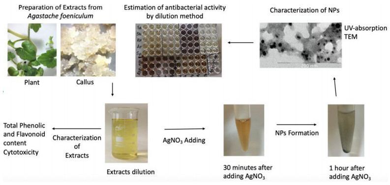
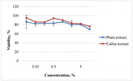
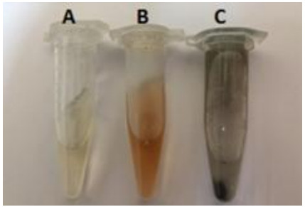
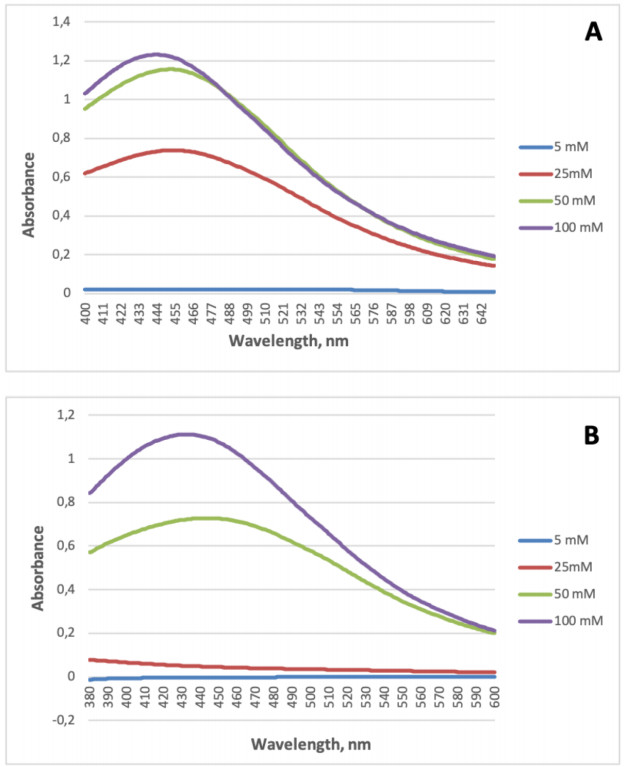
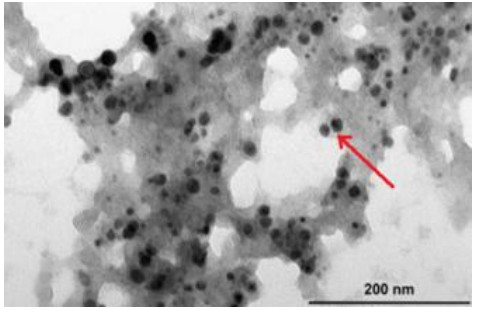
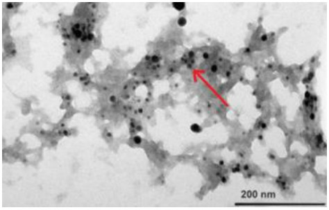
Green synthesis is an environmentally friendly, safe, affordable and low-cost method for various morphologies nanoparticles producing using bacterial, fungi or plant aqueous extracts. Antimicrobial activity of silver nanoparticles obtained from Agastache foeniculum plants and callus aqueous extracts has been demonstrated against hospital infections—Staphylococcus aureus, Staphylococcus haemolyticus, Klebsiella pneumonia and Streptococcus pneumonia. The synthesized nanoparticles were characterized by UV-spectroscopy and transmission electron microscopy. The nanoparticles had a spherical shape with an average size diameter of 19.81 ± 5.32 nm and 9.51 ± 1.55 nm for synthesis based on plant and callus extracts respectively. The minimum inhibitory concentration for the nanoparticles was 6.25 to 25.00 mg/L for all tested strains, except for Acinetobacter baumannii, which experimental cultivation conditions were inappropriate. Antioxidant capacity of the extracts expressed in total phenolic and flavonoid contents also has been shown. Total phenolic content of callus extract was 1.97 ± 0.06 mg/g in hydroquinone equivalents, which is higher than plant's extract. Supposedly biogenic nanoparticles morphology depends on the concentration of compounds with antioxidant activity in extracts. Initial extracts have proved low-cytotoxicity, indicating synthesis safety. The nanoparticles can be used as a basis for products development to prevent hospital infections spreading, including those with antibiotic resistance.
Citation: Oksana B. Polivanova, Mikhail Yu. Cherednichenko, Elena A. Kalashnikova, Rima N. Kirakosyan. In vitro antibacterial effect of silver nanoparticles synthetized using Agastache foeniculum plant and callus extracts[J]. AIMS Agriculture and Food, 2021, 6(2): 631-643. doi: 10.3934/agrfood.2021037
Green synthesis is an environmentally friendly, safe, affordable and low-cost method for various morphologies nanoparticles producing using bacterial, fungi or plant aqueous extracts. Antimicrobial activity of silver nanoparticles obtained from Agastache foeniculum plants and callus aqueous extracts has been demonstrated against hospital infections—Staphylococcus aureus, Staphylococcus haemolyticus, Klebsiella pneumonia and Streptococcus pneumonia. The synthesized nanoparticles were characterized by UV-spectroscopy and transmission electron microscopy. The nanoparticles had a spherical shape with an average size diameter of 19.81 ± 5.32 nm and 9.51 ± 1.55 nm for synthesis based on plant and callus extracts respectively. The minimum inhibitory concentration for the nanoparticles was 6.25 to 25.00 mg/L for all tested strains, except for Acinetobacter baumannii, which experimental cultivation conditions were inappropriate. Antioxidant capacity of the extracts expressed in total phenolic and flavonoid contents also has been shown. Total phenolic content of callus extract was 1.97 ± 0.06 mg/g in hydroquinone equivalents, which is higher than plant's extract. Supposedly biogenic nanoparticles morphology depends on the concentration of compounds with antioxidant activity in extracts. Initial extracts have proved low-cytotoxicity, indicating synthesis safety. The nanoparticles can be used as a basis for products development to prevent hospital infections spreading, including those with antibiotic resistance.

| [1] |
Mousavi SM, Hashemi SA, Ghasemi Y, et al. (2018) Green synthesis of silver nanoparticles toward bio and medical applications: review study. Artif Cells Nanomed Biotechnol 46: S855-S872. doi: 10.1080/21691401.2018.1517769

|
| [2] |
Zayed MF, Eisa WH, Shabaka AA (2012) Malva parviflora extract assisted green synthesis of silver nanoparticles. Spectrochim Acta Part A 98: 423−428. doi: 10.1016/j.saa.2012.08.072
|
| [3] |
Ali K, Dwivedi S, Azam A, et al. (2014) Aloe vera extract functionalized zinc oxide nanoparticles as nanoantibiotics against multi-drug resistant clinical bacterial isolates. J Colloid Interface Sci 472: 145-156. doi: 10.1016/j.jcis.2016.03.021
|
| [4] |
Samuel MS, Selvarajan E, Mathimani T, et al. (2020) Green synthesis of cobalt-oxide nanoparticle using jumbo Muscadine (Vitis rotundifolia): Characterization and photo-catalytic activity of acid Blue-74. J Photochem Photobiol 211: 112011. doi: 10.1016/j.jphotobiol.2020.112011
|
| [5] |
Surendra TV, Roopan SM (2016) Photocatalytic and antibacterial properties of phytosynthesized CeO2 NPs using Moringa oleifera peel extract. J Photochem Photobiol 161: 122-128. doi: 10.1016/j.jphotobiol.2016.05.019
|
| [6] |
Makarov VV, Makarova SS, Love AJ, et al. (2014) Biosynthesis of stable iron oxide nanoparticles in aqueous extracts of Hordeum vulgare and Rumex acetosa plants. Langmuir 30: 5982-5988. doi: 10.1021/la5011924
|
| [7] |
Husen A, Siddiqi KS (2014) Phytosynthesis of nanoparticles: concept, controversy and application. Nanoscale Res Lett 9: 229. doi: 10.1186/1556-276X-9-229
|
| [8] |
Chugh H, Sood D, Chandra I, et al. (2018) Role of gold and silver nanoparticles in cancer nano-medicine. Artif Cells Nanomed Biotechnol 46: 1210-1220. doi: 10.1080/21691401.2018.1449118
|
| [9] |
Rai PK, Kumar V, Lee S, et al. (2018) Nanoparticle-plant interaction: Implications in energy, environment, and agriculture. Environ Int 119: 1-19. doi: 10.1016/j.envint.2018.06.012
|
| [10] |
Zhao L, Lu L, Wang A, et al. (2020) Nano-biotechnology in agriculture: use of nanomaterials to promote plant growth and stress tolerance. J Agric Food Chem 68: 1935-1947. doi: 10.1021/acs.jafc.9b06615
|
| [11] |
Feng QL, Wu J, Chen GQ, et al. (2008) A mechanistic study of the antibacterial effect of silver ions on Escherichia coli and Staphylococcus aureus. J Biomed Mater Res 52: 662-668. doi: 10.1002/1097-4636(20001215)52:4<662::AID-JBM10>3.0.CO;2-3
|
| [12] |
Kim JS, Kuk E, Yu K, et al. (2007) Antimicrobial effects of silver nanoparticles. Nanomedicine 3: 95-101. doi: 10.1016/j.nano.2006.12.001
|
| [13] |
Mosaviniya M, Kikhavani T, Tanzifi M, et al. (2019) Facile green synthesis of silver nanoparticles using Crocus Haussknechtii Bois bulb extract: Catalytic activity and antibacterial properties. Colloids Interface Sci Commun 33: 100211. doi: 10.1016/j.colcom.2019.100211
|
| [14] |
Nouri A, Tavakkoli Yaraki M, Lajevardi A, et al. (2020) Ultrasonic-assisted green synthesis of silver nanoparticles using Mentha aquatica leaf extract for enhanced antibacterial properties and catalytic activity. Colloids Interface Sci Commun 35: 100252. doi: 10.1016/j.colcom.2020.100252
|
| [15] |
Jouyban A, Rahimpour E (2020) Optical sensors based on silver nanoparticles for determination of pharmaceuticals: An overview of advances in the last decade. Talanta 217: 121071. doi: 10.1016/j.talanta.2020.121071
|
| [16] |
Prosposito P, Burratti L, Venditti I (2020) Silver nanoparticles as colorimetric sensors for water pollutants. Chemosensors 8: 26. doi: 10.3390/chemosensors8020026
|
| [17] |
Wang J, Li J, Guo G, et al. (2016) Silver-nanoparticles-modified biomaterial surface resistant to staphylococcus: new insight into the antimicrobial action of silver. Sci Rep 6: 32699. doi: 10.1038/srep32699
|
| [18] |
Ghiuță I, Cristea D (2020) Silver nanoparticles for delivery purposes. Nanoeng Biomater Adv Drug Delivery 2020: 347-371. doi: 10.1016/B978-0-08-102985-5.00015-2
|
| [19] |
Acharya D, Satapathy S, Somu P, et al. (2021) Apoptotic effect and anticancer activity of biosynthesized silver nanoparticles from marine algae Chaetomorpha linum extract against human colon cancer cell HCT-116. Biol Trace Elem Res 199: 1812-1822. doi: 10.1007/s12011-020-02304-7
|
| [20] |
Zielińska S, Matkowski A (2014) Phytochemistry and bioactivity of aromatic and medicinal plants from the genus Agastache (Lamiaceae). Phytochem Rev 13: 391-416. doi: 10.1007/s11101-014-9349-1
|
| [21] |
Ainsworth EA, Gillespie KM (2007) Estimation of total phenolic content and other oxidation substrates in plant tissues using Folin-Ciocalteu reagent. Nat Protoc 2: 875-877. doi: 10.1038/nprot.2007.102
|
| [22] |
Zhishen J, Mengcheng T, Jiamming W (1999) The determination of flavonoid contents in mulberry and their scavenging effect on superoxide radicals. Food Chem 64: 555-559. doi: 10.1016/S0308-8146(98)00102-2
|
| [23] |
Bindhu MR, Umadevi M (2013) Synthesis of monodispersed silver nanoparticles using Hibiscus cannabinus leaf extract and its antimicrobial activity. Spectrochim Acta A Mol Biomol Spectrosc 101: 184-190. doi: 10.1016/j.saa.2012.09.031
|
| [24] |
Haytham MMI (2015) Green synthesis and characterization of silver nanoparticles using banana peel extract and their antimicrobial activity against representative microorganisms. J Radiat Res Appl Sci 8: 265-275. doi: 10.1016/j.jrras.2015.01.007
|
| [25] |
Wiegand I, Hilpert K, Hancock REW (2008) Agar and broth dilution methods to determine the minimal inhibitory concentration (MIC) of antimicrobial substances. Nat Protoc 3: 163-175. doi: 10.1038/nprot.2007.521
|
| [26] | Pyatenko A, Yamaguchi M, Suzuki M (2007) Synthesis of spherical silver nanoparticles with controllable sizes in aqueous solutions. J Phys Chem 111: 7910-7917. |
| [27] | Hernández-Morales L, Espinoza-Gomez H, Florez-Lopez LZ, et al. (2019) Study of the green synthesis of silver nanoparticles using a natural extract of dark or white Salvia hispanica L. seeds and their antibacterial application. Appl Surf Sci 489: 952-961. |
| [28] |
Jahan I, Erci F, Isildak I (2019) Microwave-assisted green synthesis of non-cytotoxic silver nanoparticles using the aqueous extract of Rosa santana (rose) petals and their antimicrobial activity. Anal Lett 52: 1-14. doi: 10.1080/00032719.2019.1572179
|
| [29] |
Huang J, Li Q, Sun D, et al. (2007) Biosynthesis of silver and gold nanoparticles by novel sundried Cinnamomum camphora leaf. Nanotechnol 18: 105104. doi: 10.1088/0957-4484/18/10/105104
|
| [30] |
García-Contreras R, Argueta-Figueroa L, Mejía-Rubalcava C, et al. (2011) Perspectives for the use of silver nanoparticles in dental practice. Int Dent J 61: 297-301. doi: 10.1111/j.1875-595X.2011.00072.x
|
| [31] |
Panpaliya NP, Dahake PT, Kale YJ, et al. (2019) In vitro evaluation of antimicrobial property of silver nanoparticles and chlorhexidine against five different oral pathogenic bacteria. Saudi Dent J: 31: 76-83. doi: 10.1016/j.sdentj.2018.10.004
|
| [32] |
Loo YY, Rukayadi Y, Nor-Khaizura M, et al. (2018) In vitro antimicrobial activity of green synthesized silver nanoparticles against selected gram-negative foodborne pathogens. Front Microbiol 9: 1555. doi: 10.3389/fmicb.2018.01555
|
| [33] |
Yuan YG, Peng QL, Gurunathan S (2017) Effects of silver nanoparticles on multiple drug-resistant strains of Staphylococcus aureus and Pseudomonas aeruginosa from mastitis-infected goats: an alternative approach for antimicrobial therapy. Int J Mol Sci 18: E569. doi: 10.3390/ijms18030569
|
| [34] |
Gopinath V, Priyadarshini S, Loke MF, et al. (2017) Biogenic synthesis, characterization of antibacterial silver nanoparticles and its cell cytotoxicity. Arab J Chem 10: 1107-1117. doi: 10.1016/j.arabjc.2015.11.011
|
| [35] | Salari S, Bahabadi SE, Samzadeh-Kermani A (2017) In-vitro evaluation of antioxidant and antibacterial potential of green synthesized silver nanoparticles using Prosopis farcta fruit extract. Iran J Pharm Res 18: 430-445. |
| [36] |
Garibo D, Borbón-Nuñez HA, de León JND, et al. (2020) Green synthesis of silver nanoparticles using Lysiloma acapulcensis exhibit high-antimicrobial activity. Sci Rep 10: 12805. doi: 10.1038/s41598-020-69606-7
|
Figures(6) / Tables(3)

Oksana B. Polivanova, Mikhail Yu. Cherednichenko, Elena A. Kalashnikova, Rima N. Kirakosyan. In vitro antibacterial effect of silver nanoparticles synthetized using Agastache foeniculum plant and callus extracts[J]. AIMS Agriculture and Food, 2021, 6(2): 631-643. doi: 10.3934/agrfood.2021037


 DownLoad: