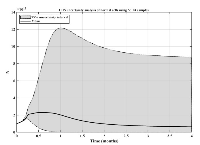
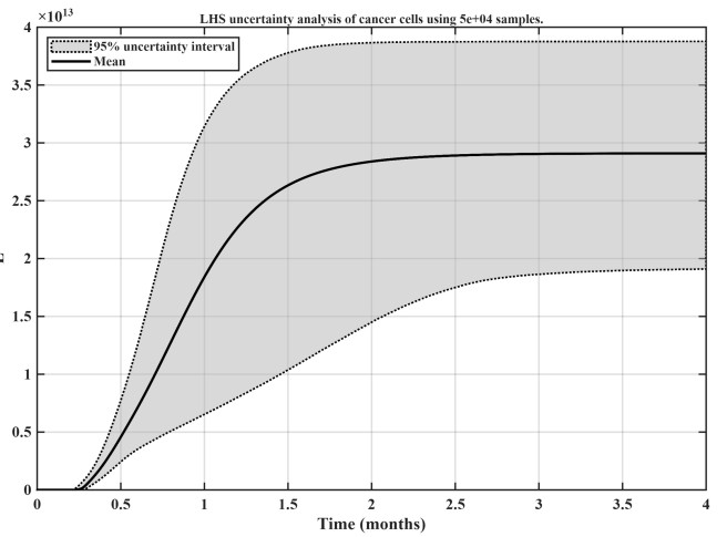
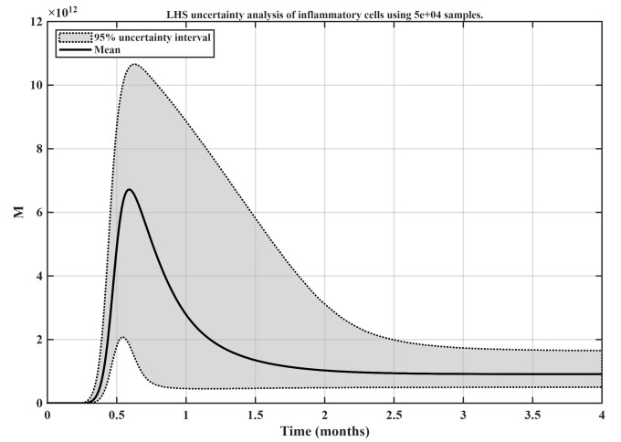
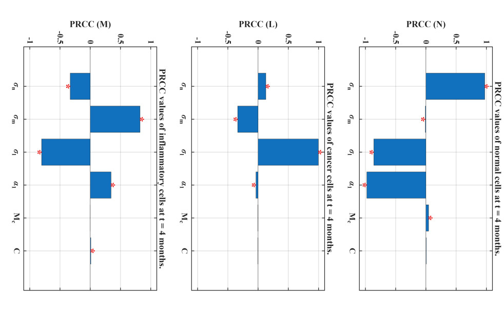
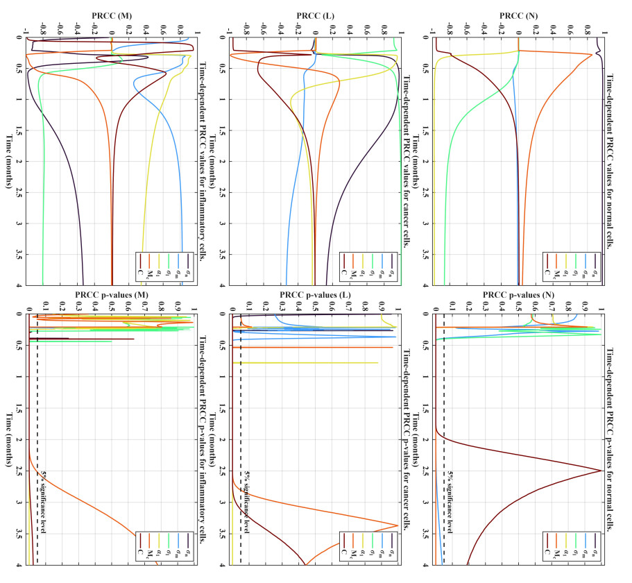
The phenomenon of inflammation is increasingly being implicated as a promoter of cancer development. Accordingly, linkages between different cancer types and inflammation are traced. Findings in the biomedical literature related to inflammatory footprints in cancer development are highlighted. Existing mathematical models, even though few, that involve inflammation and cancer are reviewed. With emphasis on the need for increased modeling activity in the area, the biomedical discussions and modeling reviews then lead into the proposal of a simple mathematical model that serves as an additional candidate model for studying cancer dynamics in an inflammatory environment. This is looked at as a way of building upon the relatively few mathematical modeling approaches that may be useful for providing insights as the biomedical studies and investigations proceed. The proposed model consists of a system of three ordinary differential equations tracking the interaction dynamics of cancer cells, inflammatory cells, and normal tissue. Subsequently, model steady states are analyzed and most importantly given necessary interpretations in biomedical contexts. To address parameter variability, uncertainty analysis using Latin Hypercube sampling is used to explore output uncertainty, while partial rank correlation coefficient (PRCC) sensitivity analysis identifies key driving parameters. PRCC analysis identifies three immunologically distinct pathways: one drives a protective anti-tumor response, promoting inflammatory cells that suppress cancer while sparing normal tissue; another drives tumor expansion and tissue damage while dampening inflammation, suggesting a pathogenic role; and a third supports normal tissue maintenance but indirectly fuels cancer growth and may limit inflammatory cells via resource competition. These findings underscore the challenge of supporting normal tissue without inadvertently fueling cancer growth.
Citation: Rachid Ouifki, Evans Afenya, Suneel Mundle. Modeling cancer dynamics in an inflammatory environment[J]. Mathematical Biosciences and Engineering, 2026, 23(8): 2429-2461. doi: 10.3934/mbe.2026088
The phenomenon of inflammation is increasingly being implicated as a promoter of cancer development. Accordingly, linkages between different cancer types and inflammation are traced. Findings in the biomedical literature related to inflammatory footprints in cancer development are highlighted. Existing mathematical models, even though few, that involve inflammation and cancer are reviewed. With emphasis on the need for increased modeling activity in the area, the biomedical discussions and modeling reviews then lead into the proposal of a simple mathematical model that serves as an additional candidate model for studying cancer dynamics in an inflammatory environment. This is looked at as a way of building upon the relatively few mathematical modeling approaches that may be useful for providing insights as the biomedical studies and investigations proceed. The proposed model consists of a system of three ordinary differential equations tracking the interaction dynamics of cancer cells, inflammatory cells, and normal tissue. Subsequently, model steady states are analyzed and most importantly given necessary interpretations in biomedical contexts. To address parameter variability, uncertainty analysis using Latin Hypercube sampling is used to explore output uncertainty, while partial rank correlation coefficient (PRCC) sensitivity analysis identifies key driving parameters. PRCC analysis identifies three immunologically distinct pathways: one drives a protective anti-tumor response, promoting inflammatory cells that suppress cancer while sparing normal tissue; another drives tumor expansion and tissue damage while dampening inflammation, suggesting a pathogenic role; and a third supports normal tissue maintenance but indirectly fuels cancer growth and may limit inflammatory cells via resource competition. These findings underscore the challenge of supporting normal tissue without inadvertently fueling cancer growth.

| [1] |
E. K. Afenya, Recovery of normal hemopoiesis in disseminated cancer therapy–a model, Math. Biosci., 172 (2001), 15–32. https://doi.org/10.1016/S0025-5564(01)00061-X doi: 10.1016/S0025-5564(01)00061-X

|
| [2] |
E. K. Afenya, S. D. Mundle, Hematologic disorders and bone marrow–peripheral blood dynamics, Math. Model. Nat. Pheno., 5 (2010), 15–27. https://doi.org/10.1051/mmnp/20105302 doi: 10.1051/mmnp/20105302
|
| [3] |
E. K. Afenya, R. Ouifki, B. I. Camara, S. D. Mundle, Mathematical modeling of bone marrow–peripheral blood dynamics in the disease state based on current emerging paradigms, part Ⅰ. Math. Biosci., 274 (2016), 83–93. https://doi.org/10.1016/j.mbs.2016.01.010 doi: 10.1016/j.mbs.2016.01.010
|
| [4] |
E. K. Afenya, R. Ouifki, S. D. Mundle, Mathematical modeling of bone marrow–peripheral blood dynamics in the disease state based on current emerging paradigms, part Ⅱ, J. Theor. Biol., 460 (2019), 37–55. https://doi.org/10.1016/j.jtbi.2018.10.008 doi: 10.1016/j.jtbi.2018.10.008
|
| [5] |
M. Al-Hajj, M. S. Wicha, A. Benito-Hernandez, S. J. Morrison, M. F. Clarke, Prospective identification of tumorigenic breast cancer cells, Proceed. Nat. Acad. Sci., 100 (2003), 3983–3988. https://doi.org/10.1073/pnas.0530291100 doi: 10.1073/pnas.0530291100
|
| [6] |
A. Amrenova, K. Suzuki, V. Saenko, S. Yamashita, N. Mitsutake, Cell competition between anaplastic thyroid cancer and normal thyroid follicular cells exerts reciprocal stress response defining tumor suppressive effects of normal epithelial tissue, PLoS ONE, 16 (2021), e0249059. https://doi.org/10.1371/journal.pone.0249059 doi: 10.1371/journal.pone.0249059
|
| [7] |
M. Andersen, Z. Sajid, R. K. Pedersen, J. Gudmand-Hoeyer, C. Ellervik, V. Skov, et al., Mathematical modelling as a proof of concept for MPNs as a human inflammation model for cancer development, PLoS ONE, 12 (2017), e0183620. https://doi.org/10.1371/journal.pone.0183620 doi: 10.1371/journal.pone.0183620
|
| [8] |
N. Behranvand, F. Nasri, R. Zolfaghari Emameh, P. Khani, A. Hosseini, J. Garssen, et al., Chemotherapy: A double-edged sword in cancer treatment, Cancer Immunol. Immunother., 71 (2022), 507–526. https://doi.org/10.1007/s00262-021-03013-3 doi: 10.1007/s00262-021-03013-3
|
| [9] |
Q. Bi, J.-Y. Wu, X.-M. Qiu, J.-D. Zhang, Z.-J. Sun, W. Wang, Tumor-associated inflammation: The tumor-promoting immunity in the early stages of tumorigenesis, J. Immunol. Res., 2022 (2022), 3128933. https://doi.org/10.1155/2022/3128933 doi: 10.1155/2022/3128933
|
| [10] |
R. L. Blaylock, Cancer microenvironment, inflammation and cancer stem cells: A hypothesis for a paradigm change and new targets in cancer control, Surg. Neurol. Int., 6 (2015), 92. https://doi.org/10.4103/2152-7806.157890 doi: 10.4103/2152-7806.157890
|
| [11] |
D. Bonnet, J. E. Dick, Human acute myeloid leukemia is organized as a hierarchy that originates from a primitive hematopoietic cell, Nat. Med., 3 (1997), 730–737. https://doi.org/10.1038/nm0797-730 doi: 10.1038/nm0797-730
|
| [12] |
S. Demaria, E. Pikarsky, M. Karin, L. M. Coussens, Y.-C. Chen, E. M. El-Omar, et al., Cancer and inflammation: Promise for biologic therapy, J. Immunother., 33 (2010), 335–351. https://doi.org/10.1097/CJI.0b013e3181d32e74 doi: 10.1097/CJI.0b013e3181d32e74
|
| [13] |
D. Dingli, F. Michor, Successful therapy must eradicate cancer stem cells, Stem Cells, 24 (2006), 2603–2610. https://doi.org/10.1634/stemcells.2006-0136 doi: 10.1634/stemcells.2006-0136
|
| [14] |
G. P. Dunn, L. J. Old, R. D. Schreiber, The Three Es of Cancer Immunoediting, Annu. Rev. Immunol., 22 (2004), 329–360. https://doi.org/10.1146/annurev.immunol.22.012703.104803 doi: 10.1146/annurev.immunol.22.012703.104803
|
| [15] |
J. L. Dunster, H. M. Byrne, J. R. King, The resolution of inflammation: A mathematical model of neutrophil and macrophage interactions, Bull. Math. Biol., 76 (2014), 1953–1980. https://doi.org/10.1007/s11538-014-9987-x doi: 10.1007/s11538-014-9987-x
|
| [16] |
American Association for Cancer Research, Early proinflammatory niches drive lung adenocarcinoma initiation, Cancer Discov., 16 (2026), OF1. https://doi.org/10.1158/2159-8290.CD-RW2025-108 doi: 10.1158/2159-8290.CD-RW2025-108
|
| [17] |
R. J Gilbertson, J. N. Rich, Making a tumour's bed: Glioblastoma stem cells and the vascular niche, Nat. Rev. Cancer, 7 (2007), 733–736. https://doi.org/10.1038/nrc2246 doi: 10.1038/nrc2246
|
| [18] |
S. I. Grivennikov, F. R. Greten, M. Karin, Immunity, inflammation, and cancer, Cell, 140 (2010), 883–899. https://doi.org/10.1016/j.cell.2010.01.025 doi: 10.1016/j.cell.2010.01.025
|
| [19] |
D. Hanahan, R. A. Weinberg, Hallmarks of Cancer: The Next Generation, Cell, 144 (2011), 646–674. https://doi.org/10.1016/j.cell.2011.02.013 doi: 10.1016/j.cell.2011.02.013
|
| [20] |
S. Hibino, T. Kawazoe, H. Kasahara, S. Itoh, T. Ishimoto, M. Sakata-Yanagimoto, et al., Inflammation-induced tumorigenesis and metastasis, Int. J. Molecular Sci., 22 (2021), 5421. https://doi.org/10.3390/ijms22115421 doi: 10.3390/ijms22115421
|
| [21] |
Y. J. Jeong, H. K. Oh, S. H. Park, J. G. Bong, Association between inflammation and cancer stem cell phenotype in breast cancer, Oncol. Lett., 15 (2018), 2380–2386. https://doi.org/10.3892/ol.2017.7607 doi: 10.3892/ol.2017.7607
|
| [22] |
M. Karin, Nuclear factor-$\kappa$b in cancer development and progression, Nature, 441 (2006), 431–436. https://doi.org/10.1038/nature04870 doi: 10.1038/nature04870
|
| [23] |
H. Kulbe, T. Hagemann, P. W. Szlosarek, F. R. Balkwill, J. L. Wilson, The inflammatory cytokine tumor necrosis factor-$\alpha$ regulates chemokine receptor expression on ovarian cancer cells, Cancer Res., 65 (2005), 10355–10362. https://doi.org/10.1158/0008-5472.CAN-05-0957 doi: 10.1158/0008-5472.CAN-05-0957
|
| [24] |
P. La Rosée, A. Horne, M. Hines, T. von B. Greenwood, R. Machowicz, N. Berliner, et al., Recommendations for the management of hemophagocytic lymphohistiocytosis in adults, Blood, 133 (2019), 2465–2477. https://doi.org/10.1182/blood.2018894618 doi: 10.1182/blood.2018894618
|
| [25] |
H. T. Lu, W. M. Ouyang, C. S. Huang, Inflammation, a key event in cancer development, Mol. Cancer Res., 4 (2006), 221–233. https://doi.org/10.1158/1541-7786.MCR-05-0261 doi: 10.1158/1541-7786.MCR-05-0261
|
| [26] |
Y. M. Ma, T. Sun, Y. X. Liu, N. Zhao, Q. Gu, D.-F. Zhang, et al., A pilot study on acute inflammation and cancer: A new balance between IFN-$\gamma$ and TGF-$\beta$ in melanoma, J. Exp. Clin. Canc. Res., 28 (2009), 1–6. https://doi.org/10.1186/1756-9966-28-23 doi: 10.1186/1756-9966-28-23
|
| [27] |
A. Mantovani, P. Allavena, A. Sica, F. Balkwill, Cancer-related inflammation, Nature, 454 (2008), 436–444. https://doi.org/10.1038/nature07205 doi: 10.1038/nature07205
|
| [28] |
S. Marino, I. B. Hogue, C. J. Ray, D. E. Kirschner, A methodology for performing global uncertainty and sensitivity analysis in systems biology, J. Theor. Biol., 254 (2008), 178–196. https://doi.org/10.1016/j.jtbi.2008.04.011 doi: 10.1016/j.jtbi.2008.04.011
|
| [29] |
M. D. McKay, R. J. Beckman, W. J. Conover, A comparison of three methods for selecting values of input variables in the analysis of output from a computer code, Technometrics, 21 (1979), 239–245. https://doi.org/10.1080/00401706.1979.10489755 doi: 10.1080/00401706.1979.10489755
|
| [30] |
S. Mohapatra, A. Guerrero, N. Rahman, K. T. Zaman, J. Wu, C. Onyeagba, et al., Novel taxol-derivative, sto-1, induces selective anti-tumor immunity and sustained remission of glioblastoma without triggering autoimmune reactions, Cells, 14 (2025), 1703. https://doi.org/10.3390/cells14211703 doi: 10.3390/cells14211703
|
| [31] |
T.-N. Nguyen, J. Clairambault, T. Jaffredo, B. Perthame, D. Salort, Adaptive dynamics of hematopoietic stem cells and their supporting stroma: a model and mathematical analysis, Math. Biosci. Eng., 16 (2019), 4818–4845. https://doi.org/10.3934/mbe.2019243 doi: 10.3934/mbe.2019243
|
| [32] | L. Norton, R. Simon, The norton-simon hypothesis revisited, Cancer Treat. Rep., 70 (1986), 163–169. |
| [33] |
F. Okada, Inflammation and free radicals in tumor development and progression, Redox Rep., 7 (2002), 357–368. https://doi.org/10.1179/135100002125001135 doi: 10.1179/135100002125001135
|
| [34] |
J. T. Ottesen, R. K. Pedersen, Z. Sajid, J. Gudmand-Hoeyer, K. O. Bangsgaard, V. Skov, et al., Bridging blood cancers and inflammation: The reduced cancitis model, J. Theor. Biol., 465 (2019), 90–108. https://doi.org/10.1016/j.jtbi.2019.01.001 doi: 10.1016/j.jtbi.2019.01.001
|
| [35] |
K. Pearson, Ⅶ. Note on regression and inheritance in the case of two parents, Proceed. Royal Soc. London, 58 (1895), 240–242. https://doi.org/10.1098/rspl.1895.0041 doi: 10.1098/rspl.1895.0041
|
| [36] |
F. Peng, A. Sinjab, Y. Dai, W. Treekitkarnmongkol, S. Yang, L. I. G. Bolanos, et al., Multimodal spatial-omics reveal co-evolution of alveolar progenitors and proinflammatory niches in progression of lung precursor lesions, Cancer Cell, 44 (2026), 321–339. https://doi.org/10.1016/j.ccell.2025.10.004 doi: 10.1016/j.ccell.2025.10.004
|
| [37] |
E. M. Pietras, Inflammation: A key regulator of hematopoietic stem cell fate in health and disease, Blood, 130 (2017), 1693–1698. https://doi.org/10.1182/blood-2017-06-780882 doi: 10.1182/blood-2017-06-780882
|
| [38] |
C. Récher, Clinical implications of inflammation in acute myeloid leukemia, Front. Oncol., 11 (2021), 623952. https://doi.org/10.3389/fonc.2021.623952 doi: 10.3389/fonc.2021.623952
|
| [39] |
T. Reya, S. J. Morrison, M. F. Clarke, I. L. Weissman, Stem cells, cancer, and cancer stem cells, Nature, 414 (2001), 105–111. https://doi.org/10.1038/35102167 doi: 10.1038/35102167
|
| [40] |
Z. Sajid, M. Andersen, J. T. Ottesen, Mathematical analysis of the cancitis model and the role of inflammation in blood cancer progression, Math. Biosci. Eng., 16 (2019), 8268–8289. https://doi.org/10.3934/mbe.2019418 doi: 10.3934/mbe.2019418
|
| [41] |
A. M. Schram, N. Berliner, How I treat hemophagocytic lymphohistiocytosis in the adult patient, Blood, 125 (2015), 2908–2914. https://doi.org/10.1182/blood-2015-01-551622 doi: 10.1182/blood-2015-01-551622
|
| [42] |
S. Shigdar, Y. Li, S. Bhattacharya, M. O'Connor, C. Pu, J. Lin, et al., Inflammation and cancer stem cells, Cancer Lett., 345 (2014), 271–278. https://doi.org/10.1016/j.canlet.2013.07.031 doi: 10.1016/j.canlet.2013.07.031
|
| [43] |
A. Sparmann, D. Bar-Sagi, Ras-induced interleukin-8 expression plays a critical role in tumor growth and angiogenesis, Cancer Cell, 6 (2004), 447–458. https://doi.org/10.1016/j.ccr.2004.09.028 doi: 10.1016/j.ccr.2004.09.028
|
| [44] | C. Spearman, The proof and measurement of association between two things, (1961). https://doi.org/10.1037/11491-005 |
| [45] | S. Wiggins, Introduction to applied nonlinear dynamical systems and chaos, Springer, 2003. |
| [46] | C. Y. Huak, Biostatistics 104: Correlational Analysis, Singapore Med. J., 44 (2003), 614–619. |
| [47] |
Y. Zhang, W. Sime, M. Juhas, A. Sjölander, Crosstalk between colon cancer cells and macrophages via inflammatory mediators and cd47 promotes tumour cell migration, Eur. J. Cancer, 49 (2013), 3320–3334. https://doi.org/10.1016/j.ejca.2013.06.005 doi: 10.1016/j.ejca.2013.06.005
|
Figures(9) / Tables(1)

Rachid Ouifki, Evans Afenya, Suneel Mundle. Modeling cancer dynamics in an inflammatory environment[J]. Mathematical Biosciences and Engineering, 2026, 23(8): 2429-2461. doi: 10.3934/mbe.2026088





 DownLoad: