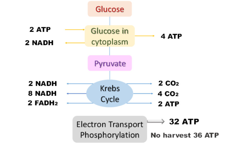
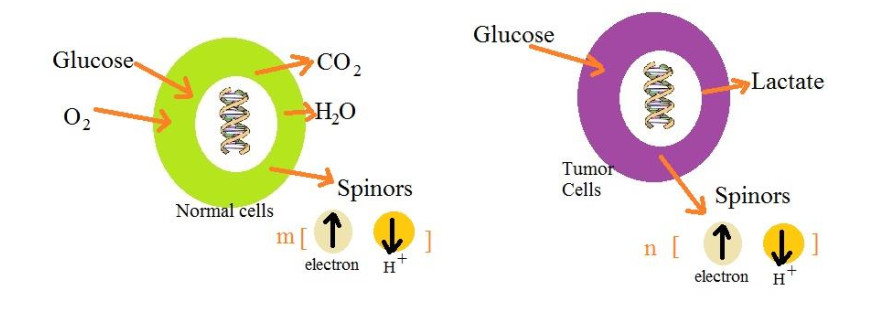
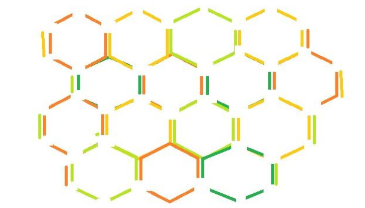
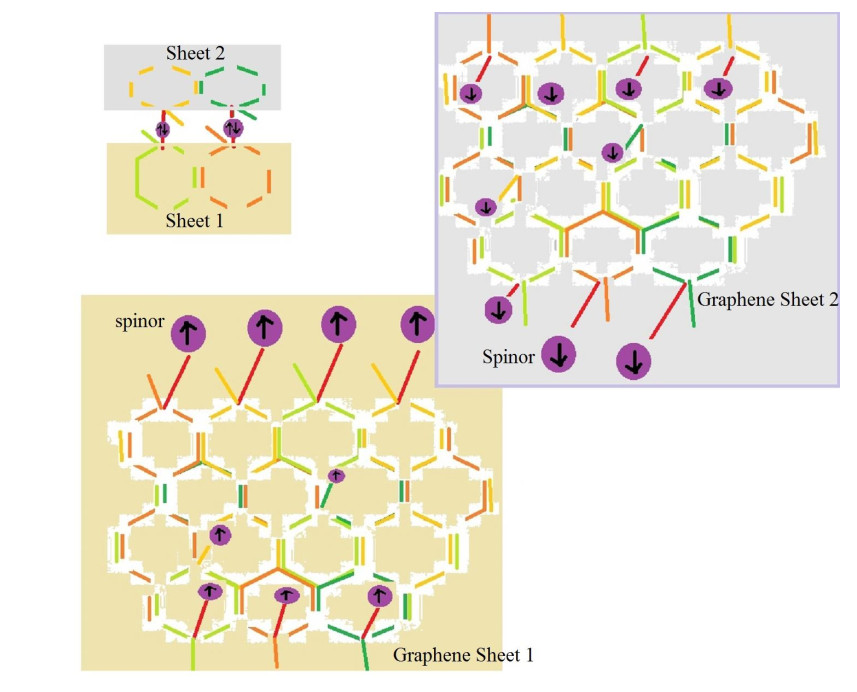
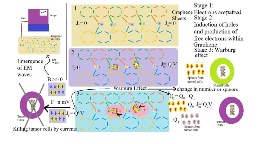
According to the Warburg effect, there are some significant differences between metabolisms, products and process of respirations of cancer cells and normal cells. For example, normal cells absorb oxygen and glucose and give water molecules, carbon dioxide, ATP molecules and some number of spinors; while cancer cells take glucose and give lactate, less number of ATP molecules and different number of spinors. Using this property, we can design a system from two graphene sheets that are connected by pairing the fourth free electrons of carbons. Then, we can break some pairs and produce some holes. The number of these holes should be equal to the number of radiated spinors by normal cells. Near a normal cell, all holes are filled and the graphene system doesn't emit any electrical current or wave. However, near a cancer cell, some extra holes or spinors remain that their motions produce some electrical currents. These currents force on cancer cell membranes and destroy them and consequently, cause the cell death. Also, these currents emit some electromagnetic waves which detectors could take them out of the human's body and consequently, they could play the main role in imaging.
Citation: Massimo Fioranelli, Hijaz Ahmad, Alireza Sepehri, Maria Grazia Roccia, Faissal Aziz. A mathematical model for imaging and killing cancer cells by using concepts of the Warburg effect in designing a Graphene system[J]. Mathematical Biosciences and Engineering, 2022, 19(3): 2985-2995. doi: 10.3934/mbe.2022137
According to the Warburg effect, there are some significant differences between metabolisms, products and process of respirations of cancer cells and normal cells. For example, normal cells absorb oxygen and glucose and give water molecules, carbon dioxide, ATP molecules and some number of spinors; while cancer cells take glucose and give lactate, less number of ATP molecules and different number of spinors. Using this property, we can design a system from two graphene sheets that are connected by pairing the fourth free electrons of carbons. Then, we can break some pairs and produce some holes. The number of these holes should be equal to the number of radiated spinors by normal cells. Near a normal cell, all holes are filled and the graphene system doesn't emit any electrical current or wave. However, near a cancer cell, some extra holes or spinors remain that their motions produce some electrical currents. These currents force on cancer cell membranes and destroy them and consequently, cause the cell death. Also, these currents emit some electromagnetic waves which detectors could take them out of the human's body and consequently, they could play the main role in imaging.

| [1] |
M. V. Liberti, J. W. Locasale, The Warburg effect: how does it benefit cancer cells?, Trends Biochem. Sci., 41 (2016), 211-218. https://doi.org/10.1016/j.tibs.2015.12.001 doi: 10.1016/j.tibs.2015.12.001

|
| [2] |
K. O. Alfarouk, Tumor metabolism, cancer cell transporters, and microenvironmental resistance, J. Enzyme Inhib. Med. Chem., 31 (2016), 859-66. https://doi.org/10.3109/14756366.2016.1140753 doi: 10.3109/14756366.2016.1140753
|
| [3] |
M. G. V. Heiden, L. C. Cantley, C. B. Thompson, Understanding the Warburg effect: the metabolic requirements of cell proliferation, Science, 324 (2009), 1029-1033. https://doi.org/10.1126/science.1160809 doi: 10.1126/science.1160809
|
| [4] |
C. Yu, J. Xue, W. Zhu, Y. Jiao, S. Zhang, J. Cao, Warburg meets non-coding RNAs: the emerging role of ncRNA in regulating the glucose metabolism of cancer cells, Tumour Biol., 36 (2015), 81-94. https://doi.org/10.1007/s13277-014-2875-z doi: 10.1007/s13277-014-2875-z
|
| [5] |
L. Li, Y. Liang, L. Kang, Y. Liu, S. Gao, S. Chen, et al., Transcriptional regulation of the Warburg effect in cancer by SIX1, Cancer Cell, 33 (2018), 368-385. https://doi.org/10.1016/j.ccell.2018.01.010 doi: 10.1016/j.ccell.2018.01.010
|
| [6] |
H. Mirzaei, M. R. Hamblin, Regulation of glycolysis by non-coding RNAs in cancer: switching on the Warburg effect, Mol. Ther. Oncolytics, 19 (2020), 218-239. https://doi.org/10.1016/j.omto.2020.10.003. doi: 10.1016/j.omto.2020.10.003
|
| [7] |
Z. Chen, W. Lu, C. Garcia-Prieto, P. Huang, The Warburg effect and its cancer therapeutic implications, J. Bioenerg. Biomembr., 39 (2007), 267-274. https://doi.org/10.1007/s10863-007-9086-x doi: 10.1007/s10863-007-9086-x
|
| [8] |
J. Pokorný, J. Pokorný, F. Borodavka, Warburg effect-damping of electromagnetic oscillations, Electromagn. Biol. Med., 36 (2017), 270-278. https://doi.org/10.1080/15368378.2017.1326933 doi: 10.1080/15368378.2017.1326933
|
| [9] |
A. R Liboff, The Warburg hypothesis and weak ELF biointeractions, Electromagn. Biol. Med., 39 (2020), 45-48. https://doi.org/10.1080/15368378.2020.1737810 doi: 10.1080/15368378.2020.1737810
|
| [10] |
T. A. Tabish, R. J. Narayan, Mitochondria-targeted graphene for advanced cancer therapeutics, Acta Biomater., 129 (2021), 43-56. https://doi.org/10.1016/j.actbio.2021.04.054 doi: 10.1016/j.actbio.2021.04.054
|
| [11] |
J. Frontiñan-Rubio, M. V. Gomez, V. J. González, M. Durán-Prado, E. Vázquez, Sublethal exposure of small few-layer graphene promotes metabolic alterations in human skin cells. Sci. Rep., 10 (2020), 18407. https://doi.org/10.1038/s41598-020-75448-0 doi: 10.1038/s41598-020-75448-0
|
| [12] |
C. Martelli, A. King, T. Simon, G. Giamas, Graphene-induced transdifferentiation of cancer stem cells as a therapeutic strategy against glioblastoma, ACS Biomater. Sci. Eng., 6 (2020), 3258-3269. https://doi.org/10.1021/acsbiomaterials.0c00197 doi: 10.1021/acsbiomaterials.0c00197
|
| [13] |
I. S. Donskyi, Y. Chen, P. Nickl, G. Guday, H. Qiao, K. Achazi, et al., Self-degrading graphene sheets for tumor therapy, Nanoscale, 12 (2020), 14222-14229. https://doi.org/10.1039/D0NR02159H doi: 10.1039/D0NR02159H
|
| [14] |
T. A. Tabish, C. J. Scotton, D. C. J. Ferguson, L. Lin, A. V. der Veen, S. Lowry, et al., Biocompatibility and toxicity of graphene quantum dots for potential application in photodynamic therapy, Nanomedicine (Lond), 13 (2018), 1923-1937. https://doi.org/10.2217/nnm-2018-0018 doi: 10.2217/nnm-2018-0018
|
| [15] |
V. Palmieri, M. Papi, Can graphene take part in the fight against COVID-19?, Nano Today, 33 (2020), 100883. https://doi.org/10.1016/j.nantod.2020.100883 doi: 10.1016/j.nantod.2020.100883
|
| [16] |
P. K. Raghav, S. Mohanty, Are graphene and graphene-derived products capable of preventing COVID-19 infection?, Med. Hypotheses, 144 (2020), 110031. https://doi.org/10.1016/j.mehy.2020.110031 doi: 10.1016/j.mehy.2020.110031
|
| [17] |
J. Sengupta, C. M. Hussain, Graphene-based field-effect transistor biosensors for the rapid detection and analysis of viruses: A perspective in view of COVID-19, Carbon Trends, 2 (2021), 100011. https://doi.org/10.1016/j.cartre.2020.100011 doi: 10.1016/j.cartre.2020.100011
|
| [18] | I. Maqbool, F. Rehman, F. Soomro, Z. Bhatti, U. Ali, A. H. Jatoi, et al., Graphene-based materials for fighting coronavirus disease 2019: challenges and opportunities. CBEN, 8 (2021), 67-77. https://doi.org/10.1002/cben.202000039 |
| [19] |
A. Rhazouani, H. Gamrani, M. E. Achaby, K. Aziz, L. Gebrati, M. S. Uddin, et al., Synthesis and toxicity of graphene oxide nanoparticles: A literature review of in vitro and in vivo studies, Biomed. Res. Int., 2021 (2021), 5518999. https://doi.org/10.1155/2021/5518999 doi: 10.1155/2021/5518999
|
| [20] |
D. Samanta, M. P. Karthikeyan, A. Banerjee, H. Inokawa, Tunable graphene nanopatch antenna design for on-chip integrated terahertz detector arrays with potential application in cancer imaging, Nanomedicine, 16 (2021), 1035-1047. https://doi.org/10.2217/nnm-2020-0386 doi: 10.2217/nnm-2020-0386
|
| [21] |
S. F. Adil, M. R. Shaik, F. A. Nasr, A. S. Alqahtani, M. Z. Ahmed, W. Qamar, et al., Enhanced apoptosis by functionalized highly reduced graphene oxide and gold nanocomposites in MCF-7 breast cancer cells, ACS Omega, 6 (2021), 15147-15155. https://doi.org/10.1021/acsomega.1c01377 doi: 10.1021/acsomega.1c01377
|
| [22] |
N. Anjum, J. He, Nonlinear dynamic analysis of vibratory behavior of a graphene nano/microelectromechanical system, Math. Meth. Appl. Sci., 2020 (2020), 1-16. https://doi.org/10.1002/mma.6699 doi: 10.1002/mma.6699
|
| [23] |
T. A. Tabish, M. R. Hamblin, Mitochondria-targeted nanoparticles (mitoNANO): An emerging therapeutic shortcut for cancer, Biomater. Biosyst., 3 (2021), 100023. https://doi.org/10.1016/j.bbiosy.2021.100023 doi: 10.1016/j.bbiosy.2021.100023
|
| [24] |
T. A. Tabish, S. Zhang, P. G. Winyard, Developing the next generation of graphene-based platforms for cancer therapeutics: The potential role of reactive oxygen species, Redox. Biol., 2018 (2018), 34-40. https://doi.org/10.1016/j.redox.2017.11.018 doi: 10.1016/j.redox.2017.11.018
|
Figures(5)

Massimo Fioranelli, Hijaz Ahmad, Alireza Sepehri, Maria Grazia Roccia, Faissal Aziz. A mathematical model for imaging and killing cancer cells by using concepts of the Warburg effect in designing a Graphene system[J]. Mathematical Biosciences and Engineering, 2022, 19(3): 2985-2995. doi: 10.3934/mbe.2022137


 DownLoad: