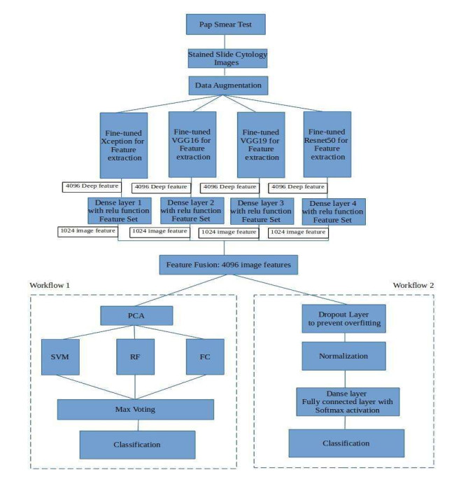
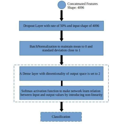
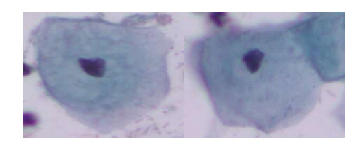
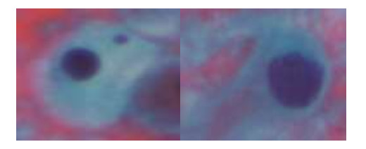
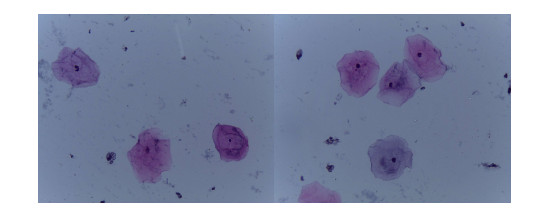
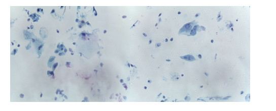
Cervical cancer is the second most commonly seen cancer in women. It affects the cervix portion of the vagina. The most preferred diagnostic test required for screening cervical cancer is the pap smear test. Pap smear is a time-consuming test as it requires detailed analysis by expert cytologists. Cytologists can screen around 100 to 1000 slides depending upon the availability of advanced equipment. Due to this reason Artificial intelligence (AI) based computer-aided diagnosis system for the classification of pap smear images is needed. There are some AI-based solutions proposed in the literature, still an effective and accurate system is under research. In this paper, the deep learning-based hybrid methodology namely DeepCyto is proposed for the classification of pap smear cytology images. The DeepCyto extracts the feature fusion vectors from pre-trained models and passes these to two workflows. Workflow-1 applies principal component analysis and machine learning ensemble to classify the pap smear images. Workflow-2 takes feature fusion vectors as an input and applies an artificial neural network for classification. The experiments are performed on three benchmark datasets namely Herlev, SipakMed, and LBCs. The performance measures of accuracy, precision, recall and F1-score are used to evaluate the effectiveness of the DeepCyto. The experimental results depict that Workflow-2 has given the best performance on all three datasets even with a smaller number of epochs. Also, the performance of the DeepCyto Workflow 2 on multi-cell images of LBCs is better compared to single cell images of other datasets. Thus, DeepCyto is an efficient method for accurate feature extraction as well as pap smear image classification.
Citation: Swati Shinde, Madhura Kalbhor, Pankaj Wajire. DeepCyto: a hybrid framework for cervical cancer classification by using deep feature fusion of cytology images[J]. Mathematical Biosciences and Engineering, 2022, 19(7): 6415-6434. doi: 10.3934/mbe.2022301
Cervical cancer is the second most commonly seen cancer in women. It affects the cervix portion of the vagina. The most preferred diagnostic test required for screening cervical cancer is the pap smear test. Pap smear is a time-consuming test as it requires detailed analysis by expert cytologists. Cytologists can screen around 100 to 1000 slides depending upon the availability of advanced equipment. Due to this reason Artificial intelligence (AI) based computer-aided diagnosis system for the classification of pap smear images is needed. There are some AI-based solutions proposed in the literature, still an effective and accurate system is under research. In this paper, the deep learning-based hybrid methodology namely DeepCyto is proposed for the classification of pap smear cytology images. The DeepCyto extracts the feature fusion vectors from pre-trained models and passes these to two workflows. Workflow-1 applies principal component analysis and machine learning ensemble to classify the pap smear images. Workflow-2 takes feature fusion vectors as an input and applies an artificial neural network for classification. The experiments are performed on three benchmark datasets namely Herlev, SipakMed, and LBCs. The performance measures of accuracy, precision, recall and F1-score are used to evaluate the effectiveness of the DeepCyto. The experimental results depict that Workflow-2 has given the best performance on all three datasets even with a smaller number of epochs. Also, the performance of the DeepCyto Workflow 2 on multi-cell images of LBCs is better compared to single cell images of other datasets. Thus, DeepCyto is an efficient method for accurate feature extraction as well as pap smear image classification.

| [1] |
M. Arbyn, E. Weiderpass, L. Bruni, S. de Sanjosé, M. Saraiya, J. Ferlay, et al., Estimates of incidence and mortality of cervical cancer in 2018: a worldwide analysis, Lancet Global Health, 8 (2020), e191-e203. https://doi.org/10.1016/S2214-109X(19)30482-6 doi: 10.1016/S2214-109X(19)30482-6

|
| [2] |
R. Hull, M. Mbele, T. Makhafola, C. Hicks, S. Wang, R. M. Reis, et al., Cervical cancer in low and middle‑income countries (Review), Oncol. Lett., 20 (2020), 2058-2074. https://doi.org/10.3892/ol.2020.11754 doi: 10.3892/ol.2020.11754
|
| [3] |
W. William, A. Ware, A. H. Basaza-Ejiri, J. Obungoloch, A pap-smear analysis tool (PAT) for detection of cervical cancer from pap-smear images, Biomed. Eng. Online, 18 (2019), 16. https://doi.org/10.1186/s12938-019-0634-5 doi: 10.1186/s12938-019-0634-5
|
| [4] | R. Li, C. Xiao, Y. Huang, H. Hassan, B. Huang, Deep learning applications in computed deep learning applications in computed tomography images for pulmonary nodule detection and diagnosis: A review, 12 (2022), 298. https://doi.org/10.3390/diagnostics12020298 |
| [5] |
H. Hassan, Z. Ren, H. Zhao, S. Huang, D. Li, S. Xiang, et al., Review and classification of AI-enabled COVID-19 CT imaging models based on computer vision tasks, Comput. Biol. Med., 141 (2022), 105123. https://doi.org/10.1016/j.compbiomed.2021.105123 doi: 10.1016/j.compbiomed.2021.105123
|
| [6] |
O. Holmström, N. Linder, H. Kaingu, N. Mbuuko, J. Mbete, F. Kinyua, et al., Point-of-care digital cytology with artificial intelligence for cervical cancer screening in a resource-limited setting, JAMA Network Open, 4 (2021). http://doi.org/10.1001/jamanetworkopen.2021.1740 doi: 10.1001/jamanetworkopen.2021.1740
|
| [7] |
B. Nithya, V. Ilango, Evaluation of machine learning based optimized feature selection approaches and classification methods for cervical cancer prediction, SN Appl. Sci., 1 (2019). https://doi.org/10.1007/s42452-019-0645-7 doi: 10.1007/s42452-019-0645-7
|
| [8] | J. Deng, W. Dong, R. Socher, L. J. Li, K. Li, F. Li, ImageNet: A large-scale hierarchical image database, in 2009 IEEE Conference on Computer Vision and Pattern Recognition, (2009), 248-255. https://doi.org/10.1109/CVPR.2009.5206848 |
| [9] |
M. Rahaman, C. Li, Y. Yao, F. Kulwa, X. Wu, X. Li, et al., DeepCervix: a deep learning-based framework for the classification of cervical cells using hybrid deep feature fusion techniques, Comput. Biol. Med., 136 (2021), 104649. https://doi.org/10.1016/j.compbiomed.2021.104649 doi: 10.1016/j.compbiomed.2021.104649
|
| [10] |
H. Basak, R. Kundu, S. Chakraborty, N. Das, Cervical cytology classification using PCA and GWO enhanced deep features selection, SN Comput. Sci., 2 (2021), 369. https://doi.org/10.1007/s42979-021-00741-2 doi: 10.1007/s42979-021-00741-2
|
| [11] |
T. Chankong, N. Theera-Umpon, S. Auephanwiriyakul, Automatic cervical cell segmentation and classification in Pap smears, Comput. Methods Programs Biomed, , 113 (2014), 539-556. https://doi.org/10.1016/j.cmpb.2013.12.012 doi: 10.1016/j.cmpb.2013.12.012
|
| [12] |
W. William, A. Ware, A. H. Basaza-Ejiri, J. Obungoloch, A pap-smear analysis tool (PAT) for detection of cervical cancer from pap-smear images, BioMed. Eng. OnLine, 18 (2019), 16. https://doi.org/10.1186/s12938-019-0634-5 doi: 10.1186/s12938-019-0634-5
|
| [13] | J. Byriel, Neuro-fuzzy classification of cells in cervical smears, Master's Thesis, Technical University of Denmark, 1999. |
| [14] |
L. Zhang, H. Kong, C. T. Chin, S. Liu, X. Fan, T. Wang, et al., Automation-assisted cervical cancer screening in manual liquid-based cytology with hematoxylin and eosin staining, Cytometry, 85 (2014), 214-230. https://doi.org/10.1002/cyto.a.22407 doi: 10.1002/cyto.a.22407
|
| [15] |
Y. Marinakis, G. Dounias, J. Jantzen, Pap smear diagnosis using a hybrid intelligent scheme focusing on genetic algorithm based feature selection and nearest neighbor classification, Comput. Biol. Med., 39 (2009), 69-78. https://doi.org/10.1016/j.compbiomed.2008.11.006 doi: 10.1016/j.compbiomed.2008.11.006
|
| [16] |
L. Zhang, L. Lu, I. Nogues, R. M. Summers, S. Liu, J. Yao, DeepPap: Deep convolutional networks for cervical cell classification, IEEE J. Biomed. Health Inf., 21 (2017), 1633-1643. https://doi.org/10.1109/JBHI.2017.2705583 doi: 10.1109/JBHI.2017.2705583
|
| [17] |
C. Shorten, T. M. Khoshgoftaar, A survey on image data augmentation for deep learning, J. Big Data, 6 (2019), 60. https://doi.org/10.1186/s40537-019-0197-0 doi: 10.1186/s40537-019-0197-0
|
| [18] | R. Hataya, J. Zdenek, K. Yoshizoe, H. Nakayama, Faster autoaugment: Learning augmentation strategies using backpropagation, in Computer Vision-ECCV 2020-16th European Conference, 2020, 12370 (2020). https://doi.org/10.1007/978-3-030-58595-2_1 |
| [19] |
R. Ogawa, T. Kido, T. Kido, T. Mochizuki, Effect of augmented datasets on deep convolutional neural networks applied to chest radiographs, Clin. Radiol., 74 (2019), 697-701. https://doi.org/10.1016/j.crad.2019.04.025 doi: 10.1016/j.crad.2019.04.025
|
| [20] |
T. Kaur, T. K. Gandhi, Deep convolutional neural networks with transfer learning for automated brain image classification, Mach. Vision Appl., 31 (2020), 20. https://doi.org/10.1007/s00138-020-01069-2 doi: 10.1007/s00138-020-01069-2
|
| [21] | F. Chollet, Xception: Deep learning with depthwise separable convolutions, in Proceedings of the IEEE Conference on Computer Vision and Pattern Recognition, (2017), 1251-1258. https://doi.org/10.48550/arXiv.1610.02357 |
| [22] | K. Simonyan, A. Zisserman, Very deep convolutional networks for large-scale image recognition, preprint, arXiv: 1409.1556. |
| [23] | K. He, X. Zhang, S. Ren, J. Sun, Deep residual learning for image recognition, In Proceedings of the IEEE Conference on Computer Vision and Pattern Recognition, (2016), 770-778. |
| [24] | D. Kingma, J. Ba, Adam: A method for stochastic optimization, preprint, arXiv: 1412.6980v9. |
| [25] |
J. V. Kriti, R. Agarwal, Deep feature extraction and classification of breast ultrasound images, Multimed Tools Appl., 79 (2020), 27257-27292. https://doi.org/10.1007/s11042-020-09337-z doi: 10.1007/s11042-020-09337-z
|
| [26] |
S. P. Mishra, U. Sarkar, S. Taraphder, S. Datta, D. P. Swain, R. Saikhom, et al., Principal component analysis, Int. J. Livest. Res., 7 (2017), 60-78. https://doi.org/10.5455/ijlr.20170415115235 doi: 10.5455/ijlr.20170415115235
|
| [27] |
J. Cervantes, F. Garcia-Lamont, L. Rodríguez-Mazahua, A. Lopez, A comprehensive survey on support vector machine classification: Applications, challenges and trends, Neurocomputing, 408 (2020), 189-215. https://doi.org/10.1016/j.neucom.2019.10.118 doi: 10.1016/j.neucom.2019.10.118
|
| [28] |
L. Breiman, Random forests, Mach. Learn., 45 (2001), 5-32. https://doi.org/10.1023/A:1010933404324 doi: 10.1023/A:1010933404324
|
| [29] |
S. H. S. Basha, S. R. Dubey, V. Pulabaigari, S. Mukherjee, Impact of fully connected layers on performance of convolutional neural networks for image classification, Neurocomputing, 378 (2020), 112-119. https://doi.org/10.1016/j.neucom.2019.10.008 doi: 10.1016/j.neucom.2019.10.008
|
| [30] |
A. Maćkiewicz, W Ratajczak, Principal components analysis (PCA), Comput. Geosci., 19 (1993), 303-342. https://doi.org/10.1016/0098-3004(93)90090-R doi: 10.1016/0098-3004(93)90090-R
|
| [31] | E. Bisong, Introduction to Scikit-learn. in Building Machine Learning and Deep Learning Models on Google Cloud Platform, USA press, Berkeley, CA, 2019. https://doi.org/10.1007/978-1-4842-4470-8_18 |
| [32] | M. E. Wall, A. Rechtsteiner, L. M. Rocha, Singular value decomposition and principal component analysis, in A Practical Approach to Microarray Data Analysis, Springer, Boston, MA, (2003), 91-109. https://doi.org/10.1007/0-306-47815-3_5 |
| [33] |
N. Halko, P. G. Martinsson, J. A. Tropp, Finding structure with randomness: Probabilistic algorithms for constructing approximate matrix decompositions, SIAM Rev., 53 (2011), 217-288. https://doi.org/10.1137/090771806 doi: 10.1137/090771806
|
| [34] | sklearn decomposition PCA, scikit-learn documentation, 2022. Available from: https://scikit-learn.org/stable/modules/generated/sklearn.decomposition.PCA.html. |
| [35] |
A. S. Assiri, S. Nazir, S. A. Velastin, Breast tumor classification using an ensemble machine learning method, J. Imaging, 6 (2020), 39. https://doi.org/10.3390/jimaging6060039 doi: 10.3390/jimaging6060039
|
| [36] | J. Jantzen, J. Norup, G. Dounias, B. Bjerregaard, Pap-smear benchmark data for pattern classification, Nat. Inspired Smart Inf. Syst., 2005 (2005), 1-9. |
| [37] | Pap-Smear Databases and Related Studies, 2022. Available from: http://mde-lab.aegean.gr/index.php/downloads. |
| [38] | M. E. Plissiti, P. Dimitrakopoulos, G. Sfikas, C. Nikou, O. Krikoni, A. Charchanti, Sipakmed: A new dataset for feature and image based classification of normal and pathological cervical cells in Pap smear images, in IEEE International Conference on Image Processing (ICIP) 2018, (2018), 3144-3148. https://doi.org/10.1109/ICIP.2018.8451588 |
| [39] | SIPakMed Database, 2022. Available from: https://www.cs.uoi.gr/~marina/sipakmed.html. |
| [40] |
E. Hussain, L. B. Mahanta, H. Borah, C. R. Das, Liquid based-cytology Pap smear dataset for automated multi-class diagnosis of pre-cancerous and cervical cancer lesions, Data Brief, 30 (2020), 105589. https://doi.org/10.1016/j.dib.2020.105589 doi: 10.1016/j.dib.2020.105589
|
| [41] | Liquid based cytology pap smear images for multi-class diagnosis of cervical cancer, 2022. Available from: https://data.mendeley.com/datasets/zddtpgzv63/4. |
| [42] | Google colab, Google colaboratory, 2022. Available from: https://research.google.com/colaboratory/. |
| [43] |
A. Tharwat, Classification assessment methods, Appl. Comput. Inf., 17 (2020), 168-192. https://doi.org/10.1016/j.aci.2018.08.003 doi: 10.1016/j.aci.2018.08.003
|
Figures(8) / Tables(13)

Swati Shinde, Madhura Kalbhor, Pankaj Wajire. DeepCyto: a hybrid framework for cervical cancer classification by using deep feature fusion of cytology images[J]. Mathematical Biosciences and Engineering, 2022, 19(7): 6415-6434. doi: 10.3934/mbe.2022301


 DownLoad: