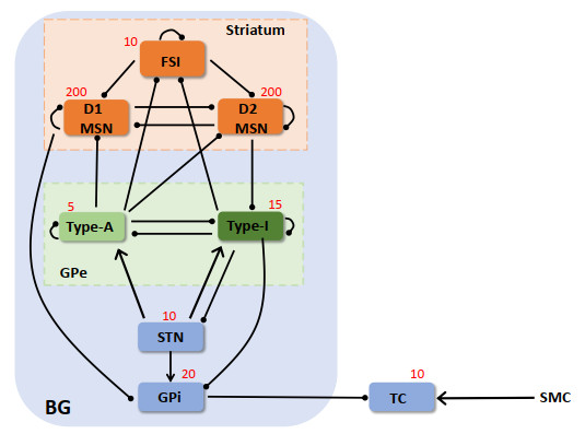
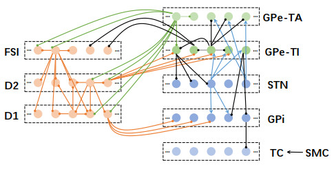
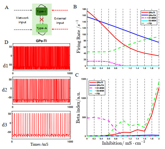
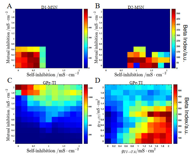
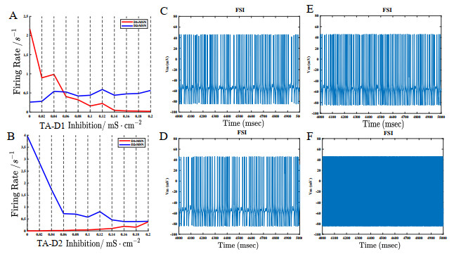
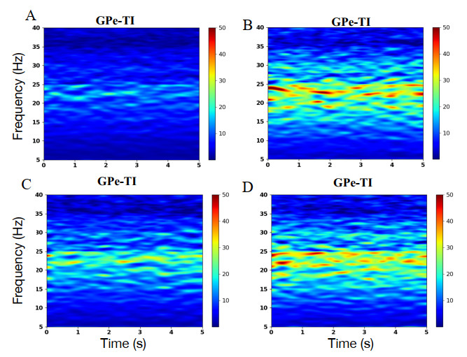
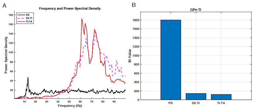
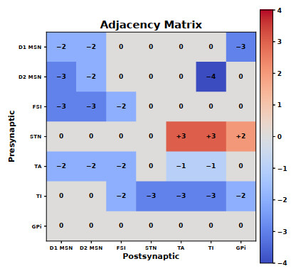
Pathological $ \beta $-band oscillations (13–35 Hz) in the basal ganglia (BG) are strongly associated with Parkinson's disease (PD). Recent evidence shows that subpopulations of external globus pallidus (GPe) neurons exhibit distinct responses to pathological conditions, and that their inhibitory feedback to the striatum strongly shape BG dynamics, features often overlooked in conventional models. To address this, we developed an extended BG network using a modified Hodgkin–Huxley framework, incorporating two GPe subclasses, arkypallidal (TA) and prototypical (TI), along with striatal medium spiny neurons (MSNs) and fast-spiking interneurons (FSIs). Simulations revealed that mutual inhibition within the GPe drives TI neurons from tonic firing into $ \beta $-bursting, retrogradely suppressing striatal activity through the GPe–FSI–MSN loop and disrupting direct/indirect pathway balance. We further show that GPe-TA projections exert strong inhibitory control over striatal populations, and that reducing MSN M-current reproduces $ \beta $ oscillations that propagate downstream. Blocking D2 MSN $ \rightarrow $ GPe-TI and GPe-TI $ \rightarrow $ GPe-TA synapses restores normal TI firing. Our results emphasize the role of GPe heterogeneity in pathological oscillations and suggest circuit-level therapeutic strategies for PD.
Citation: Zihan Li, Xia Shi, Bei Bai. The effect of the feedback inhibition of heterogeneous external globus pallidus on beta oscillations in an extended basal ganglia network[J]. Electronic Research Archive, 2025, 33(10): 6070-6095. doi: 10.3934/era.2025270
Pathological $ \beta $-band oscillations (13–35 Hz) in the basal ganglia (BG) are strongly associated with Parkinson's disease (PD). Recent evidence shows that subpopulations of external globus pallidus (GPe) neurons exhibit distinct responses to pathological conditions, and that their inhibitory feedback to the striatum strongly shape BG dynamics, features often overlooked in conventional models. To address this, we developed an extended BG network using a modified Hodgkin–Huxley framework, incorporating two GPe subclasses, arkypallidal (TA) and prototypical (TI), along with striatal medium spiny neurons (MSNs) and fast-spiking interneurons (FSIs). Simulations revealed that mutual inhibition within the GPe drives TI neurons from tonic firing into $ \beta $-bursting, retrogradely suppressing striatal activity through the GPe–FSI–MSN loop and disrupting direct/indirect pathway balance. We further show that GPe-TA projections exert strong inhibitory control over striatal populations, and that reducing MSN M-current reproduces $ \beta $ oscillations that propagate downstream. Blocking D2 MSN $ \rightarrow $ GPe-TI and GPe-TI $ \rightarrow $ GPe-TA synapses restores normal TI firing. Our results emphasize the role of GPe heterogeneity in pathological oscillations and suggest circuit-level therapeutic strategies for PD.

| [1] |
G. S. Yi, J. Wang, B. Deng, X. L. Wei, Complexity of resting-state eeg activity in the patients with early-stage Parkinson's disease, Cognit. Neurodyn., 11 (2017), 147–160. https://doi.org/10.1007/s11571-016-9415-z doi: 10.1007/s11571-016-9415-z

|
| [2] | L. V. Kalia, A. E. Lang, Parkinson's disease, Lancet, 386 (2015), 896–912. https://doi.org/10.1016/S0140-6736(14)61393-3 |
| [3] |
J. Jankovic, Parkinson's disease: clinical features and diagnosis, J. Neurol. Neurosurg. Psychiatry, 79 (2008), 368–376. https://doi.org/10.1136/jnnp.2007.131045 doi: 10.1136/jnnp.2007.131045
|
| [4] |
A. Cuk, T. Bezdan, L. Jovanovic, M. Antonijevic, M. Stankovic, V. Simic, et al., Tuning attention based long-short term memory neural networks for parkinson's disease detection using modified metaheuristics, Sci. Rep., 14 (2024), 4309. https://doi.org/10.1038/s41598-024-54680-y doi: 10.1038/s41598-024-54680-y
|
| [5] |
P. Jiang, J. R. Scarpa, V. D. Gao, M. H. Vitaterna, A. Kasarskis, F. W. Turek, Parkinson's disease is associated with dysregulations of a dopamine-modulated gene network relevant to sleep and affective neurobehaviors in the striatum, Sci. Rep., 9 (2019), 4808. https://doi.org/10.1038/s41598-019-41248-4 doi: 10.1038/s41598-019-41248-4
|
| [6] |
M. Lindahl, I. K. Sarvestani, Ö. Ekeberg, J. H. Kotaleski, Signal enhancement in the output stage of the basal ganglia by synaptic short-term plasticity in the direct, indirect, and hyperdirect pathways, Front. Comput. Neurosci., 7 (2013), 76. https://doi.org/10.3389/fncom.2013.00076 doi: 10.3389/fncom.2013.00076
|
| [7] |
R. L. Albin, A. B. Young, J. B. Penney, The functional anatomy of basal ganglia disorders, Trends Neurosci., 12 (1989), 366–375. https://doi.org/10.1016/0166-2236(89)90074-X doi: 10.1016/0166-2236(89)90074-X
|
| [8] |
O. V. Popovych, P. A. Tass, Adaptive delivery of continuous and delayed feedback deep brain stimulation-a computational study, Sci. Rep., 9 (2019), 10585. https://doi.org/10.1038/s41598-019-47036-4 doi: 10.1038/s41598-019-47036-4
|
| [9] |
V. L. Corbit, T. C. Whalen, K. T. Zitelli, S. Y. Crilly, J. E. Rubin, A. H. Gittis, Pallidostriatal projections promote $\beta$ oscillations in a dopamine-depleted biophysical network model, J. Neurosci., 36 (2016), 5556–5571. https://doi.org/10.1523/JNEUROSCI.0339-16.2016 doi: 10.1523/JNEUROSCI.0339-16.2016
|
| [10] |
N. Mallet, A. Pogosyan, A. Sharott, J. Csicsvari, J. Bolam, P. Brown, et al., Disrupted dopamine transmission and the emergence of exaggerated beta oscillations in subthalamic nucleus and cerebral cortex, J. Neurosci., 28 (2008), 4795–4806. https://doi.org/10.1523/JNEUROSCI.0123-08.2008 doi: 10.1523/JNEUROSCI.0123-08.2008
|
| [11] |
J. Dong, S. Hawes, J. Wu, W. Le, H. Cai, Connectivity and functionality of the globus pallidus externa under normal conditions and Parkinson's disease, Front. Neural Circuits, 15 (2021), 645287. https://doi.org/10.3389/fncir.2021.645287 doi: 10.3389/fncir.2021.645287
|
| [12] |
M. DeLong, Activity of pallidal neurons during movement, J. Neurophysiol., 34 (1971), 414–427. https://doi.org/10.1152/jn.1971.34.3.414 doi: 10.1152/jn.1971.34.3.414
|
| [13] |
R. Albin, A. Reiner, K. Anderson, L. Dure, B. Handelin, R. Balfour, et al., Preferential loss of striato-external pallidal projection neurons in presymptomatic Huntington's disease, Ann. Neurol., 31 (1992), 425–430. https://doi.org/10.1002/ana.410310412 doi: 10.1002/ana.410310412
|
| [14] |
N. Mallet, A. Pogosyan, L. Marton, J. Bolam, P. Brown, P. Magill, Parkinsonian beta oscillations in the external globus pallidus and their relationship with subthalamic nucleus activity, J. Neurosci., 28 (2008), 14245–14258. https://doi.org/10.1523/JNEUROSCI.4199-08.2008 doi: 10.1523/JNEUROSCI.4199-08.2008
|
| [15] |
N. Mallet, B. Micklem, P. Henny, M. Brown, C. Williams, J. Bolam, et al., Dichotomous organization of the external globus pallidus, Neuron, 74 (2012), 1075–1086. https://doi.org/10.1016/j.neuron.2012.04.027 doi: 10.1016/j.neuron.2012.04.027
|
| [16] |
Z. Abecassis, B. Berceau, P. Win, D. García, H. Xenias, Q. Cui, et al., Npas1$^{\pm}$ -nkx2.1$^{\pm}$ neurons are an integral part of the cortico-pallido-cortical loop, J. Neurosci., 40 (2020), 743–768. https://doi.org/10.1523/JNEUROSCI.1199-19.2019 doi: 10.1523/JNEUROSCI.1199-19.2019
|
| [17] |
A. Abdi, N. Mallet, F. Y. Mohamed, A. Sharott, P. D. Dodson, K. C. Nakamura, et al., Prototypic and arkypallidal neurons in the dopamine-intact external globus pallidus, J. Neurosci., 35 (2015), 6667–6688. https://doi.org/10.1523/JNEUROSCI.4662-14.2015 doi: 10.1523/JNEUROSCI.4662-14.2015
|
| [18] |
K. Glajch, D. Kelver, D. Hegeman, Q. Cui, H. Xenias, E. Augustine, et al., Npas1$^{\pm}$ pallidal neurons target striatal projection neurons, J. Neurosci., 36 (2016), 5472–5488. https://doi.org/10.1523/JNEUROSCI.1720-15.2016 doi: 10.1523/JNEUROSCI.1720-15.2016
|
| [19] |
A. Kravitz, B. Freeze, P. Parker, K. Kay, M. Thwin, K. Deisseroth, et al., Regulation of parkinsonian motor behaviours by optogenetic control of basal ganglia circuitry, Nature, 466 (2010), 622–626. https://doi.org/10.1038/nature09159 doi: 10.1038/nature09159
|
| [20] |
A. C. Kreitzer, Physiology and pharmacology of striatal neurons, Annu. Rev. Neurosci., 32 (2009), 127–147. https://doi.org/10.1146/annurev.neuro.051508.135422 doi: 10.1146/annurev.neuro.051508.135422
|
| [21] |
A. Rădulescu, J. Herron, C. Kennedy, A. Scimemi, Global and local excitation and inhibition shape the dynamics of the cortico-striatal-thalamo-cortical pathway, Sci. Rep., 7 (2017), 7608. https://doi.org/10.1038/s41598-017-07527-8 doi: 10.1038/s41598-017-07527-8
|
| [22] |
R. So, A. Kent, W. Grill, Relative contributions of local cell and passing fiber activation and silencing to changes in thalamic fidelity during deep brain stimulation and lesioning: a computational modeling study, J. Comput. Neurosci., 32 (2012), 499–519. https://doi.org/10.1007/s10827-011-0366-4 doi: 10.1007/s10827-011-0366-4
|
| [23] |
D. E. Oorschot, Total number of neurons in the neostriatal, pallidal, subthalamic, and substantia nigral nuclei of the rat basal ganglia: a stereological study using the cavalieri and optical disector methods, J. Comp. Neurol., 366 (1996), 580–599. https://doi.org/10.1002/(SICI)1096-9861(19960318)366:4<580::AID-CNE3>3.0.CO;2-0 doi: 10.1002/(SICI)1096-9861(19960318)366:4<580::AID-CNE3>3.0.CO;2-0
|
| [24] |
A. H. Gittis, G. B. Hang, E. S. LaDow, L. R. Shoenfeld, B. V. Atallah, S. Finkbeiner, et al., Rapid target-specific remodeling of fast-spiking inhibitory circuits after loss of dopamine, Neuron, 71 (2011), 858–868. https://doi.org/10.1016/j.neuron.2011.06.035 doi: 10.1016/j.neuron.2011.06.035
|
| [25] |
T. Koós, J. M. Tepper, Inhibitory control of neostriatal projection neurons by gabaergic interneurons, Nat. Neurosci., 2 (1999), 467–472. https://doi.org/10.1038/8138 doi: 10.1038/8138
|
| [26] |
M. Galarreta, S. Hestrin, Electrical synapses between gaba-releasing interneurons, Nat. Rev. Neurosci., 2 (2001), 425–433. https://doi.org/10.1038/35077566 doi: 10.1038/35077566
|
| [27] |
K. E. Sabol, D. B. Neill, S. A. Wages, W. H. Church, J. B. Justice, Dopamine depletion in a striatal subregion disrupts performance of a skilled motor task in the rat, Brain Res., 335 (1985), 33–43. https://doi.org/10.1016/0006-8993(85)90273-2 doi: 10.1016/0006-8993(85)90273-2
|
| [28] | K. T. Zitelli, Cell-Type Specific Plasticity at Intrapallidal Synapses in a Mouse Model of Parkinson's Disease, Master's thesis, University of Pittsburgh, 2016, Available from: https://core.ac.uk/download/pdf/78482375.pdf. |
| [29] |
C. Miguelez, S. Morin, A. Martinez, M. Goillandeau, E. Bezard, B. Bioulac, et al., Altered pallido-pallidal synaptic transmission leads to aberrant firing of globus pallidus neurons in a rat model of Parkinson's disease, J. Physiol., 590 (2012), 5861–5875. https://doi.org/10.1113/jphysiol.2012.241331 doi: 10.1113/jphysiol.2012.241331
|
| [30] |
F. Fujiyama, T. Nakano, W. Matsuda, T. Furuta, J. Udagawa, T. Kaneko, A single-neuron tracing study of arkypallidal and prototypic neurons in healthy rats, Brain Struct. Funct., 221 (2016), 4733–4740. https://doi.org/10.1007/s00429-015-1152-2 doi: 10.1007/s00429-015-1152-2
|
| [31] |
M. Cazorla, F. de Carvalho, M. Chohan, M. Shegda, N. Chuhma, S. Rayport, et al., Dopamine D2 receptors regulate the anatomical and functional balance of basal ganglia circuitry, Neuron, 81 (2014), 153–164. https://doi.org/10.1016/j.neuron.2013.10.041 doi: 10.1016/j.neuron.2013.10.041
|
| [32] |
K. J. Mastro, R. S. Bouchard, H. A. K. Holt, A. H. Gittis, Transgenic mouse lines subdivide external segment of the globus pallidus (gpe) neurons and reveal distinct gpe output pathways, J. Neurosci., 34 (2014), 2087–2099. https://doi.org/10.1523/JNEUROSCI.4646-13.2014 doi: 10.1523/JNEUROSCI.4646-13.2014
|
| [33] |
M. McCarthy, C. Moore-Kochlacs, X. Gu, E. Boyden, X. Han, N. Kopell, Striatal origin of the pathologic beta oscillations in Parkinson's disease, PNAS, 108 (2011), 11620–11625. https://doi.org/10.1073/pnas.1107748108 doi: 10.1073/pnas.1107748108
|
| [34] |
S. Damodaran, R. C. Evans, K. T. Blackwell, Synchronized firing of fast-spiking interneurons is critical to maintain balanced firing between direct and indirect pathway neurons of the striatum, J. Neurophysiol., 111 (2014), 836–848. https://doi.org/10.1152/jn.00382.2013 doi: 10.1152/jn.00382.2013
|
| [35] |
J. A. Wolf, J. T. Moyer, M. T. Lazarewicz, D. Contreras, M. Benoit-Marand, P. O'Donnell, et al., Nmda/ampa ratio impacts state transitions and entrainment to oscillations in a computational model of the nucleus accumbens medium spiny projection neuron, J. Neurosci., 25 (2005), 9080–9095. https://doi.org/10.1523/JNEUROSCI.2220-05.2005 doi: 10.1523/JNEUROSCI.2220-05.2005
|
| [36] |
A. Saunders, K. Huang, B. Sabatini, Globus pallidus externus neurons expressing parvalbumin interconnect the subthalamic nucleus and striatal interneurons, PLoS One, 11 (2016), e0149798. https://doi.org/10.1371/journal.pone.0149798 doi: 10.1371/journal.pone.0149798
|
| [37] |
V. Hernandez, D. Hegeman, Q. Cui, D. Kelver, M. Fiske, K. Glajch, et al., Parvalbumin$^{\pm}$ neurons and npas1$^{\pm}$ neurons are distinct neuron classes in the mouse external globus pallidus, J. Neurosci., 35 (2015), 11830–11847. https://doi.org/10.1523/JNEUROSCI.4672-14.2015 doi: 10.1523/JNEUROSCI.4672-14.2015
|
| [38] |
M. Nomura, T. Fukai, T. Aoyagi, Synchrony of fast-spiking interneurons interconnected by gabaergic and electrical synapses, Neural Comput., 15 (2003), 2179–2198. https://doi.org/10.1162/089976603322297340 doi: 10.1162/089976603322297340
|
| [39] |
P. Dodson, J. Larvin, J. Duffell, F. Garas, N. Doig, N. Kessaris, et al., Distinct developmental origins manifest in the specialized encoding of movement by adult neurons of the external globus pallidus, Neuron, 86 (2015), 501–513. https://doi.org/10.1016/j.neuron.2015.03.007 doi: 10.1016/j.neuron.2015.03.007
|
| [40] |
C. Deister, R. Dodla, D. Barraza, H. Kita, C. Wilson, Firing rate and pattern heterogeneity in the globus pallidus arise from a single neuronal population, J. Neurophysiol., 109 (2013), 497–506. https://doi.org/10.1152/jn.00677.2012 doi: 10.1152/jn.00677.2012
|
| [41] |
Q. Cui, A. Pamukcu, S. Cherian, I. Chang, B. Berceau, H. Xenias, et al., Dissociable roles of pallidal neuron subtypes in regulating motor patterns, J. Neurosci., 41 (2021), 4036–4059. https://doi.org/10.1523/JNEUROSCI.2210-20.2021 doi: 10.1523/JNEUROSCI.2210-20.2021
|
| [42] |
C. Gunay, J. Edgerton, D. Jaeger, Channel density distributions explain spiking variability in the globus pallidus: a combined physiology and computer simulation database approach, J. Neurosci., 28 (2008), 7476–7491. https://doi.org/10.1523/JNEUROSCI.4198-07.2008 doi: 10.1523/JNEUROSCI.4198-07.2008
|
| [43] | P. Moolchand, S. Jones, M. Frank, Biophysical and architectural mechanisms of subthalamic theta under response conflict, J. Neurosci., 2022. https://doi.org/10.1101/2021.11.10.468101 |
| [44] |
D. Terman, J. E. Rubin, A. C. Yew, C. J. Wilson, Activity patterns in a model for the subthalamopallidal network of the basal ganglia, J. Neurosci., 22 (2002), 2963–2976. https://doi.org/10.1523/JNEUROSCI.22-07-02963.2002 doi: 10.1523/JNEUROSCI.22-07-02963.2002
|
| [45] |
J. Bahuguna, A. Aertsen, A. Kumar, Existence and control of go/no-go decision transition threshold in the striatum, PLoS Comput. Biol., 11 (2015), e1004233. https://doi.org/10.1371/journal.pcbi.1004233 doi: 10.1371/journal.pcbi.1004233
|
| [46] |
A. Aristieta, M. Barresi, S. A. Lindi, G. Barrière, G. Courtand, B. de la Crompe, et al., A disynaptic circuit in the globus pallidus controls locomotion inhibition, Curr. Biol., 31 (2021), 707–721. https://doi.org/10.1016/j.cub.2020.11.019 doi: 10.1016/j.cub.2020.11.019
|
| [47] |
N. Mallet, R. Schmidt, D. Leventhal, F. Chen, N. Amer, T. Boraud, et al., Arkypallidal cells send a stop signal to striatum, Neuron, 89 (2016), 308–316. https://doi.org/10.1016/j.neuron.2015.12.017 doi: 10.1016/j.neuron.2015.12.017
|
| [48] |
A. H. Gittis, J. D. Berke, M. D. Bevan, C. S. Chan, N. Mallet, M. M. Morrow, et al., New roles for the external globus pallidus in basal ganglia circuits and behavior, J. Neurosci., 34 (2014), 15178–15183. https://doi.org/10.1523/JNEUROSCI.3252-14.2014 doi: 10.1523/JNEUROSCI.3252-14.2014
|
| [49] |
M. Lindahl, J. H. Kotaleski, Untangling basal ganglia network dynamics and function: Role of dopamine depletion and inhibition investigated in a spiking network model, eNeuro, 3 (2016), ENEURO.0156–16.2016. https://doi.org/10.1523/ENEURO.0156-16.2016 doi: 10.1523/ENEURO.0156-16.2016
|
| [50] |
M. Ketzef, G. Silberberg, Differential synaptic input to external globus pallidus neuronal subpopulations in vivo, Neuron, 109 (2021), 516–529. https://doi.org/10.1016/j.neuron.2020.11.006 doi: 10.1016/j.neuron.2020.11.006
|
| [51] |
P. Brown, Oscillatory nature of human basal ganglia activity: relationship to the pathophysiology of Parkinson's disease, Mov. Disord., 18 (2003), 357–363. https://doi.org/10.1002/mds.10358 doi: 10.1002/mds.10358
|
| [52] |
A. Ortone, A. A. Vergani, M. Ahmadipour, R. Mannella, A. Mazzoni, Dopamine depletion leads to pathological synchronization of distinct basal ganglia loops in the beta band, PLoS Comput. Biol., 19 (2023), e1010645. https://doi.org/10.1371/journal.pcbi.1010645 doi: 10.1371/journal.pcbi.1010645
|
| [53] |
X. Wang, Y. Yu, F. Han, Q. Wang, Beta-band bursting activity in computational model of heterogeneous external globus pallidus circuits, Commun. Nonlinear Sci. Numer. Simul., 110 (2022), 106388. https://doi.org/10.1016/j.cnsns.2022.106388 doi: 10.1016/j.cnsns.2022.106388
|
| [54] |
X. Wang, Y. Yu, F. Han, Q. Wang, Dynamical mechanism of parkinsonian beta oscillation in a heterogeneous subthalamopallidal network, Nonlinear Dyn., 111 (2023), 10505–10527. https://doi.org/10.1007/s11071-023-08381-2 doi: 10.1007/s11071-023-08381-2
|
| [55] |
M. A. J. Lourens, H. G. Meijer, T. Heida, E. Marani, S. A. van Gils, The pedunculopontine nucleus as an additional target for deep brain stimulation, Neural Networks, 24 (2011), 617–630. https://doi.org/10.1016/j.neunet.2011.03.007 doi: 10.1016/j.neunet.2011.03.007
|
| [56] |
Y. Yu, H. Zhang, L. Zhang, Q. Wang, Dynamical role of pedunculopntine nucleus stimulation on controlling Parkinson's disease, Physica A, 525 (2019), 834–848. https://doi.org/10.1016/j.physa.2019.04.016 doi: 10.1016/j.physa.2019.04.016
|
Figures(11) / Tables(6)
Zihan Li, Xia Shi, Bei Bai. The effect of the feedback inhibition of heterogeneous external globus pallidus on beta oscillations in an extended basal ganglia network[J]. Electronic Research Archive, 2025, 33(10): 6070-6095. doi: 10.3934/era.2025270


 DownLoad: