Obstructive sleep apnea syndrome (OSAS) is characterized by recurrent upper airway collapse during sleep, yet the relative contributions of soft palate versus tongue collapse remain poorly quantified.
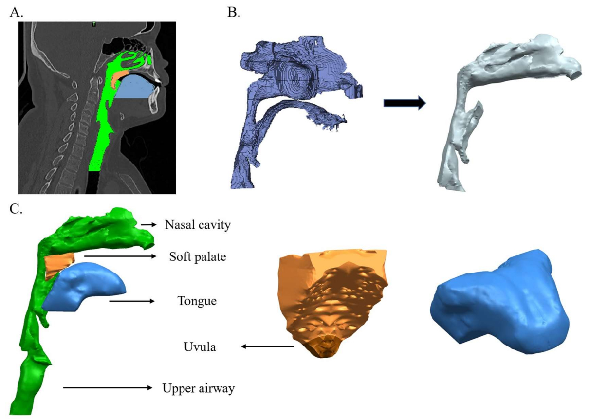
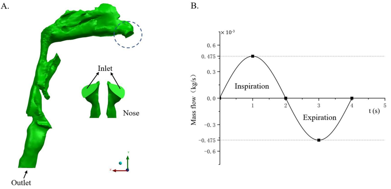
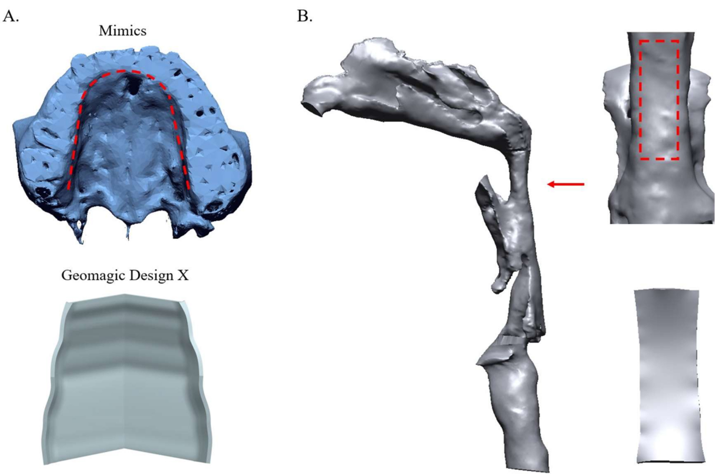
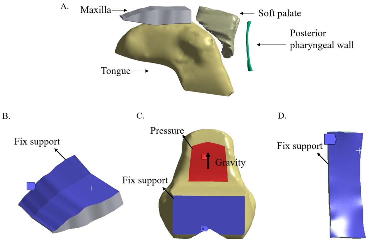
Three-dimensional numerical models of the upper airway, soft palate, and tongue were reconstructed from computed tomography (CT) images. A transient bidirectional fluid-structure interaction (FSI) method was employed to determine the ventilation pressure required to maintain a prescribed inspiratory flow across progressively reduced artificial support loads applied to the soft palate and tongue (0–100% of the calibrated reference loads).
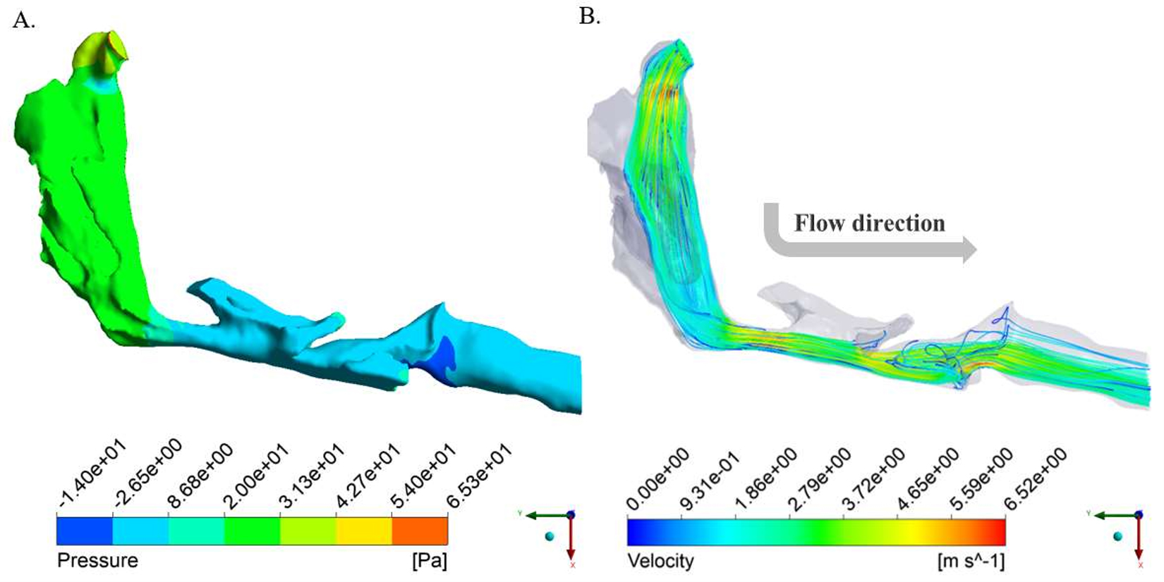
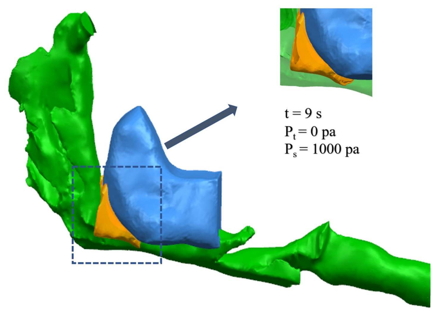
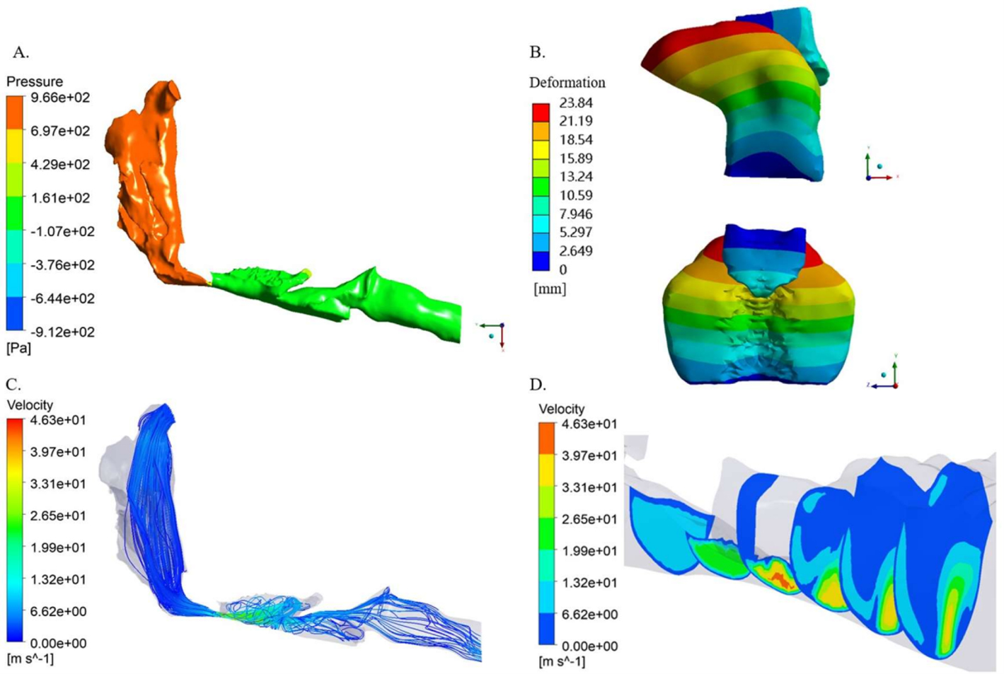
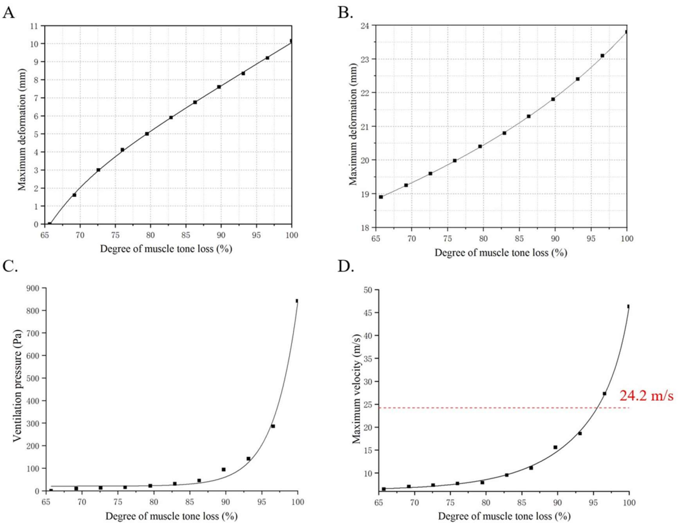
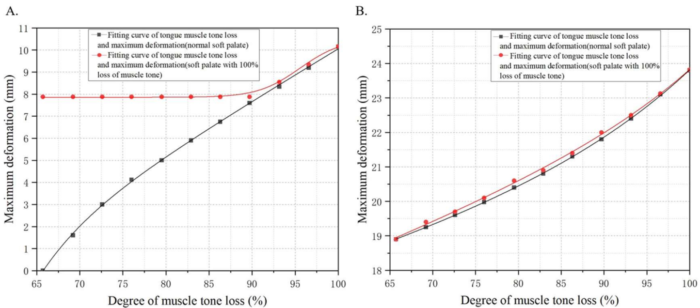
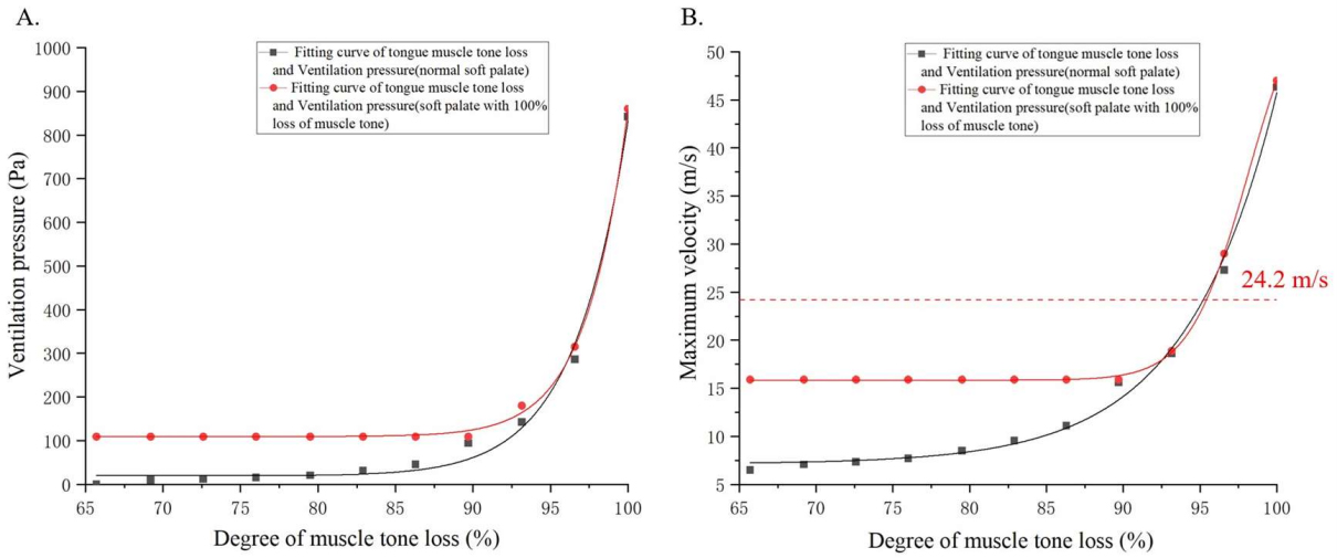
Isolated soft palate collapse exerted minimal effect on the required ventilation pressure. Obstruction severity was predominantly governed by tongue behavior: Below approximately 93% reduction of the reference tongue supported load, and airflow dynamics remained largely unchanged (maximum velocity ≤ 15.6 m/s, ventilation pressure ≤ 94 Pa). Beyond this level, appreciable tongue–soft palate compression was established and a marked nonlinear transition occurred: The ventilation pressure required to maintain the prescribed flow increased by approximately 806% (to 852 Pa), and the maximum velocity reached 46.3 m/s. Joint collapse analysis confirmed that tongue behavior, rather than soft palate status, is the primary determinant of flow obstruction in this model.
In this single-subject model and under the selected artificial support loads, appreciable tongue–soft palate compression produces a marked nonlinear increase in the pressure required to maintain a prescribed flow, occurring at approximately 93% reduction of the reference tongue support load. This contact transition is model-specific and should not be interpreted as a universal neuromuscular threshold. The findings provide proof-of-concept evidence for a biomechanical mechanism of tongue-driven obstruction that warrants validation in multi-subject, clinically characterized cohorts.
Citation: Zhi Guo Zhang, Mao Ning Wei, Yun-Ying Guo, Wei Xu. Effects of soft palate and tongue collapse on upper airway flow field under progressive muscle tone loss: a fluid-structure interaction study[J]. AIMS Bioengineering, 2026, 13(3): 366-393. doi: 10.3934/bioeng.2026018
Obstructive sleep apnea syndrome (OSAS) is characterized by recurrent upper airway collapse during sleep, yet the relative contributions of soft palate versus tongue collapse remain poorly quantified.
Three-dimensional numerical models of the upper airway, soft palate, and tongue were reconstructed from computed tomography (CT) images. A transient bidirectional fluid-structure interaction (FSI) method was employed to determine the ventilation pressure required to maintain a prescribed inspiratory flow across progressively reduced artificial support loads applied to the soft palate and tongue (0–100% of the calibrated reference loads).
Isolated soft palate collapse exerted minimal effect on the required ventilation pressure. Obstruction severity was predominantly governed by tongue behavior: Below approximately 93% reduction of the reference tongue supported load, and airflow dynamics remained largely unchanged (maximum velocity ≤ 15.6 m/s, ventilation pressure ≤ 94 Pa). Beyond this level, appreciable tongue–soft palate compression was established and a marked nonlinear transition occurred: The ventilation pressure required to maintain the prescribed flow increased by approximately 806% (to 852 Pa), and the maximum velocity reached 46.3 m/s. Joint collapse analysis confirmed that tongue behavior, rather than soft palate status, is the primary determinant of flow obstruction in this model.
In this single-subject model and under the selected artificial support loads, appreciable tongue–soft palate compression produces a marked nonlinear increase in the pressure required to maintain a prescribed flow, occurring at approximately 93% reduction of the reference tongue support load. This contact transition is model-specific and should not be interpreted as a universal neuromuscular threshold. The findings provide proof-of-concept evidence for a biomechanical mechanism of tongue-driven obstruction that warrants validation in multi-subject, clinically characterized cohorts.
three-dimensional
apnea-hypopnea index
continuous positive airway pressure
computed tomography
fluid-structure interaction
magnetic resonance imaging
obstructive sleep apnea syndrome

| [1] |
Benjafield AV, Ayas NT, Eastwood PR, et al. (2019) Estimation of the global prevalence and burden of obstructive sleep apnoea: a literature-based analysis. Lancet Respir Med 7: 687-698. https://doi.org/10.1016/S2213-2600(19)30198-5 
|
| [2] |
Diamond JA, Ismail H (2021) Obstructive sleep apnea and cardiovascular disease. Clin Geriatr Med 37: 445-456. https://doi.org/10.1016/j.cger.2021.04.006
|
| [3] |
Weaver TE, Grunstein RR (2008) Adherence to continuous positive airway pressure therapy: the challenge to effective treatment. Proc Am Thorac Soc 5: 173-178. https://doi.org/10.1513/pats.200708-119MG
|
| [4] |
Shukla RK, Srivastav VK, Paul AR, et al. (2020) Fluid structure interaction studies of human airways. Sadhana 45: 229. https://doi.org/10.1007/s12046-020-01460-9
|
| [5] |
Mihaescu M, Murugappan S, Kalra M, et al. (2008) Large eddy simulation and reynolds-averaged navier–stokes modeling of flow in a realistic pharyngeal airway model: an investigation of obstructive sleep apnea. J Biomech 41: 2279-2288. https://doi.org/10.1016/j.jbiomech.2008.04.013
|
| [6] |
Hebbink RHJ, Wessels BJ, Hagmeijer R, et al. (2023) Computational analysis of human upper airway aerodynamics. Med Biol Eng Comput 61: 541-553. https://doi.org/10.1007/s11517-022-02716-8
|
| [7] |
Wang Y, Wang J, Liu Y, et al. (2012) Fluid-structure interaction modeling of upper airways before and after nasal surgery for obstructive sleep apnea. Int J Numer Methods Biomed Eng 28: 528-546. https://doi.org/10.1002/cnm.1486
|
| [8] |
Faizal WM, Ghazali NNN, Khor CY, et al. (2020) Computational fluid dynamics modelling of human upper airway: a review. Comput Methods Programs Biomed 196: 105627. https://doi.org/10.1016/j.cmpb.2020.105627
|
| [9] |
Ashraf W, Jacobson N, Popplewell N, et al. (2022) Fluid-structure interaction modelling of the upper airway with and without obstructive sleep apnea: a review. Med Biol Eng Comput 60: 1827-1849. https://doi.org/10.1007/s11517-022-02592-2
|
| [10] |
Huang Y, White DP, Malhotra A (2007) Use of computational modeling to predict responses to upper airway surgery in obstructive sleep apnea. Laryngoscope 117: 648-653. https://doi.org/10.1097/MLG.0b013e318030ca55
|
| [11] |
Zhao M, Barber T, Cistulli P, et al. (2013) Computational fluid dynamics for the assessment of upper airway response to oral appliance treatment in obstructive sleep apnea. J Biomech 46: 142-150. https://doi.org/10.1016/j.jbiomech.2012.10.033
|
| [12] |
Kim SK, Na Y, Kim JI, et al. (2013) Patient specific CFD models of nasal airflow: overview of methods and challenges. J Biomech 46: 299-306. https://doi.org/10.1016/j.jbiomech.2012.11.022
|
| [13] |
Kimbell JS, Frank DO, Laud P, et al. (2013) Changes in nasal airflow and heat transfer correlate with symptom improvement after surgery for nasal obstruction. J Biomech 46: 2634-2643. https://doi.org/10.1016/j.jbiomech.2013.08.007
|
| [14] | Yue S, Liu HG (2021) Research progress of obstructive sleep apnea hypoventilation syndrome and upper airway dilator muscle. Chin J Tuberc Respir Dis 44: 661-664. https://doi.org/10.3760/cma.j.cn112147-20200721-00827 |
| [15] |
Younes M (2003) Contributions of upper airway mechanics and control mechanisms to severity of obstructive apnea. Am J Respir Crit Care Med 168: 645-658. https://doi.org/10.1164/rccm.200302-201OC
|
| [16] |
Vroegop AV, Vanderveken OM, Boudewyns AN, et al. (2014) Drug-induced sleep endoscopy in sleep-disordered breathing: report on 1,249 cases. Laryngoscope 124: 797-802. https://doi.org/10.1002/lary.24479
|
| [17] | Zhang Y, He SF (2023) Analysis of changes in chin and tongue muscle electromyography in patients with obstructive sleep apnea-hypopnea syndrome with different sleep staging. Chin Community Doctors 39: 111-113. |
| [18] |
Lin CL, Tawhai MH, McLennan G, et al. (2007) Characteristics of the turbulent laryngeal jet and its effect on airflow in the human intra-thoracic airways. Respir Physiol Neurobiol 157: 295-309. https://doi.org/10.1016/j.resp.2007.02.006
|
| [19] |
Wang YL, McDonald JP, Liu YH, et al. (2014) Analysis of the dynamic changes in the soft palate and uvula in obstructive sleep apnea-hypopnea using ultrafast magnetic resonance imaging. Genet Mol Res 13: 8596-8608. https://doi.org/10.4238/2014.January.24.16
|
| [20] |
Carberry JC, Jordan AS, White DP, et al. (2016) Upper airway collapsibility (Pcrit) and pharyngeal dilator muscle activity are sleep stage dependent. Sleep 39: 511-521. https://doi.org/10.5665/sleep.5516
|
| [21] |
Bafkar O, Cajas JC, Calmet H, et al. (2020) Impact of sleeping position, gravitational force & effective tissue stiffness on obstructive sleep apnoea. J Biomech 104: 109715. https://doi.org/10.1016/j.jbiomech.2020.109715
|
| [22] |
Pirnar J, Dolenc-Groselj L, Fajdiga I, et al. (2015) Computational fluid-structure interaction simulation of airflow in the human upper airway. J Biomech 48: 3685-3691. https://doi.org/10.1016/j.jbiomech.2015.08.017
|
| [23] |
Huang RH, Li XP, Rong QG (2013) Control mechanism for the upper airway collapse in patients with obstructive sleep apnea syndrome: a finite element study. Sci China Life Sci 56: 366-372. https://doi.org/10.1007/s11427-013-4448-6
|
| [24] | Kang W, Xu P, Bu WP, et al. (2022) Mechanical testing of biological soft tissue and related theoretical research. Acta Armamentarii 43: 2164-2171. https://doi.org/10.12382/bgxb.2022.0444 |
| [25] |
Haddad SMH, Dhaliwal SS, Rotenberg BW, et al. (2020) Estimation of the hyperelastic parameters of fresh human oropharyngeal soft tissues using indentation testing. J Mech Behav Biomed Mater 108: 103798. https://doi.org/10.1016/j.jmbbm.2020.103798
|
| [26] |
Haddad SMH, Dhaliwal SS, Rotenberg BW, et al. (2018) Estimation of the Young's moduli of fresh human oropharyngeal soft tissues using indentation testing. J Mech Behav Biomed Mater 86: 352-358. https://doi.org/10.1016/j.jmbbm.2018.07.004
|
| [27] |
Pelteret JP, Reddy BD (2012) Computational model of soft tissues in the human upper airway. Int J Numer Methods Biomed Eng 28: 111-132. https://doi.org/10.1002/cnm.1487
|
| [28] | Wang Q, Wang XM, Wang XQ, et al. (2019) In vitro oral soft friction study based on simulated surface of human tongue. The 16th Annual Meeting of the Chinese Society of Food Science and Technology and the 10th China-US Food Industry High Level Forum . |
| [29] |
Gupta JK, Lin CH, Chen Q (2010) Characterizing exhaled airflow from breathing and talking. Indoor Air 20: 31-39. https://doi.org/10.1111/j.1600-0668.2009.00623.x
|
| [30] |
Mylavarapu G, Murugappan S, Mihaescu M, et al. (2009) Validation of computational fluid dynamics methodology used for human upper airway flow simulations. J Biomech 42: 1553-1559. https://doi.org/10.1016/j.jbiomech.2009.03.035
|
| [31] | Yang YF, Zhang X, Chen XM (2012) Denervation change of tensor veli palati in obstructive sleep apnea-hypopnea syndrome. J Clin Otorhinolaryngol Head Neck Surg 26: 977-979, 983. https://doi.org/10.13201/j.issn.1001-1781.2012.21.011 |
| [32] |
Kajee Y, Pelteret JP, Reddy BD (2013) The biomechanics of the human tongue. Int J Numer Methods Biomed Eng 29: 492-514. https://doi.org/10.1002/cnm.2531
|
| [33] |
Cid-Verdejo R, Domínguez Gordillo AA, Sánchez-Romero EA, et al. (2023) Diagnostic accuracy of a portable electromyography and electrocardiography device to measure sleep bruxism in a sleep apnea population. Clocks Sleep 5: 717-733. https://doi.org/10.3390/clockssleep5040047
|
| [34] |
Cid-Verdejo R, Chávez Farías C, Martínez-Pozas O, et al. (2024) Instrumental assessment of sleep bruxism: A systematic review and meta-analysis. Sleep Med Rev 74: 101906. https://doi.org/10.1016/j.smrv.2024.101906
|
| [35] |
Ismail AMA, Gadallah NGM, Elfahl AMA (2026) Apnea-hypopnea index, blood pressure, daytime sleepiness, and sleeping quality in obstructive sleep apnea: randomized-controlled responses to Shitali respiratory training. Respir Med 252: 108608. https://doi.org/10.1016/j.rmed.2025.108608
|
| [36] | Ismail AMA, Khattab MNM, Gadallah NGM, et al. (2026) Effect of inspiratory muscle training on fatigue, aerobic capacity, pulmonary functions, and quality of life in adolescents with familial Mediterranean fever: a randomized controlled trial. Ir J Med Sci . https://doi.org/10.1007/s11845-026-04360-9 |
| [37] | Taha MM, Alothman SA, Alawafi R, et al. (2026) Knowledge, attitude, and practice of allied healthcare professionals about lifestyle medicine for diabetes: a cross-sectional study. Work 2026: 10519815261435992. https://doi.org/10.1177/10519815261435992 |
| [38] |
Elfahl AM, Elfeky HM, Ismail AMA (2026) Pilates versus gyrotonic expansion system on chemotherapy-induced peripheral neuropathy and fall risk. Explore 22: 103475. https://doi.org/10.1016/j.explore.2026.103475
|
| [39] |
Justribó-Manion G, Sánchez-Romero EA, Cuenca-Zaldivar JN, et al. (2026) Active conservative interventions for obstructive sleep apnea: an umbrella review and meta-meta-analysis of systematic reviews. Sleep Med 138: 108665. https://doi.org/10.1016/j.sleep.2025.108665
|
Figures(15) / Tables(4)

Zhi Guo Zhang, Mao Ning Wei, Yun-Ying Guo, Wei Xu. Effects of soft palate and tongue collapse on upper airway flow field under progressive muscle tone loss: a fluid-structure interaction study[J]. AIMS Bioengineering, 2026, 13(3): 366-393. doi: 10.3934/bioeng.2026018




 DownLoad: