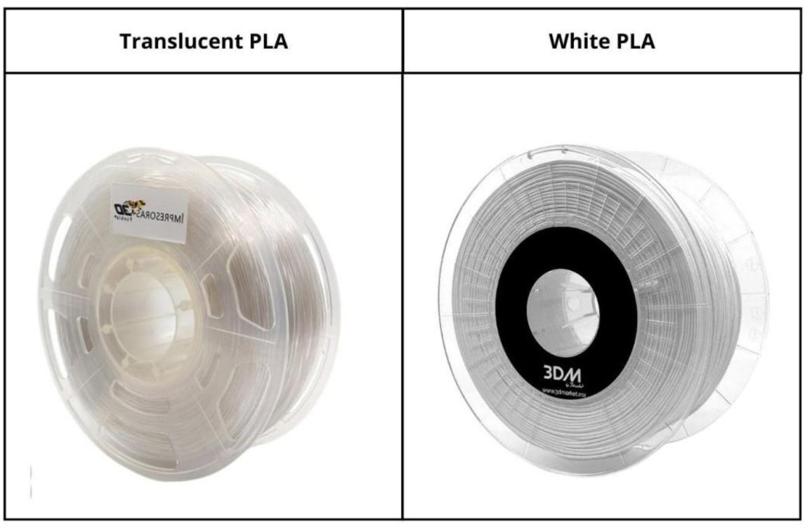
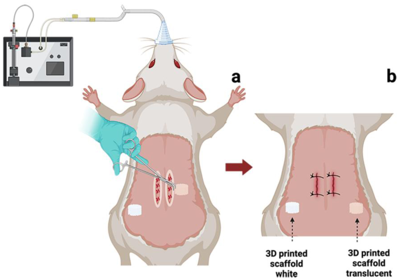
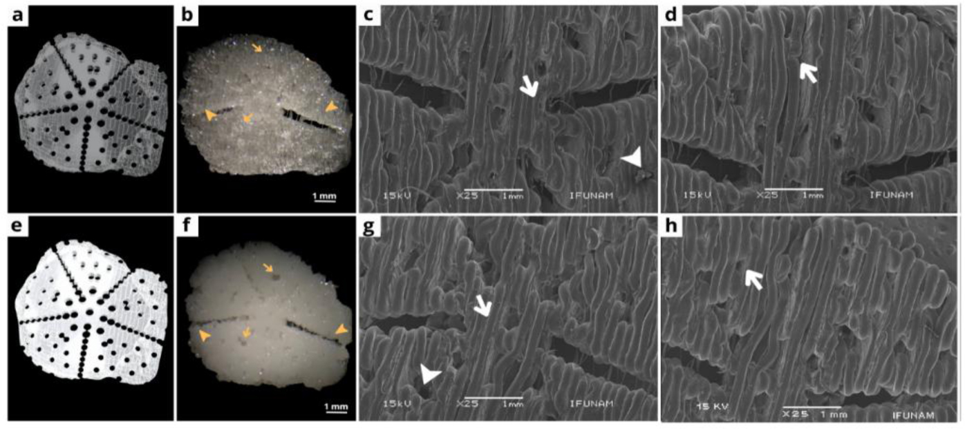
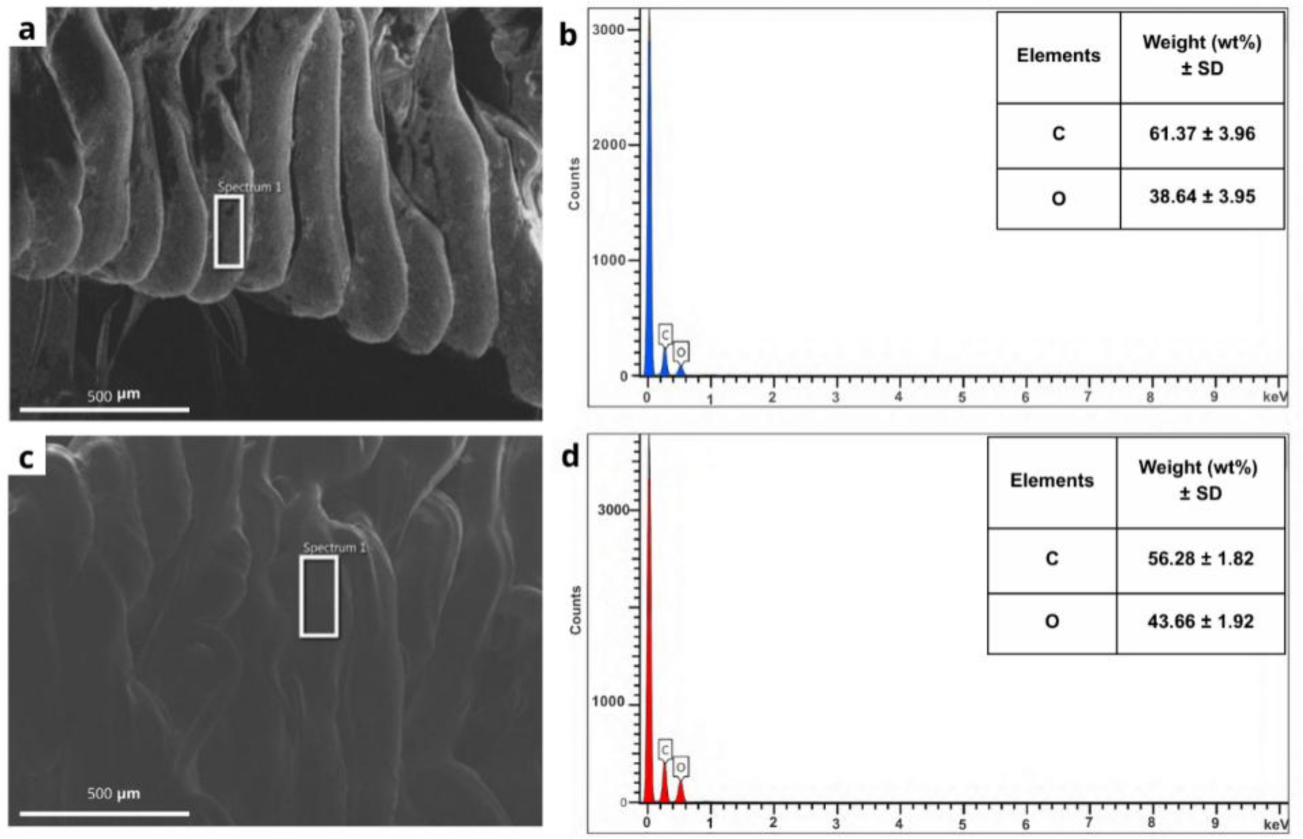
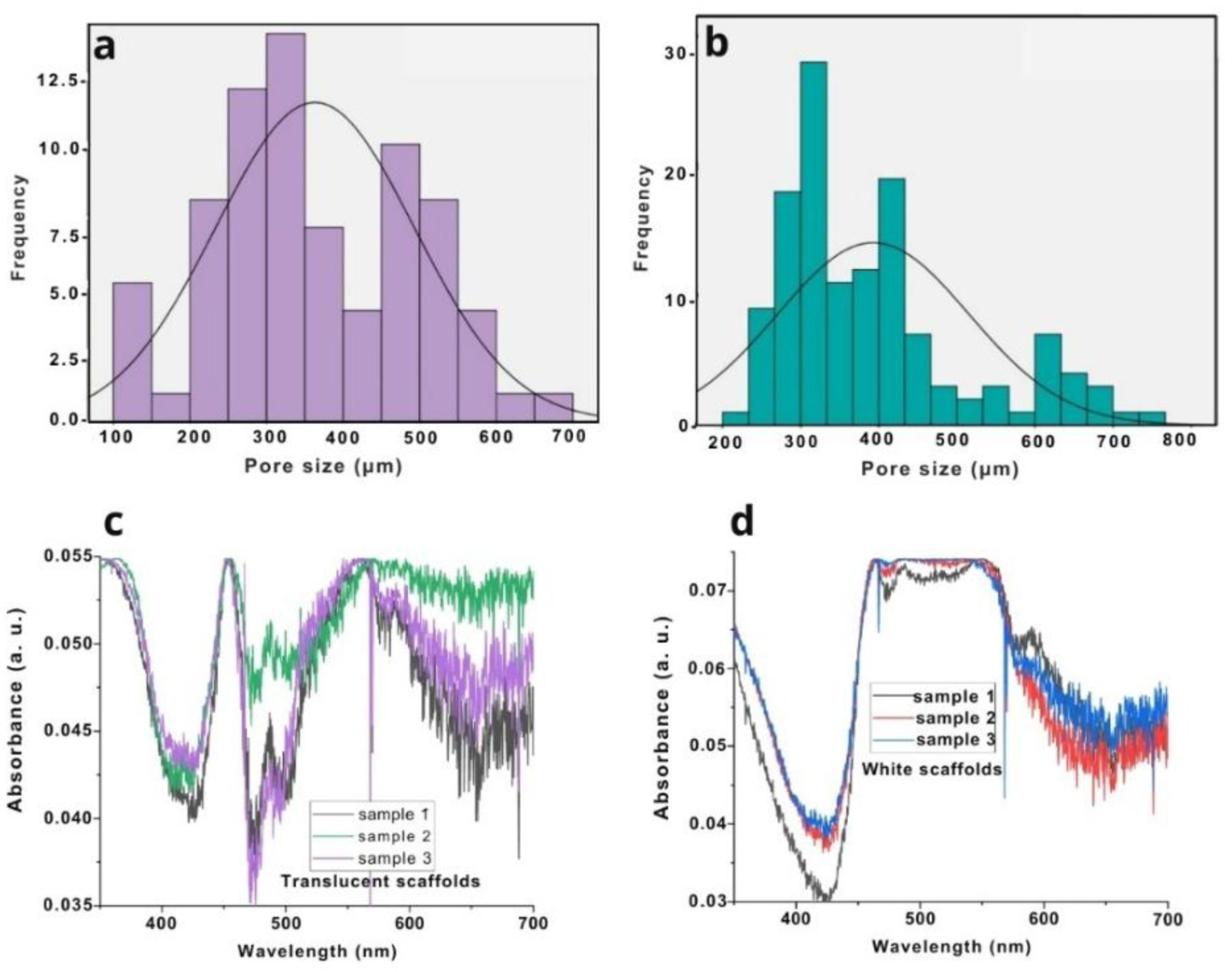
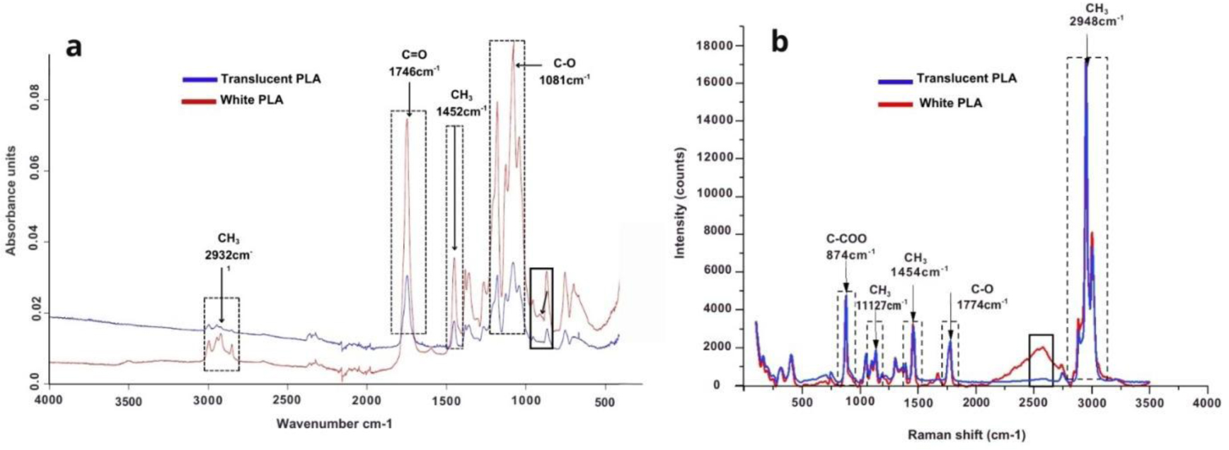
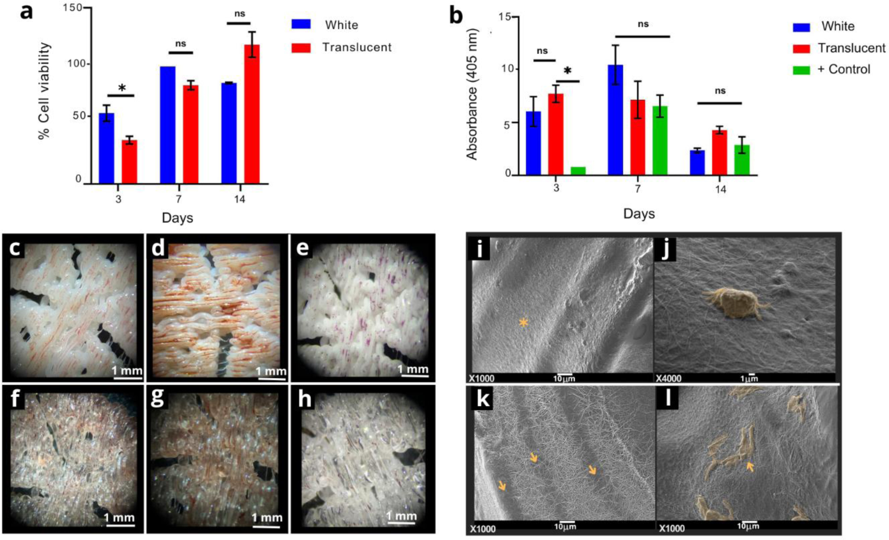
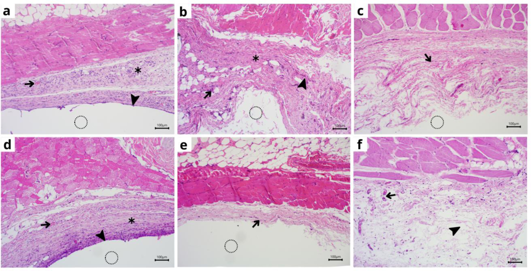
Polylactic acid (PLA) is a widely used polymer in tissue engineering due to its biocompatibility, biodegradability, and easy processing. However, to this day, white and translucent PLA are used indistinctly in the field though there are several studies that establish changes in the materials properties in the presence or absence of pigments. Here, we proposed an in vitro and in vivo methodology with 3D-printed scaffolds to further examine how their surface structure and cellular response vary. We observed surface structure, pore size, and porosity, and conducted in vitro cell assays. In vivo, male Wistar rats were used to collect histological samples and analyze the inflammatory response. DS revealed that the translucent scaffold contained 61.37 ± 3.96 wt% carbon and 38.64 ± 3.95 wt% oxygen and measured 8.96 ± 0.85 mm in diameter and 0.97 ± 0.3 mm in width, while the white scaffold showed 56.28 ± 1.82 wt% carbon and 43.66 ± 1.92 wt% oxygen and measured a diameter of 9.15 ± 0.56 mm and width of 1.01 ± 0.2 mm. The white PLA scaffold showed better structure and 67% porosity with larger pores compared to the translucent scaffold, which had 53% porosity and smaller pores. Both types were accepted by fetal osteoblast cells, but the translucent scaffold was associated with large foreign-body cells. Moreover, PLA's pigment increases surface tension, which helps the material maintain its shape as it cools and produces a better surface structure.
Citation: Mariana Nataly Carbajal-Casique, Lucía Pérez-Sánchez, Marco Antonio Alvarez-Perez, Gerardo Daniel Rayo-López, Jorge-Alejandro Reyes-Esqueda, Francisco Marichi-Rodríguez, Janeth Serrano Bello. Influence of white pigment on the properties of polylactic acid scaffolds for 3D printing: in vitro and in vivo analysis[J]. AIMS Bioengineering, 2025, 12(2): 225-248. doi: 10.3934/bioeng.2025011
Polylactic acid (PLA) is a widely used polymer in tissue engineering due to its biocompatibility, biodegradability, and easy processing. However, to this day, white and translucent PLA are used indistinctly in the field though there are several studies that establish changes in the materials properties in the presence or absence of pigments. Here, we proposed an in vitro and in vivo methodology with 3D-printed scaffolds to further examine how their surface structure and cellular response vary. We observed surface structure, pore size, and porosity, and conducted in vitro cell assays. In vivo, male Wistar rats were used to collect histological samples and analyze the inflammatory response. DS revealed that the translucent scaffold contained 61.37 ± 3.96 wt% carbon and 38.64 ± 3.95 wt% oxygen and measured 8.96 ± 0.85 mm in diameter and 0.97 ± 0.3 mm in width, while the white scaffold showed 56.28 ± 1.82 wt% carbon and 43.66 ± 1.92 wt% oxygen and measured a diameter of 9.15 ± 0.56 mm and width of 1.01 ± 0.2 mm. The white PLA scaffold showed better structure and 67% porosity with larger pores compared to the translucent scaffold, which had 53% porosity and smaller pores. Both types were accepted by fetal osteoblast cells, but the translucent scaffold was associated with large foreign-body cells. Moreover, PLA's pigment increases surface tension, which helps the material maintain its shape as it cools and produces a better surface structure.

| [1] | Lutzweiler G, Ndreu Halili A, Engin Vrana N (2020) The overview of porous, bioactive scaffolds as instructive biomaterials for tissue regeneration and their clinical translation. Pharmaceutics 12: 602. https://doi.org/10.3390/pharmaceutics12070602 |
| [2] | Ikada Y (2006) Challenges in tissue engineering. J R Soc Interface 3: 589-601. https://doi.org/10.1098/rsif.2006.0124 |
| [3] | Ko HS, Lee S, Lee D, et al. (2020) Mechanical properties and bioactivity of poly(Lactic acid) composites containing poly(glycolic acid) fiber and hydroxyapatite particles. Nanomaterials 11: 1-13. https://doi.org/10.3390/nano11010249 |
| [4] | Messimer SL, Rocha Pereira T, Patterson AE, et al. (2019) Full-density fused deposition modeling dimensional error as a function of raster angle and build orientation: large dataset for eleven materials. J Manufd MaterProcess 3: 6. https://doi.org/10.3390/jmmp3010006 |
| [5] | Wickramasinghe S, Do T, Tran P (2020) FDM-based 3D printing of polymer and associated composite: a review on mechanical properties, defects and treatments. Polymers 12: 1529. https://doi.org/10.3390/polym12071529 |
| [6] | Howard D, Buttery LD, Shakesheff KM, et al. (2008) Tissue engineering: strategies, stem cells and scaffolds. J Anat 213: 66-72. https://doi.org/10.1111/j.1469-7580.2008.00878.x |
| [7] | Marin E (2023) History of dental biomaterials: biocompatibility, durability and still open challenges. Herit Sci 11: 207. https://doi.org/10.1186/s40494-023-01046-8 |
| [8] | Raghunath J, Rollo J, Sales KM, et al. (2007) Biomaterials and scaffold design: key to tissue-engineering cartilage. Biotechnol Appl Biochem 46: 73-84. https://doi.org/10.1042/BA20060134 |
| [9] | Tymrak BM, Kreiger M, Pearce JM (2014) Mechanical properties of components fabricated with open-source 3-D printers under realistic environmental conditions. Mater Des 58: 242-246. https://doi.org/10.1016/j.matdes.2014.02.038 |
| [10] | Ranakoti L, Gangil B, Bhandari P, et al. (2023) Promising role of polylactic acid as an ingenious biomaterial in scaffolds, drug delivery, tissue engineering, and medical implants: research developments, and prospective applications. Molecules 28: 485. https://doi.org/10.3390/molecules28020485 |
| [11] | Pérez-Sánchez L, Ortiz de la O MA, Álvarez-Pérez MA, et al. (2024) Standardization of 3D printing parameters to control the size and shape of pores in polylactic acid scaffolds. MedComm Biomate Appl 3: e74. https://doi.org/10.1002/mba2.74 |
| [12] | Caramês JMM, Vieira FA, Caramês GB, et al. (2022) Guided bone regeneration in the edentulous atrophic maxilla using deproteinized bovine bone mineral (DBBM) combined with platelet-rich fibrin (PRF)—a prospective study. J Clin Med 11: 894. https://doi.org/10.3390/jcm11030894 |
| [13] | Suniya NK, Verma AK (2023) A review on optimization of process parameters of fused deposition modeling. Res Eng Struct Mater 9: 631-659. https://doi.org/10.17515/resm2022.520ma0909 |
| [14] | Zhang K, Fan Y, Dunne N, et al. (2018) Effect of microporosity on scaffolds for bone tissue engineering. Regen Biomater 5: 115-124. https://doi.org/10.1093/rb/rby001 |
| [15] | Böttcher-Haberzeth S, Biedermann T, Reichmann E (2010) Tissue engineering of skin. Burns 36: 450-460. https://doi.org/10.1016/j.burns.2009.08.016 |
| [16] | Zhao R, Yang R, Cooper PR, et al. (2018) Bone grafts and substitutes in dentistry: a review of current trends and developments. Molecules 26: 3007. https://doi.org/10.3390/molecules26103007 |
| [17] | Farah S, Anderson DG, Langer R (2016) Physical and mechanical properties of PLA, and their functions in widespread applications — a comprehensive review. Adv Drug Deliv Rev 107: 367-392. https://doi.org/10.1016/j.addr.2016.06.012 |
| [18] | O'Brien FJ (2011) Biomaterials & scaffolds for tissue engineering. Mater Today 14: 88-95. https://doi.org/10.1016/S1369-7021(11)70058-X |
| [19] | Soares JB, Finamor J, Silva FP, et al. (2018) Analysis of the influence of polylactic acid (PLA) colour on FDM 3D printing temperature and part finishing. Rapid Prototyp J 24: 1305-1316. https://doi.org/10.1108/RPJ-09-2017-0177 |
| [20] | Valerga AP, Batista M, Salguero J, et al. (2018) Influence of PLA filament conditions on characteristics of FDM parts. Materials 11: 1322. https://doi.org/10.3390/ma11081322 |
| [21] | Frunzaverde D, Cojocaru V, Ciubotariu CR, et al. (2022) The influence of the printing temperature and the filament color on the dimensional accuracy, tensile strength, and friction performance of FFF-printed PLA specimens. Polymers 14: 1978. https://doi.org/10.3390/polym14101978 |
| [22] | Wittbrodt B, Pearce JM (2015) The effects of PLA color on material properties of 3-D printed components. Addit Manuf 8: 110-116. https://doi.org/10.1016/j.addma.2015.09.006 |
| [23] | Joseph TM, Kallingal A, Suresh AM, et al. (2023) 3D printing of polylactic acid: recent advances and opportunities. Int J Adv Manuf Technol 125: 1015-1035. https://doi.org/10.1007/s00170-022-10795-y |
| [24] | Liao C, Li Y, Tjong SC (2020) Polyetheretherketone and its composites for bone replacement and regeneration. Polymers 12: 2858. https://doi.org/10.3390/polym12122858 |
| [25] | Ramot Y, Haim-Zada M, Domb AJ, et al. (2016) Biocompatibility and safety of PLA and its copolymers. Adv Drug Deliv Rev 107: 153-162. https://doi.org/10.1016/j.addr.2016.03.012 |
| [26] | Pavan Kalyan B, Kumar L (2022) 3D printing: applications in tissue engineering, medical devices, and drug delivery. Aaps PharmSciTech 23: 92. https://doi.org/10.1208/s12249-022-02242-8 |
| [27] | Revati R, Abdul Majid MS, Ridzuan MJM, et al. (2017) Mechanical, thermal and morphological characterisation of 3D porous Pennisetum purpureum/PLA biocomposites scaffold. Mater Sci Eng C 75: 752-759. https://doi.org/10.1016/j.msec.2017.02.127 |
| [28] | Fillingham Y, Jacobs J (2016) Bone grafts and their substitutes. Bone Joint J 98-B: 6-9. https://doi.org/10.1302/0301-620X.98B.36350 |
| [29] | Beyerle A, Schulz H, Kissel T, et al. (2009) Screening strategy to avoid toxicological hazards of inhaled nanoparticles for drug delivery: the use of a-quartz and nano zinc oxide particles as benchmark. J Phys Conf Ser 151: 012034. https://doi.org/10.1088/1742-6596/151/1/012034 |
| [30] | Gendviliene I, Simoliunas E, Alksne M, et al. (2021) Effect of extracellular matrix and dental pulp stem cells on bone regeneration with 3D printed PLA/HA composite scaffolds. Eur Cell Mater 41: 204-215. https://doi.org/10.22203/eCM.v041a15 |
| [31] | Pant S, Thomas S, Loganathan S, et al. (2022) 3D bioprinted poly(lactic acid)/mesoporous bioactive glass based biomimetic scaffold with rapid apatite crystallization and in-vitro Cytocompatability for bone tissue engineering. Int J Biol Macromol 217: 979-997. https://doi.org/10.1016/j.ijbiomac.2022.07.202 |
| [32] | Kubelka P, Munk F (1931) An article on optics of paint layers. Tech Phys 12: 593-601. https://doi.org/10.4236/jamp.2021.911188 |
| [33] | Nanjundeswaraswamy TS, Divakar S (2021) Determination of sample size and sampling methods in applied research. Proc Eng Sci 3: 25-32. https://doi.org/10.24874/PES03.01.003 |
| [34] | Onuma K, Watanabe M, Sasaki N (2024) The grimace scale: a useful tool for assessing pain in laboratory animals. Exp Anim 73: 234-245. https://doi.org/10.1538/expanim.24-0010 |
| [35] | Liu YC, Lo GJ, Shyu VBH, et al. (2023) Surface modification of polylactic acid bioscaffold fabricated via 3D printing for craniofacial bone tissue engineering. Int J Mol Sci 24: 17410. https://doi.org/10.3390/ijms242417410 |
| [36] | Zhang X, Chen JL, Xing F, et al. (2022) Three-dimensional printed polylactic acid and hydroxyapatite composite scaffold with urine-derived stem cells as a treatment for bone defects. J Mater Sci Mater Med 33: 71. https://doi.org/10.1007/s10856-022-06686-z |
| [37] | Valerga AP, Batista M, Puyana R, et al. (2017) Preliminary study of PLA wire colour effects on geometric characteristics of parts manufactured by FDM. Procedia Manuf 13: 924-931. https://doi.org/10.1016/j.promfg.2017.09.161 |
| [38] | Beauson J, Schillani G, Van der Schueren L, et al. (2022) The effect of processing conditions and polymer crystallinity on the mechanical properties of unidirectional self-reinforced PLA composites. Compos Part A Appl Sci Manuf 152: 106668. https://doi.org/10.1016/j.compositesa.2021.106668 |
| [39] | Mitchell MK, Hirt DE (2015) Degradation of PLA fibers at elevated temperature and humidity. Polym Eng Sci 55: 1652-1660. https://doi.org/10.1002/pen.24003 |
| [40] | Laske S, Ziegler W, Kainer M, et al. (2015) Enhancing the temperature stability of PLA by compounding strategies. Polym Eng Sci 55: 2849-2858. https://doi.org/10.1002/pen.24176 |
| [41] | Coppola B, Cappetti N, Di Maio L, et al. (2018) 3D printing of PLA/clay nanocomposites: influence of printing temperature on printed samples properties. Materials 11: 1947. https://doi.org/10.3390/ma11101947 |
| [42] | Adamczyk A, Długoń E (2012) The FTIR studies of gels and thin films of Al2O3–TiO2 and Al2O3–TiO2–SiO2 systems. Spectrochim Acta A Mol Biomol Spectrosc 89: 11-17. https://doi.org/10.1016/j.saa.2011.12.018 |
| [43] | Wang L, Xie G, Mi X, et al. (2023) Surface-modified TiO2@SiO2 nanocomposites for enhanced dispersibility and optical performance to apply in the printing process as a pigment. ACS Omega 8: 20116-20124. https://doi.org/10.1021/acsomega.3c02679 |
| [44] | Ciliveri S, Bandyopadhyay A (2022) Influence of strut-size and cell-size variations on porous Ti6Al4V structures for load-bearing implants. J Mech Behav Biomed Mater 126: 105023. https://doi.org/10.1016/j.jmbbm.2021.105023 |
| [45] | Karageorgiou V, Kaplan D (2005) Porosity of 3D biomaterial scaffolds and osteogenesis. Biomaterials 26: 5474-5491. https://doi.org/10.1016/j.biomaterials.2005.02.002 |
| [46] | Valenzuela-Villela KS, García-Casillas PE, Chapa-González C (2020) Progress of the 3D printing of medical devices. Rev Mex Ing Biomed 41: 151-66. https://doi.org/10.17488/RMIB.41.1.12 |
| [47] | Zhao Y, Cai Y, Wang W, et al. (2025) Periosteum-bone inspired hierarchical scaffold with endogenous piezoelectricity for neuro-vascularized bone regeneration. Bioact Mater 44: 339-353. https://doi.org/10.1016/j.bioactmat.2024.10.020 |
| [48] | Wang CY, Qin ZX, Wei Y, et al. (2023) The immunomodulatory effects of RNA-based biomaterials on bone regeneration. Acta Biomater 162: 32-43. https://doi.org/10.1016/j.actbio.2023.03.031 |
| [49] | Song JH, Gu JT, Dang GP, et al. (2023) The immunomodulatory effects of DNA-conjugated collagen scaffolds on bone healing. Chem Eng J 474: 145318. https://doi.org/10.1016/j.cej.2023.145318 |
| [50] | Amnael Orozco-Díaz C, Moorehead R, Reilly GC, et al. (2020) Characterization of a composite polylactic acid-hydroxyapatite 3D-printing filament for bone-regeneration. Biomed Phys Eng Express 6: 025007. https://doi.org/10.1088/2057-1976/ab73f8 |
| [51] | Delgado-Calle J, Sañudo C, Sánchez-Verde L, et al. (2011) Epigenetic regulation of alkaline phosphatase in human cells of the osteoblastic lineage. Bone 49: 830-838. https://doi.org/10.1016/j.bone.2011.06.006 |
| [52] | Bernar A, Gebetsberger JV, Bauer M, et al. (2022) Optimization of the alizarin red s assay by enhancing mineralization of osteoblasts. Int J Mol Sci 24: 723. https://doi.org/10.3390/ijms24010723 |
| [53] | Wo J, Huang SS, Wu DY, et al. (2020) The integration of pore size and porosity distribution on Ti-6A1-4V scaffolds by 3D printing in the modulation of osteo-differentation. J Appl Biomater Funct Mater 18: 228080002093465. https://doi.org/10.1177/2280800020934652 |
| [54] | Abdul Samat A, Abdul Hamid ZA, Jaafar M, et al. (2023) Investigation of the in vitro and in vivo biocompatibility of a three-dimensional printed thermoplastic polyurethane/polylactic acid blend for the development of tracheal scaffolds. Bioengineering 10: 394. https://doi.org/10.3390/bioengineering10040394 |
| [55] | Moreira AC, Fernandes CP, de Oliveira MV, et al. (2021) The effect of pores and connections geometries on bone ingrowth into titanium scaffolds: an assessment based on 3D microCT images. Biomed Mater 16: 065010. https://doi.org/10.1088/1748-605X/ac246b |
| [56] | Pérez-Sánchez L, Ortiz de la O MA, González-Alva P, et al. (2021) In vivo study on bone response to 3D-printed constructs designed from microtomographic images. J Mater Eng Perform 30: 5005-5012. https://doi.org/10.1007/s11665-021-05585-8 |
| [57] | Aronin CEP, Sadik KW, Lay AL, et al. (2009) Comparative effects of scaffold pore size, pore volume, and total void volume on cranial bone healing patterns using microsphere-based scaffolds. J Biomed Mater Res A 89: 632-641. https://doi.org/10.1002/jbm.a.32015 |
Figures(8)

Mariana Nataly Carbajal-Casique, Lucía Pérez-Sánchez, Marco Antonio Alvarez-Perez, Gerardo Daniel Rayo-López, Jorge-Alejandro Reyes-Esqueda, Francisco Marichi-Rodríguez, Janeth Serrano Bello. Influence of white pigment on the properties of polylactic acid scaffolds for 3D printing: in vitro and in vivo analysis[J]. AIMS Bioengineering, 2025, 12(2): 225-248. doi: 10.3934/bioeng.2025011


 DownLoad: