Research article Topical Sections

Cardiomyotic induction and proliferation of dental stem cells on electrospun scaffolds

  • Stem cells from human exfoliated deciduous teeth (SHED) are a unique source of stem cells because they are relatively easy to obtain and harvest from discarded teeth. While they could differentiate into neurons, adipocytes, osteoblasts, and myocytes, little is known whether they can differentiate into cardiomyocytes. The potential use of a biomaterial scaffold to deliver cardiomyocytes to the site of damaged heart tissue for cellular therapy is an attractive concept. Gelatin-B and poly-(lactic-co-glycolic acid) (PLGA) were selected for the present study. The gelatin-B and PLGA scaffolds were constructed using an electro-spinning technique. SHED cells proliferate in both PLGA scaffolds and gelatin-B scaffolds and maintain adequate viability as determined with calcein-AM staining and DNA quantification. SHED cells were treated with a predetermined optimized cardio-treatment protocol. qPCR analysis of the cardiomyotic genes, MEF2.C, Cx-43, TNNT2.C, Nkx2.5, and GATA-4, showed that SHED cells differentiated on PLGA significantly up-regulated these cardiogenic markers compared to SHED cells cultured in control media . In summary, we demonstrate the growth and cardiomyogenesis of SHED cells on electrospun gelatin and PLGA scaffolds. Further development of our research concepts for cardiovascular regeneration using in vivo research and clinical trials, could allow the development of therapies involving the delivery of cardiomyocytes differentiated from SHED using electrospun scaffolds to the site of damaged heart tissue.

    Citation: Pornchai Kittivarakarn, Matthew Penna, Zenith Acosta, Daniel Pelaez, Ramon Montero, Fotios M. Andreopoulos, Herman S. Cheung. Cardiomyotic induction and proliferation of dental stem cells on electrospun scaffolds[J]. AIMS Bioengineering, 2016, 3(2): 139-155. doi: 10.3934/bioeng.2016.2.139

    Related Papers:

    [1] Shaoyung Chen, Hsinyi Mao, Pinhua Tu, Naichen Cheng, Jiashing Yu . Fabrication of decellularized adipose tissue/alginate composite microspheres with pASCs encapsulation for tissue engineering. AIMS Bioengineering, 2017, 4(3): 351-365. doi: 10.3934/bioeng.2017.3.351
    [2] Carlos M. Carballosa, Jordan M. Greenberg, Herman S. Cheung . Expression and function of nicotinic acetylcholine receptors in stem cells. AIMS Bioengineering, 2016, 3(3): 245-263. doi: 10.3934/bioeng.2016.3.245
    [3] Mark Weingarten, Nathan Akhavan, Joshua Hanau, Yakov Peter . A Place to Call Home: Bioengineering Pluripotential Stem Cell Cultures. AIMS Bioengineering, 2015, 2(2): 15-28. doi: 10.3934/bioeng.2015.2.15
    [4] Yihan Zhang . Manufacture of complex heart tissues: technological advancements and future directions. AIMS Bioengineering, 2021, 8(1): 73-92. doi: 10.3934/bioeng.2021008
    [5] Daniel Pelaez, John H. Michel, Herman S. Cheung . Growth on elastic silicone substrate elicits a partial myogenic response in periodontal ligament derived stem cells. AIMS Bioengineering, 2016, 3(4): 515-527. doi: 10.3934/bioeng.2016.4.515
    [6] Daria Wehlage, Hannah Blattner, Al Mamun, Ines Kutzli, Elise Diestelhorst, Anke Rattenholl, Frank Gudermann, Dirk Lütkemeyer, Andrea Ehrmann . Cell growth on electrospun nanofiber mats from polyacrylonitrile (PAN) blends. AIMS Bioengineering, 2020, 7(1): 43-54. doi: 10.3934/bioeng.2020004
    [7] Izgen Karakaya, Nuran Ulusoy . Basics of dentin-pulp tissue engineering. AIMS Bioengineering, 2018, 5(3): 162-178. doi: 10.3934/bioeng.2018.3.162
    [8] Joseph M. Chambers, Robert A. McKee, Bridgette E. Drummond, Rebecca A. Wingert . Evolving technology: creating kidney organoids from stem cells. AIMS Bioengineering, 2016, 3(3): 305-318. doi: 10.3934/bioeng.2016.3.305
    [9] Poonam Sharma, Shalise Burch, Tejasvi Peesay, Susan M. Hamilla, Adam H. Hsieh, Carlos Luna Lopez . Downregulation of vimentin intermediate filaments affect human mesenchymal stem cell adhesion and formation of cellular projections. AIMS Bioengineering, 2020, 7(4): 272-288. doi: 10.3934/bioeng.2020023
    [10] Yung-Hua Li, Xingxing Huang, Xiao-Lin Tian . Recent advances in dental biofilm: impacts of microbial interactions on the biofilm ecology and pathogenesis. AIMS Bioengineering, 2017, 4(3): 335-350. doi: 10.3934/bioeng.2017.3.335
  • Stem cells from human exfoliated deciduous teeth (SHED) are a unique source of stem cells because they are relatively easy to obtain and harvest from discarded teeth. While they could differentiate into neurons, adipocytes, osteoblasts, and myocytes, little is known whether they can differentiate into cardiomyocytes. The potential use of a biomaterial scaffold to deliver cardiomyocytes to the site of damaged heart tissue for cellular therapy is an attractive concept. Gelatin-B and poly-(lactic-co-glycolic acid) (PLGA) were selected for the present study. The gelatin-B and PLGA scaffolds were constructed using an electro-spinning technique. SHED cells proliferate in both PLGA scaffolds and gelatin-B scaffolds and maintain adequate viability as determined with calcein-AM staining and DNA quantification. SHED cells were treated with a predetermined optimized cardio-treatment protocol. qPCR analysis of the cardiomyotic genes, MEF2.C, Cx-43, TNNT2.C, Nkx2.5, and GATA-4, showed that SHED cells differentiated on PLGA significantly up-regulated these cardiogenic markers compared to SHED cells cultured in control media . In summary, we demonstrate the growth and cardiomyogenesis of SHED cells on electrospun gelatin and PLGA scaffolds. Further development of our research concepts for cardiovascular regeneration using in vivo research and clinical trials, could allow the development of therapies involving the delivery of cardiomyocytes differentiated from SHED using electrospun scaffolds to the site of damaged heart tissue.


    1. Introduction

    Heart disease is the leading cause of death in the United States [1]. Coronary artery disease is the most common heart disease, caused by limited or lack of blood flow to various parts of the heart. Limited blood flow can result in damage to heart tissues and further heart complications.

    Some researchers have identified a need to harvest and differentiate stem cells into cardiomyocytes as a potential novel therapy for accomplishing cardiovascular regeneration. However, harvesting donor stem cells and inducing their differentiation into a functional cell type is a difficult task. There are several stem cell-types that have been used for cardiac repair therapy including human embryonic stem cells (hESC) and adult stem cells, e.g. bone marrow, adipose tissue, and umbilical cord blood [2]. The use of hESC brings with it controversies surrounding creating, using, and destroying potential human embryos; and while mesenchymal stem cells are less controversial there is a large variety of cell-types to choose from with varying degrees of multipotency.

    In this experiment cells from human exfoliated deciduous teeth (SHED) are used in a cardiomyogenic media to assess cardio induction. The media used for cardiomyogenic induction contains cardiogenol-C, H2O2, oxytocin, ascorbic acid, BMP-2, bFGF, IgF-1, and 5-azacytidine dissolved in culture media. Cardiogenol-C is a synthetic chemical compound which reportedly induces cardiomyogenic differentiation in mouse P19 embryonic stem cells at the concentration of 0.25 µM. Cardiogenol-C induced differentiation showed positive up-regulation of Nkx-2.5 and GATA-4 [3]. Furthermore, at a concentration above 25 µM, Cardiogenol-C showed cellular toxicity. Hydrogen peroxide has been shown to enhance cardiomyocyte-like gene expressions of GATA-4, Nkx-2.5, and MEF-2C in P19 mESC as well [4]. From ex vivo experiments Jankowski, et al. showed that oxytocin is involved in the developmental formation of coronary vessels [5]. Treating mouse P19 embryonic stem cells with 10−7 M of oxytocin induces differentiation of the mESC into cardiomyocytes [6]. Furthermore, 5-azacytidine has been shown to have the capability to cause differentiation of hMSCs into cardiomyocyte in vitro [7]. In hMSCs treated with 5-azacytidine, GATA-4 and connexin-43 gene expressions were highly expressed along with positive staining of Nkx-2.5 [8].

    Along with cardio-related transcription factors, connexin gap junctions are integral to heart tissues and are thus important to induce. Using neonatal rat myocytes, Doble, et al., showed the importance of bFGF in the regulation of connexin-43, which is a necessary protein in the human rat gap junctions. bFGF also plays a key role after myocardial injury by stimulating the growth and proliferation of cardiac tissue [9]. Another growth factor found in freshly isolated cardiomyocytes of neonatal rats is insulin-like growth factor 1 (IgF-1) [10]. Even though IgF-1 is produced mainly in the liver, it is able to travel through the blood stream and eventually reach the heart. IgF-1 has been shown to induce cardiomyocyte hypertrophy in vitro [10,11]. Cardiac hypertrophy is mediated through several signaling pathways, one of which involves the growth factors IgF-1, FGF, and PDGF [12].

    Inducing cardio-specific transcription factors and structural components are important steps culminating in rhythmically beating cells. Ling-Ling, et al. induced beating cells with cardio-specific genes expressions in mESC using ascorbic acid [13]. Bone morphogenetic protein 2 (BMP-2) have also been shown to induce rhythmic beating, along with expressions of Nkx-2.5, GATA-4, and ventricular myosin heavy chain in explant tissue culture of chicken embryos [14]. It must be stated that there are other members of the BMP family, i.e. BMP-4 and BMP-7, which will cause expressions of the Nkx-2.5, GATA-4 and vMHC as well.

    Alongside the cardio differentiation, the biomaterial scaffold employed is a significant aspect of the experiment. Electrospinning was used to synthesize the necessary scaffolds for the experiment. Electro-spinning is a simple fabrication method of preparing nano-fibrous scaffolds with tunable fiber dimensions. A charged polymeric solution is introduced within an electric field through a syringe pump and polymer filaments are formed on an electrically conductive collector plate. The size and pattern of the deposited nanofibers depends on the strength of the electric field, the needle to collector distance, the concentration and flow rate of the polymeric solution and the polarity of the collecting plate. A custom made electro-spinning apparatus has been developed in our laboratories that allows for automated control of the flow rate, electric field potential, needle to collector distance and collector polarity through a graphical user interface. Electro-spinning is proven to be an effective method to fabricate highly organized scaffolds that could conform to a wide range of polymers (e.g. cellulose, chitin, dextrose, collagen, gelatin), provide spatial control over scaffold architecture, as well as easily tailoring cytokine release profiles [15,16].

    A hypothesized course of direction is to use adult stem cells extracted from SHED and induce in vitro cardiomyocyte differentiation, which can later be transplanted into the damaged heart region for cellular therapy. This study aims to show that SHED cells can attach and differentiate into cardiomyocytes on an electrospun scaffold. If proven, this technique can be employed to combat scarred and damaged heart tissue by directly transplanting the cardiomyocyte-differentiated SHED cells on an electrospun scaffold to the site of tissue damage. Not only will the scaffold provide an environment for the differentiating cells to attach, proliferate, and differentiate on, but over time the biomaterial scaffold will slowly degrade away leaving only a new, healthy layer of cardiac tissue.

    SHED cells are harvested from the dental pulp of “milk teeth”. The natural transition from baby teeth to adult permanent teeth is a dynamic process that involves the development and eruption of permanent teeth coordinated with the re-absorption of the roots of the deciduous teeth [17]. SHED cells are adult stem cells with limited differentiation capabilities, unlike embryonic stem cells, which have pluripotent differentiation abilities. SHED cells have been shown to be capable of differentiating into neurons, adipocytes, odontoblasts [17], osteoblasts [18], and myocytes [19].

    Although SHED cells have been induced to differentiate into a variety of cellular tissues, cardiomyocyte differentiation has yet to be demonstrated. The overall goal is to achieve cytokine-induced cardiomyogenic differentiation of SHED cells on electrospun scaffolds that can be tested in vitro to relieve, or repair, damaged cardiomyocytes caused by heart disease.

    To achieve the overall objective two aims are being assessed:

    1) Determine the optimal electrospun scaffold between either gelatin-B and PLGA scaffolds for cell adhesion and proliferation.

    2) Induce cardiomyogenic differentiation of SHED cells using a pre-determined optimal cardiomyocyte-inducing protocol on the two aforementioned electrospun scaffolds.

    2. Materials and Methods

    2.1. Electrospinning scaffolds

    2.1.1. Gelatin-B

    Gelatin-B (Sigma-Aldrich, St. Louis, MO) is derived from denatured bovine collagen. The scaffold was formed by dissolving 10% w/v gelatin-B in 1, 1, 1, 3, 3, 3-hexafluoro-2-propanol (HFIP) solvent and then loaded into a 5 mL syringe. The electro-spinning process used a 22 gauge blunt needle attached to a 15 kV electrical supply to charge and eject the polymer solution. gelatin-B was extruded at a rate of 4 mL/hr. Crosslinking of gelatin-B scaffold was performed using glutaraldehyde vapor (Sigma-Aldrich, St. Louis, MO, Glutaraldehyde Solution Grade II 25% in H2O) for 1hr in room temperature, followed by a 100mM glycine wash for an hour to block any unreacted aldehyde groups.

    2.1.2. PLGA

    Poly-(lactic-co-glycolic acid) nanofibrous scaffolds were electro-spun from a polymer solution of 10% poly-(DL-lactide-co-glycolide) (Sigma-Aldrich, St. Louis, MO) in HFIP. A voltage of 12 kV was applied to the 22 gauge blunt needle while being extruded from the syringe pump at 4 mL/hr.

    2.2. In-vitro SHED cell expansion

    SHED cells provided by the National Institute of Dental and Craniofacial Research (NIDCR), Bethesda, MD were isolated using the technique described by Miura, et al. [17]. The SHED cells were cultured and passaged when the cells reached 80% confluency in High-Glucose Common Culture Media (HG-CCM), composed of high glucose DMEM (Invitrogen, Carlsbad, CA), supplemented with 10% Heat-Inactivated Fetal Bovine Serum (HI-FBS) (Invitrogen, Carlsbad, CA), 1% Penicillin/Streptomycin (Invitrogen, Carlsbad, CA), 0.1% Amphotericin B (HyClone, Waltham, MA). The cells were cultured in an incubator at 37 °C and 5% CO2. The cells were detached from the flask using Trypsin-EDTA, frozen and stored in 10% dimethyl sulfoxide (DMSO) (Sigma-Aldrich, St. Louis, MO) in HG-CCM. SHED cells used in the experiment underwent four culture passages.

    2.3. Proliferation study

    2.3.1. Viability using calcein-AM staining

    The first aim of the experiment was to show that SHED cells can be seeded and grown on gelatin-B and PLGA scaffolds, compared to tissue culture plastic well plates (TCP). Initially, SHED cells are seeded at a density of 2,000 cells/cm2 onto, gelatin-B (n = 3), and PLGA scaffolds (n = 3). The gelatin-B and PLGA scaffolds are placed in the well plates on top of a base layer of 1% fibrin diluted in distilled water to ensure that the SHED cells only attach to the electrospun scaffold. The cells were seeded in 1 mL of HG-CCM per well and incubated at 37 °C and 5% CO2. The media was aspirated and changed every two days. After two and seven days from initial seeding, staining for viable cells (measured by esterase activity) was performed to determine the attachment and proliferation capabilities of the SHED cells on the electrospun scaffolds. Staining was accomplished using a 4 µM Calcein Acetoxymethyl (AM) solution in PBS and incubated for 30mins in a light protected environment. After incubation samples were imaged with a fluorescence microscope.

    2.3.2. Quantitative analysis

    To obtain a quantitative cell count on the scaffolds, quantification of DNA correlating to cell population was utilized. SHED cells were seeded onto gelatin-B (n = 3) and PLGA (n = 3) scaffolds at 10,000 cells/sample. The gelatin-B and PLGA scaffolds were cut into 10mm diameter circles and placed on a layer of 1% fibrin gel similarly to the viability experiments. The samples were incubated at 37°C and 5% CO2. Additionally, media changes of HG-CCM every other day was performed after initial seeding for the following 11 days.

    2.3.3. DNA extraction and isolation

    Trypsin-EDTA was used to detach the cells from the PLGA, and gelatin-B scaffolds on days 3, 5, 7, 9, and 11 after initial seeding. To ensure that only cells that are attached to the gelatin-B and PLGA scaffolds are collected, trypsinization of the cells on gelatin-B and PLGA scaffolds were done in a 1.7 mL tube instead of in the 24-well plate.

    After the cells were detached from the scaffolds, DNA extraction and isolation was performed. Using Gentra® Puregene® Cell Kit (Qiagen, Valencia, CA) and protocol for cultured cells, DNA was extracted and isolated from the samples. Additionally, RNase A Solution was included in the process to remove RNA. After DNA extraction was completed a solid, white pellet was visible. 10 µL of DNA Hydration Solution was added and then placed in a 65°C oven for 1 hour to re-dissolve the DNA pellet. Subsequently, the re-dissolved DNA was analyzed and quantified using a NanoDrop (Thermo-Fisher Scientific, Waltham, MA) with ND-100 software. DNA Hydration Solution was used as to normalize the samples. From quantification of DNA, cell population can be determined from known correlation between DNA mass and cell population from literature and experiment.

    2.4. Cardio-differentiation on electrospun scaffolds

    2.4.1. Cardio-differentiation treatments

    To test the efficacy of differentiation of SHED cells on the gelatin-B and PLGA scaffolds compared to that of TCP, the SHED cells were treated with a cardiomyocyte-inducing media. SHED cells were seeded on TCP at 2,000 cells/cm2; gelatin-B and PLGA scaffolds on a base layer of 1% fibrin gel at 6,000 cells/cm2. Three times more SHED cells were seeded onto the electrospun scaffolds since only a fraction of the initially seeded cells actually attach to the scaffolds as determined by the quantitative proliferation experiment.

    After two days of incubation at 37°C and 5% CO2 with HG-CCM, cells were viewed under light microscopy to ensure that cells were attached and proliferating normally. After confirming the SHED cells were attached and proliferated to ~30% confluency, the HG-CCM was aspirated out and either cardio- media or control HG-CCM was added. The experiment called for a nine-day treatment period with media changes every two days.

    Cardio-media is a cocktail mixture of 250 nM cardiogenol-C (Sigma-Aldrich), 10 µM hydrogen peroxide (Sigma-Aldrich), 100nM oxytocin (EMD Biosciences, Darmstadt, Germany), 100 µM ascorbic acid (Sigma-Aldrich), 10 ng/mL BMP-2 (Invitrogen), 50 ng/mL bFGF (Invitrogen), 5 ng/mL IgF-1 (Invitrogen), and 10 µM 5-azacytidine (Sigma-Aldrich) diluted in HG-CCM media.

    2.4.2. mRNA extraction and isolation

    After the nine-day period treatment, mRNA was extracted from the samples using TRIzol LS (Invitrogen, Carlsbad, CA). For the samples on TCP 0.5mL of TRIzol was added directly to the samples and incubated for 5 mins. For samples of gelatin-B and PLGA, 0.5 mL of TRIzol was added to 1.7 mL microcentrifuge tubes. The gelatin-B and PLGA scaffolds were then transferred into the 1.7 mL tubes of TRIzol using sterile tweezers and left to incubate in room temperature for 5 mins. mRNA isolation was performed according to TRIzol’s mRNA extraction protocol.

    2.4.3. mRNA quantification

    After isolating the mRNA from the samples, normalizing of the mRNA was done using the NanoDrop. Furthermore, mRNA samples with a 260/280 ratio of only 1.6 and greater was used to synthesize cDNA.

    2.4.4. Reverse transcription to synthesize cDNA

    Using the rehydrated mRNA that was previously quantified, synthesis of complimentary DNA was achieved by utilizing the High Capacity cDNA Reverse Transcription Kit (Applied Biosystems, Carlsbad, CA) and the supplied protocol. RT-PCR was performed on a GeneAmp PCR System 8700 Thermocycler (Applied Biosystems, Carlsbad, CA). The cDNA was then diluted 1:5.

    2.4.5. Real-time polymerase chain reaction

    Primers were designed using known gene sequences from NIH’s GeneBank. The DNA sequences from the coding region of the genes were then copied into OLIGO v6.7 and the primers were screened to exclude strong hairpins, and internal and external duplexes, as outlined in the OLIGO Handbook. All the primers were designed to have a melting point of 60 °C. Primers were ordered from Operon (Huntsville, AL).

    Cardiomyogenic gene expressions of the samples were quantified using a real-time polymerase chain reaction using SYBR Green Master Mix (Applied Biosystems, Carlsbad, CA) and following the company’s protocol.

    Following qPCR using Stratagene Mx3000P (Santa Clara, CA), analysis of relative gene expressions of the target genes, MEF2.C, Cx-43, TNNT2.C, Nkx2.5, and GATA-4, were normalized relative to the GAP-DH. Using CT values collected from the qPCR analysis, the relative gene expression quantity (RQ) was obtained using the delta-delta-CT method, where the reference is GAP-DH, control is samples treated with only HG-CCM, treated are samples treated with cardio-cocktail media, and E is the efficiency calculated to be 2 from the standard curve of the primers [20]:

    @RQ = \frac{{E_{Target}^{{\text{\Delta }}{C_T}{{\left( {Control - Treated} \right)}_{Target}}}}}{{E_{Reference}^{{\text{\Delta }}{C_T}{{\left( {Control - Treated} \right)}_{Reference}}}}}@

    2.5. Immunohistochemistry analysis

    Samples that were analyzed via immunohistochemistry were fixed in 10% neutral-buffered formalin by first rinsing the cells with PBS two times at room temperature followed by fixation in 10% formalin in PBS for 10 min at room temperature. Samples were then washed 3X in PBS containing 0.05% v/v Tween 20 (Sigma). Immunohistochemistry of nuclear localized antigens required further solubilization of the nuclear membrane in 0.1% Triton X-100 (Sigma) for 5 min. All samples were blocked using 1% BSA (containing 0.05% Tween 20) in PBS for 1 hr at room temperature. Immunostaining was carried out using the following primary antibodies and dilutions in 0.5 mg/mL BSA in PBS: GATA4 - 1:500, Nkx2.5 - 1:100, and Anti-Cardiac Troponin 1 (cTnT) - 1:250. All primary antibodies were purchased from Abcam (Abcam, Cambridge, MA). Cells were left incubating in the primary antibodies overnight. Following incubation with primary antibodies, the samples were washed 2X in Tween 20-containing PBS and then incubated with the appropriate secondary antibodies for two hours in the absence of light. The following secondary antibodies were applied diluted 1:200 in PBS: GATA4- Anti-Rabbit IgG (Rhodamine), Nkx2.5- Anti-Rabbit IgG (FITC), and cTnT- Anit-Rabbit IgG (FITC). All secondary antibodies were purchased from Santa Cruz (Santa Cruz Biotechnology, Santa Cruz CA).

    Following secondary antibody incubation, samples were again washed with PBS 2X and counterstained for DAPI. DAPI counterstaining was performed concurrently with mounting of the samples by means of the VectaShield hardest mounting media (Vector Laboratories, Burlingame CA) following the published protocol. Samples were then imaged using an inverted fluorescent microscope with the appropriate filters.

    2.6. Statistical analysis

    Gene expression: Values from three independent experiments were pooled for the analysis. A one-way ANOVA with a Tukey post-test was performed for each gene expression value using the statistical software SigmaPlot (Systat&Mystat Products, Chicago IL) and p values were obtained through this method. Statistical significance was determined as p ≤ 0.05.

    3. Results

    3.1. SEM analysis of electrospun scaffolds

    Figure 1 and Figure 2 display the scanning electron microscope (SEM) results for gelatin-B and PLGA, respectively. Figure 1A shows un-cross-linked, single fibers of gelatin-B created using electrospinning. Here it is seen that the electrospinning method has a high degree of precision, able to create fibers of very similar diameter. Figure 1B is a crosslinked gelatin-B scaffold showing high-porosity and crosslinking. Figure 1C is a cross-section image of a crosslinked gelatin-B scaffold. The thickness of the scaffold can be easily manipulated, as well as the diameter and degree of crosslinking. This manipulability is ideal for cellular adhesion.

    Figure 1. A) SEM image of un-cross-linked, single fibers of gelatin-B B) SEM image of crosslinked gelatin-B scaffold. C) SEM cross-section image of a crosslinked gelatin-B scaffold.

    The SEM images of the scaffolds shown in Figure 2 were made from PLGA fibers. Figure 2A shows the cross-linked PLGA fibers. Figure 2B provides an SEM image at higher magnification that better show the porosity and interconnectivity of the fibers.

    Figure 2. A) SEM image of PLGA fibers. B) SEM image of PLGA at higher magnification.

    3.2. Viability assessment

    Live staining of SHED cells after two days (Figure 3) and seven days (Figure 4) of HG-CCM shows that SHED cells attached and proliferated on gelatin-B and PLGA scaffolds. After Day 7, SHED cells grew to confluency on both scaffolds. A quantitative measurement proved to be unreliable due to IPLab software’s segmentation and analyzing algorithms which were not adequately detecting live cells because of the interference caused by the scaffolds. This inability to accurately quantify cell population led to using total mass of DNA to quantify SHED cell population growth.

    Figure 3. Live staining two days after initial seeding of SHED p4 cells onto Tissue culture. A) TCP. B) Gel-B. C) PLGA.
    Figure 4. Live staining seven days after initial seeding of SHED p4 cells onto Tissue culture. A) TCP. B) Gel-B. C) PLGA.

    3.3. Proliferation quantification

    DNA quantification is employed to quantify SHED attachment because we found this technique to be more feasible and accurate in quantifying cells on the given scaffolds compared to cell viability assays. To acquire population size of the SHED cells in the experiment, a determination of quantity of DNA in a SHED cell was required. In knowing the mass of DNA in one SHED cell, we are then able to determine not only fold increases in population but also quantify the approximate cell population. DNA mass per SHED cell was determined by quantifying DNA from a known amount of SHED cells, which were counted manually using a hemocytometer. It was determined that each SHED cell contained approximately 3.30 picogram of DNA; this was similar to Qiagen’s 6.0 pg of DNA per animal cell as stated in Qiagen’s PureGene protocol manual [21].

    Quantitative cell population for an eleven-day span were extrapolated and compared between gelatin-B and PLGA (Figure 5). The data is presented in the form of mean ± standard deviation.

    Figure 5. Histogram showing the undifferentiated SHED cell population size on gelatin-B (blue), and PLGA (red) scaffolds over a period of eleven days.

    The results in Figure 5 alongside Figure 4 suggest that the SHED cells are able to maintain viability on each scaffold. The results in Figure 5 suggest that the SHED cells are better able to proliferate on gelatin-B compared to PLGA.

    3.4. qPCR relative gene fold expression

    qPCR was performed and relative gene expression was calculated using RQ equation (Figure 6). Triplicate CT data were analyzed for outliers using Grubb’s test for outliers, followed by averaging the triplicate CT for each sample. The three averaged CT values for each test group were averaged before performing ΔΔCT calculations to normalize to GAP-DH and control groups.

    Figure 6. qPCR Relative cardiac gene fold expression of SHED cells after 9 days of cardio-treatments. (*: p < 0.05, n = 3).

    Since the averaging of the samples within the group occurred before ΔΔCT, a different method to calculate error bars was required. Using standard error of means (SE) directly was impossible since the standard deviations were calculated before ΔΔCT calculations. Upper and lower error bars had to be calculated to account for logarithmic differences to the relative quantity (RQ).

    Magnitude of upper error:

    @{2^{ - \left( {\Delta \Delta {C_T} - SE} \right)}} - RQ@

    Magnitude of lower error:

    @RQ - {2^{ - \left( {\Delta \Delta {C_T} + SE} \right)}}@

    Statistical analysis was performed to compare gene fold increase of cardio-media treated samples in comparison to the HG-CCM treated controls for gelatin-B, PLGA, and TCP. Gene expression analysis shows a significant cardio-inductive response in SHED cells cultured on the scaffolds as well as on TCP. Cells grown on PLGA scaffolds demonstrated a statistically significant fold increase in all cardiac-specific transcription factors (GATA4, MEF2C, and Nkx2.5) as well as cardiac structural and functional proteins connexin 43 (Cx43) and cardiac troponin T (TNNT2) when compared to SHED cells cultured in control media. While both Cx43 and TNNT2 were significantly upregulated in cardio-treated cells on gelatin-B substrates, these cells only show a statistically significant upregulation of one (MEF2C) out of the three cardiac-specific transcription factors explored. Similarly, cells grown on tissue culture plastic show some cardiac response in terms of gene expression but failed to significantly upregulate Cx43 and Nkx2.5 when treated with cardio-inductive media. Regardless, these results show that under the right conditions, SHED cells are capable of undergoing cardiomyogenesis and that this can be achieved on various scaffold materials.

    3.5. Immunohistochemistry analysis

    Both gelatin-B and PLGA scaffolds auto-phosphorylated and created a very high background for any meaningful immunhistochemical analysis of cardiac protein deposited in the scaffolds. To demonstrate the capability of cardiomyotic differentiation, the SHED cells were cultured with the aforementioned cardio-treament. The results of the immunohistochemical staining are displayed in Figure 7.

    Figure 7. Immunohistochemical analysis of several cardiomyogenic gene markers. A) Expression of GATA4 for cardio-treated SHED cells. B) Expression of GATA4 for control SHED cells C) Expression of NKX2.5 for cardio-treated SHED cells. D) Expression of NKX2.5 for control SHED cells. E) Expression of cTnT for cardio-treated SHED cells. F) Expression of cTnT for control SHED cells.

    Immunohistochemical analysis of cardiogenic induced SHED cells shows positive staining for cardio-specific transcription factors GATA 4 and NkX2.5. Additionally, positive staining is found with the cardiogenic structural protein cTnT. The cardio-treated SHED cells appear more positive than that of the HG-CCM treated controls.

    4. Discussion

    This study assesses the capability of inducing cardiomyotic differentiation of SHED cells through exposure to chemical compounds and growth factors, while at the same time trying to determine the effect of scaffold properties on cell attachment, proliferation, and differentiation. The study is composed of two aims: a) assess the ability of SHED cells attaching and proliferating on gelatin-B and PLGA electrospun scaffolds, and b) assess whether SHED cells are able to differentiate into cardiomyocytes on gelatin-B and PLGA scaffolds.

    4.1. SHED cells & biomaterials for cardiac repair

    Coronary heart disease will continue to be a matter of importance for years to come. One way to restore damaged heart tissue would be to use the patient’s own cells to repair damaged cardiac tissue. It is generally agreed that transplanted cells play a large role through the paracrine effect by creating a favorable environment for cell recovery, enhancing angiogenesis, and limiting ventricular modeling [22].

    Even though transplanted cardiomyocyte-like SHED cells may not directly become the new cardiac tissue, these cells may have an effect on the local environment of transplantation which would be conducive for the heart’s progenitor cells to repair the damage tissue. Past transplantation of stem cells into the heart through intracoronary delivery has shown to improve acute myocardial infarctions [23]. However, only a small percentage of the delivered cells, ~1.3 to 2.6%, were detected in the infracted myocardium; the majority of the cells were found in the liver and spleen [24]. A biomaterial scaffold, such as a PLGA patch covered with cardiomyocyte-like SHED cells, will allow for a precise delivery of therapeutic cells to the infracted area and better enhance cardiac tissue repair. Being able to concentrate the majority of transplanted cells to the targeted area and minimize any movement will greatly reduce the number of cells required for treatment. Thus, equating to less stem cell needing to be extracted and cultured, and lowering the likelihood of cell migration to other parts of the body. SHED cell treatments would be an autologous transplantation thus reducing or negating any immune reaction or rejection. It is important to note that gelatin-B is a bovine-based scaffold. Further, HG-CCM media contains fetal bovine serum (FBS), thus new approaches must be investigated to overcome potential immune reactions.

    PLGA scaffold is shown to be adequate for cell attachment, proliferation, and very capable of providing a suitable environment for cardiomyotic differentiation of SHED cells. PLGA is biocompatible and biodegradable within human tissue, and most importantly the mechanical strength and degradation time can be altered very easily during the electro-spinning process and with the use of co-polymers.

    4.2. Proliferation study

    The proliferation study used qualitative staining assessment and DNA quantification to assess the ability of the two electrospun scaffolds to promote attachment and proliferation of SHED cells. Both electrospun scaffolds allowed attachment of SHED cell. The background fluorescence of the scaffolds from the viability assay led to difficulties in quantifying the SHED cells with typical imaging software. Thus, we needed to lift the SHED cells from the scaffolds before quantification. We extracted the DNA and correlated this with an experimentally determined weight of SHED cells. Though this approach is not a typical cell quantification method, it provided an effective and cost-efficient manner to quantify the SHED cells on the scaffold.

    An important note from the proliferation experiment is that after one day (24hrs) SHED cells were adequately attached to all the scaffolds. According to Figure 5 approximately one-third of the initially seeded 10,000 SHED cells attached to the gelatin-B and PLGA scaffolds one day after seeding. This is a considerable loss and should be accounted for in future studies.

    Clinical use of gelatin-B and PLGA scaffolds would ultimately require either a significantly higher seeding density of SHED cells, or a longer proliferation period, to allow for greater cell population before transplantation. The difference between using gelatin-B or PLGA scaffolds are minimal when evaluating cell attachment and proliferation data, but when assessing the ability to induce differentiation the PLGA scaffold has a significant advantage.

    4.3. Induction of cardiomyocyte differentiation

    Through qPCR data acquisition, cardio-specific gene expressions of SHED cells exposed to cardio-media on both gelatin-B and PLGA scaffolds were analyzed. Cardio-media treated SHED cells on PLGA scaffolds resulted in significant up-regulation compared to the control group in all five cardio specific genes (MEF2.c, Cx-43, TNNT2.c, Nkx-2.5, GATA-4). Compared to SHED cells cultured in control media, SHED cells on the gelatin-B scaffolds treated with the cardio-media expressed statistically significant increased levels of three out of the five cardio-specific genes tested (CX43, TNNT2, and MEF2C). This illustrates that the PLGA scaffold may provide a better environment for the SHED cells to differentiate compared to gelatin-B. This should be considered for future cellular transplantation therapy studies. For the gene expression experiment, the sample number used (n = 3) was chosen because the cardiomyotic induction was a complex and expensive process. However, with this sample number, statistically significant gene expression results were obtained. Further reinforcing the effect of the cardio treatment used in this study is the immunohistochemical analysis. Positive staining for GATA 4, NkX2.5, and cTnT was found in cardio-treated SHED cells.

    The use of growth factors and proteins in the cardio-media induces a higher degree of cardiomyogenesis. Growth factors are important in inducing cardiomyocyte-like cells from SHED cells; these growth factors were shown to be present in fresh cardiac tissues of neonatal rat and chicken. Growth factors, either directly or indirectly, are involved in the development of new cardiac tissue. But in some cases, such as bFGF, levels of growth factors differ depending on the stage of the developing heart. For early stages of development, as in this experiment, bFGF is integral to early cardiomyocyte differentiation. But if we were to look at the effects of bFGF on terminally differentiated cardiomyocyte protein levels, such as of contractile troponin, bFGF does not create higher levels of troponin [25]. It is important to note that although all of the genes being tested are linked to cardiomyogenesis, GATA-4 is also believed to regulate embryogenesis as well [26].

    5. Conclusion

    There is a great need to understand SHED cell’s growth and differentiation capabilities. SHED cells can lead to a cell line that is ethically acceptable, non-invasive to acquire, and has the potential to differentiate to a variety of tissues which can be employed in regenerative tissue engineering. Every person will lose deciduous teeth as a child to make room for adult permanent teeth, and this makes it an excellent source to easily collect, isolate, and store these multipotent stem cells if the situation arises to treat injuries in the future.

    Furthermore, the use of an optimized biomaterial scaffold, such as PLGA, will need to be further analyzed and studied in vivo to test for its ability to function as a delivery system for cell transplantation. Even though the mechanical structure, e.g. pore size, porosity, degradation time, can be easily modified, further tests should be done to find the optimal specifications for certain transplantation situations.

    Acknowledgements

    This project was supported by the Veterans Affairs (VA) Merit Review and VA Senior Research Career Scientist Award programs.

    Conflict of Interest

    All authors declare no conflict of interest in this paper.

    [1] Heron M, Hoyert D, Murphy S, et al. (2009) Deaths: Final data for 2006. National Vital Statistics Reports 57: 14.
    [2] Segers V, Lee R (2008) Stem-cell therapy for cardiac disease. Nature 45: 937–942.
    [3] Wu X, Ding S, Ding Q, et al. (2004) Small molecules that induce cardiomyogenesis in embryonic stem cells. J Am Chem Soc 126: 1590–1591. doi: 10.1021/ja038950i
    [4] Buggisch M, Ateghang B, Ruhe C, et al. (2007) Stimulation of ES-cell-derived cardiomyogenesis and neonatal cardiac cell proliferation by reactive oxygen species and NADPH oxidase. J Cell Sci: 885–894.
    [5] Jankowski M, Danalache B, Wang D, et al. (2004) Oxytocin in cardiac ontogeny. P Natl Acad Sci U S A 35: 13074–13079.
    [6] Paquin J, Danalache B, Jankowski M, et al. (2002) Oxytocin induces differentiation of P19 embryonic stem cells to cardiomyocytes. P Natl Acad Sci U S A 99: 9550–9555.
    [7] Antonitsis P, Ioannidou-Papagiannaki E, Kaidoglou A, et al. (2008) Cardiomyogenic potential of human adult bone marrow mesenchymal stem cells in vitro. J Thorac Cardiovasc Surg 56: 77–82.
    [8] Labovsky V, Hofer E, Feldman L, et al. (2010) Cardiomyogenic differentiation of human bone marrow mesenchymal cells: role of cardiac extract from neonatal rat cardiomyocytes. Differentiation 79: 93–101.
    [9] Doble B, Kardami E (1995) Basic fibroblast growth factor stimulates connexin-43 expression and intercellular communication of cardiac fibroblasts. Mol Cell Biochem 143: 81–87.
    [10] Long C (1996) Autocrine and paracrine regulation of myocardial cell growth in vitro: the TGF beta paradigm. Trends Cardiovasc Med 6: 217–226.
    [11] Ito H, Hiroe M, Tsujino M, et al. (1993) Insulin-like growth factor-I induces hypertrophy with enhanced expression of muscle specific genes in cultured rat cardiomyocytes. Circulation: Am Heart J 87: 1715–1721.
    [12] Hefti M, Harder B, Eppendberger H, et al. (1997) Signaling pathways in cardiac myocyte hypertrophy. J Mol Cell Cardiol 29: 2873–2892.
    [13] Ling-Ling E, Zhao Y, Guo X, et al. (2005) Enrichment of cardiomyocytes derived from mouse embryonic stem cells. J Heart Lung Transplant 25: 664–674.
    [14] Schultheiss T, Burch J, Lassar A (1997) A role for bone morphogenetic proteins in the induction of cardiac myogenesis. Genes Dev 11: 451–462.
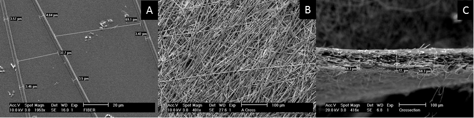
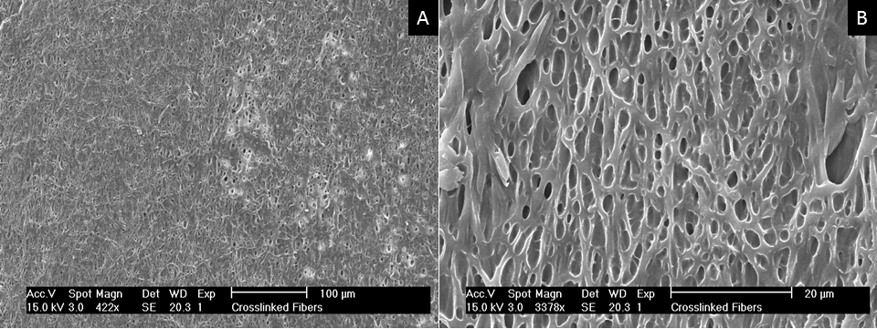
    [15] Hiep N, Lee B (2010). Electro-spinning of PLGA/PCL blends for tissue engineering and their biocompatibility. J Mater Sci Mater Med 21: 1969–1978.
    [16] Ifkovitz J, Sundararaghavan H, Burdick J (2009) Electrospinning fibrous polymer scaffolds for tissue engineering and cell culture. J Vis Exp 32.
    [17] Miura M, Gronthos S, Zhao M, et al. (2003) SHED: Stem cells from human exfoliated deciduous teeth. P Natl Acad Sci U S A 100: 5807–5812.
    [18] Huang G, Gronthos S, Shi S (2009) Mesenchymal stem cells derived from dental tissues vs. those from other sources: their biology and role in regenerative medicine. J Dent Res 88: 792–806.
    [19] Kerkis I, Kerkis A, Dozortsez D, et al. (2006) Isolation and characterization of a population of immature dental pulp stem cells expressing oct-4 and other embyronic stem cell markers. Cells Tissues Organs: 105–116.
    [20] Pfaffl M (2001). A new mathematical model for relative quantification in real-time RT-PCR. Nucleic Acids Res 29: 45.
    [21] QIAGEN (2011). Gentra® Puregene® Handbook, 3rd Eds.
    [22] Hamamoto H, Gorman J, Ryan L, et al. (2009). Allogeneic mesenchymal precursor cell therapy to limit remodeling after myocardial. Ann Thorac Surg. 87: 794–801.
    [23] Brignier A, Gewirtz A (2010) Embryonic and adult stem cell therapy. J Allergy Clin Immunol: 336–344.
    [24] Hofmann M, Wollert K, Meyer G, et al. (2005) Monitoring of bone marrow cell homing into the infarcted human myocardium. Circulation 111: 2198–2202. doi: 10.1161/01.CIR.0000163546.27639.AA
    [25] Velez C, Aranega E, Melguizo C, et al. (1994) Modulation of contractile protein troponin-T in chick myocardial cells by basic fibroblast growth factor and platelet-derived growth factor during development. J Cardiovasc Pharmacol 24: 906–913. doi: 10.1097/00005344-199424060-00007
    [26] Lourenco D, Brauner R, Rybczynska M, et al. (2011). Loss-of-function mutation in GATA4 causes anomalies of human testicular development. P Natl Acad Sci U S A 108: 1597–1602.
  • This article has been cited by:

    1. Iwona Deszcz, Daniele Torella, Stem Cell-Based Therapy and Cell-Free Therapy as an Alternative Approach for Cardiac Regeneration, 2023, 2023, 1687-9678, 1, 10.1155/2023/2729377
  • Reader Comments
  • © 2016 the Author(s), licensee AIMS Press. This is an open access article distributed under the terms of the Creative Commons Attribution License (http://creativecommons.org/licenses/by/4.0)
通讯作者: 陈斌, bchen63@163.com
  • 1. 

    沈阳化工大学材料科学与工程学院 沈阳 110142

  1. 本站搜索
  2. 百度学术搜索
  3. 万方数据库搜索
  4. CNKI搜索

Metrics

Article views(5764) PDF downloads(1156) Cited by(1)

Article outline

Figures and Tables

Figures(7)

/

DownLoad:  Full-Size Img  PowerPoint
Return
Return

Catalog