Citation: Merja Lusa, Jenna Knuutinen, Malin Bomberg. Uptake and reduction of Se(IV) in two heterotrophic aerobic Pseudomonads strains isolated from boreal bog environment[J]. AIMS Microbiology, 2017, 3(4): 798-814. doi: 10.3934/microbiol.2017.4.798
[1] | Merja Lusa, Jukka Lehto, Malin Bomberg . The uptake of Ni2+ and Ag+ by bacterial strains isolated from a boreal nutrient-poor bog. AIMS Microbiology, 2016, 2(2): 120-137. doi: 10.3934/microbiol.2016.2.120 |
[2] | Neda Askari, Hassan Momtaz, Elahe Tajbakhsh . Acinetobacter baumannii in sheep, goat, and camel raw meat: virulence and antibiotic resistance pattern. AIMS Microbiology, 2019, 5(3): 272-284. doi: 10.3934/microbiol.2019.3.272 |
[3] | Andrey A. Belov, Vladimir S. Cheptsov, Elena A. Vorobyova . Soil bacterial communities of Sahara and Gibson deserts: Physiological and taxonomical characteristics. AIMS Microbiology, 2018, 4(4): 685-710. doi: 10.3934/microbiol.2018.4.685 |
[4] | Arnab Bhowmik, Mara Cloutier, Emily Ball, Mary Ann Bruns . Underexplored microbial metabolisms for enhanced nutrient recycling in agricultural soils. AIMS Microbiology, 2017, 3(4): 826-845. doi: 10.3934/microbiol.2017.4.826 |
[5] | José Rodrigo da-Silva, Ana Alexandre, Clarisse Brígido, Solange Oliveira . Can stress response genes be used to improve the symbiotic performance of rhizobia?. AIMS Microbiology, 2017, 3(3): 365-382. doi: 10.3934/microbiol.2017.3.365 |
[6] | Eman Koshlaf, Andrew S Ball . Soil bioremediation approaches for petroleum hydrocarbon polluted environments. AIMS Microbiology, 2017, 3(1): 25-49. doi: 10.3934/microbiol.2017.1.25 |
[7] | Alexandra Díez-Méndez, Raul Rivas . Improvement of saffron production using Curtobacterium herbarum as a bioinoculant under greenhouse conditions. AIMS Microbiology, 2017, 3(3): 354-364. doi: 10.3934/microbiol.2017.3.354 |
[8] | Farzad Rahmati . Characterization of Lactobacillus, Bacillus and Saccharomyces isolated from Iranian traditional dairy products for potential sources of starter cultures. AIMS Microbiology, 2017, 3(4): 815-825. doi: 10.3934/microbiol.2017.4.815 |
[9] | Jessica Bannon, Mohammad Melebari, Cleso Jordao Jr, Carlos G Leon-Velarde, Keith Warriner . Incidence of Top 6 shiga toxigenic Escherichia coli within two Ontario beef processing facilities: Challenges in screening and confirmation testing. AIMS Microbiology, 2016, 2(3): 278-291. doi: 10.3934/microbiol.2016.3.278 |
[10] | Anil K. Persad, Michele L. Williams, Jeffrey T. LeJeune . Rapid loss of a green fluorescent plasmid in Escherichia coli O157:H7. AIMS Microbiology, 2017, 3(4): 872-884. doi: 10.3934/microbiol.2017.4.872 |
79Se is one of the high priority radionuclides in the long-term biosphere safety assessment of spent nuclear fuel [1]. In addition, considerable amounts of stable selenium enter the environment via anthropogenic activities including coal combustion, mining, refining of sour crude oils and agricultural irrigation of seleniferous soils [2,3,4,5]. The biological effects of selenium make it a particular hazard for environmental releases and both its environmental mobility and biological effects are mainly controlled by its chemical speciation [6,7,8]. Although selenium is an essential micronutrient for animals and humans, the higher valence states (selenate, Se(Ⅵ)O42– and selenite, Se(Ⅳ)O32–) are toxic at elevated concentrations [9,10] The toxic character of these oxyanionic compounds is related to their oxidant capacity [11]. Selenite is a highly reactive species of selenium and reacts in particular with thiol groups found in glutathione (GSH). This explains, at least in part, its toxic character [11]. Glutathione and selenite spontaneously react to produce several selenium-containing compounds, including selenodiglutathione, glutathioselenol, hydrogen selenide and elemental selenium [12]. This reaction produces also highly toxic oxygen species like H2O2 and O2– [13]. The reduction of the soluble oxyanions selenate and selenite converts selenium into the less toxic, insoluble Se0. In the environment, selenate and selenite reduction is mainly mediated by microorganisms [14,15,16], although slow abiotic reduction of selenium oxyanions can also occur under highly reducing conditions in the presence of Fe(Ⅱ, Ⅲ) oxides [17]. Some anaerobic and phototrophic aerobic selenium respiring bacteria have previously shown to be able to use selenium oxyanions as terminal electron acceptors precipitating insoluble elemental Se0 particles [11,18,19,20,21,22,23]. Following reduction, both intracellular and extracellular selenium granules have been found in phylogenetically and physiologically distinct bacteria like Chromatium vinosum, Desulfovibrio desulfuricans, Sulfospirillum barnesii, Bacillus selenitireducens, Selenihalanaerobacter shriftii, Shewanella oneidensis MR-1, Paenibacillus selenitireducens sp. nov., and Cupriavidus metallidurans CH34 (formerly Ralstonia metallidurans CH34) [22,24,25]. Microbial reduction of selenium oxyanions produces red elemental selenium with crystalline or amorphous structures and the elemental Se0 particles formed by Se-respiring Sulfurospirillum barnesii, Bacillus selenitireducens and Selenihalanaerobacter shriftii have been shown to be structurally distinctive from elemental selenium formed by chemical synthesis [22,26]. In addition there are indications that these different species of Se-reducing bacteria produce Se0 biominerals with different atomic structures [22]. However, while Se-reduction has been shown to be an environmentally important process in diverse terrestrial and aquatic environments, the mechanisms of Se0 biomineralization are poorly understood and the molecular factors behind Se reduction reactions have not been identified [27].
Previously, we found the two heterotrophic aerobic Pseudomonas strains, used in this study, to remove 75Se(Ⅳ)O32– from solutions under different nutrient conditions [28]. In this study, the factors affecting the uptake and reduction of Se oxyanions by these two Pseudomonas strains, PS-0-L and T5-6-Ⅰ isolated from a boreal bog environment [29], was examined using batch experiments, transmission electron microscopy (TEM) and energy dispersive X-ray spectroscopy (EDX). In addition, SDS-PAGE was used to study the proteins expressed in these bacteria in the presence of both selenium oxyanions Se(Ⅳ)/Se(Ⅵ) as well as other anionic macronutrients (sulphate (SO42–), sulphite (SO32–), nitrate (NO3–), nitrite (NO2–)).
The two Pseudomonas sp. strains PS-0-L and T5-6-Ⅰ (Figure 1) used in this study were isolated from the peat of Lastensuo Bog in June 2013 and identified using 16S rRNA gene sequencing as described in Lusa et al. [29]. Both rod shaped oxidase-negative and catalase-positive strains belonged to the genus Pseudomonas and stained Gram negative. The utilization of substrates by these two strains was tested using the RapIDTM ONE and RapIDTM NF Plus systems (Remell) [29] and both strains T5-6-Ⅰ and PS-0-L had similar phenotypic biochemical characteristics, with the exception of the capability to hydrolyse acrylamide; while T5-6-Ⅰ could hydrolyse γ-glutamyl β-naphthylamide and Pyrrolidine-β-naphthylamide, PS-0-L could not. Both strains were able to reduce nitrate and hydrolyze arginine, fatty acids and acrylamids. The sequences have been deposited in Genbank under accession numbers KP100424 and KP100425.
Isolated bacterial strains were cultured aerobically on sterile PCA growth plates (PCA, Merckoplate®) at 20 ℃ in the dark and the strains were recultivated on new plates weekly. A batch method utilizing both radioactive tracer and stable selenium salts was used in following experiments. In radioactive tracer experiments Na275Se(Ⅳ)O3 with 2.7 × 10⁻⁸ M stable Se(Ⅳ) carrier was used. In non-radioactive experiments Na2Se(Ⅳ)O3 and Na2Se(Ⅵ)O4 salts were used.
To study the transport systems present in these bacteria different nutrient and incubation conditions were used. As active transport systems are typically affected by glucose and various inhibitors, we tested the effect of glucose and L-cysteine on Se(Ⅳ) uptake using medium A (0.5% peptone + 0.25% yeast extract + 4 µM phosphate buffer) to which 0.1% glucose was added or medium B (1% Yeast extract + 4 µM phosphate buffer) to which 100–5000 µM L-cysteine was added. Control tests were also prepared with both media (A and B) without the addition of glucose or L-cysteine. The cell growth after L-cysteine addition was monitored by measuring cell density at 660 nm. As the used strains reach mid and late stationary phase after 4 to 7 days incubation, cell density was measured at t = 0 and after 7 days of incubation. The possible toxic effects of Se(Ⅳ) were tested by increasing Se(Ⅳ) carrier concentration in the 1% Tryptone medium from 0.3 mM to 60 mM (medium C, in 4 µM phosphate buffer). The effect of Se(Ⅳ) carrier concentration on the following 75Se(Ⅳ) uptake was monitored after 24 h of incubation. pH influence on Se(Ⅳ) uptake by Pseudomonas sp. strain PS-0-L was in addition tested by adjusting the pH of 1% Yeast extract solution to between 4 and 7.2, which represents the naturally occurring pH in the boreal bog environment, with 1 M HCl/1 M NaOH (medium D).
Bacterial colonies from the PCA plates were moved into sterile 0.01 M phosphate buffer using a sterile loop and bacterial mass was added until the turbidity of the solution corresponded to a McFarland standard No.6, which corresponds to an approximate cell density of 18 × 108 Cells/mL. The suspensions were weighted and 2 ml of this suspension was added to 5 ml of medium A–C, after which 200 Bq of 75[Se(Ⅳ)O32–]-label per suspensionwas added. All tests were conducted using triplicate samples. The suspensions were incubated at 4 ℃, 20 ℃ or 37 ℃ depending on the set of experiments in the dark for 7 days, after which the suspensions were filtered through a 0.2 µm sterile membrane filter and the activity of the resulting solutions was measured using a NaI(Tl)-gammaspectrometer (Wizard® automatic gammacounter, PerkinElmer). In addition, suspensions without added bacteria were prepared accordingly and measured to assure that no sorption or precipitation of 75Se(Ⅳ) occurred on laboratory equipment, filters or nutrient broth solutions. The uptake of 75Se(Ⅳ) induced by the bacterial cells was calculated from the difference between initial and final (after filtration) Se(Ⅳ) concentration in the solution and expressed as correlation coefficient (L kg–1 DW):
[Kd=Ai−AfAf×Vm] | (1) |
where Ai (Bq L–1) and Af (Bq L–1) are the initial activity and final activity concentration of the solution, V (L) is the solution volume, and m (kg DW) is the sample mass at t = 0. All calculations were performed using dry mass determined at 105 ℃ and the uncertainty of the measurements was expressed as standard deviation of three parallel determinations.
The role of macronutrients on selenite uptake was examined using solutions E1–E14 (Table 1), in which 3–6 µM of SeO32–, SeO42–, NO3–, NO2–, SO42– and/or SO32– were added in 1% Tryptone solution, pH 7 in phosphate buffer, depending on the test series. E-solutions were used both in the tracer experiments with 75Se(Ⅳ) as well as in the protein induction and TEM experiments using stable NaSe(Ⅳ)O3 or NaSe(Ⅵ)O4 as described below.
Macronutrient solutions | Selenite solutions | Selenate solutions | |||
E1 | 6 mM NO2– | E5 | 6 mM SeO32– | E10 | 6 mM SeO42– |
E2 | 6 mM NO3– | E6 | 3 mM SeO32– + 3 mM NO2– | E11 | 3 mM SeO42– + 3 mM NO2– |
E3 | 6 mM SO32– | E7 | 3 mM SeO32– + 3 mM NO3– | E12 | 3 mM SeO42– + 3 mM NO3– |
E4 | 6 mM SO42– | E8 | 3 mM SeO32– + 3 mM SO32– | E13 | 3 mM SeO42– + 3 mM SO32– |
E9 | 3 mM SeO32– + 3 mM SO42– | E14 | 3 mM SeO42– + 3 mM SO42– |
The stress responses generated by Se(Ⅳ) or Se(Ⅵ)O42– addition were examined to identify potentially important proteins involved in Se(Ⅳ) metabolism and reduction. Bacterial cultures under aerobic conditions at 4 ℃ or 20 ℃ in the dark were treated with 0–6 mM Se(Ⅳ) or 0–6 mM Se(Ⅵ) and 0–3 mM NO2–/NO3–/SO32–/SO42– (media E1–E14, Table 1). The two Pseudomonas sp. strains used in our study are relatively slowly growing boreal strains and according to the growth curves they reach mid-exponential growth phase after 1–2 days incubation and stationary growth phase at 4–7 days (data not shown). Therefore incubation times of 24 h (exponential phase) and 7 days (late stationary phase) were used in the experiments. After the incubation period, a 100 µl aliquot sample was used for dilution plating on PCA plates to ensure the viability of the bacterial cells after Se(Ⅳ) treatment. The soluble protein fractions from control untreated, SeO32–/SeO42–/NO2–/NO3–/SO32–/SO42– treated and SeO32–/SeO42– + NO2–/NO3–/SO32–/SO42– treated cells were isolated using the BugBusterTM method according to the manufacturer's instructions and isolated proteins were separated with SDS-PAGE using pre-cast ClearPageTM SDS 12% gels with constant voltage of 175 V for 45 minutes. SDS-PAGE samples were prepared following the manufacturer's instructions for ClearPageTM gels, using 1 µL of 10× DTT per 10 µL of sample volume. TEO-Tricine (ClearPAGE SDS Running Buffer) was used as running buffer. The proteins were visualized using the Blue BANDITTM protein stain.
TEM (Transmission Electron Microscopy) and EDX (Energy Dispersive X-Ray Spectroscopy) (Electron Microscopy Unit, Institute of Biotechnology, University of Helsinki) were used to identify intra-and extracellular Se0 granules in bacterial cells grown with 6–60 mM Se(Ⅳ) or Se(Ⅵ)O42–. Cells were grown at 20 ℃ and an incubation time of seven days was used. After 7 days incubation an 1 mL subsample of the growing suspension was taken and the cells were fixed in 1 mL of 5% glutaraldehyde. Cells were dehydrated through an ethanol series and then embedded in Taab hard epon and polymerized at 60 ℃. Thin sections were cut using Leica ultracut UCT ultramicrotome (Leica Mikrosysteme GmbH, Austria) and collected on single-slot copper grids. Section thickness of 60 nm was used for the morphological examinations and 90 nm for elemental analysis. After double staining with uranyl acetate and lead citrate, the sections were examined using FEI Tecnai F20 at 200 kV under standard operating conditions. EDX was performed for unstained samples at 120 kV using a (live) counting time of 20 s.
We tested the effect of glucose (medium A) and L-cysteine (medium B) on the uptake of SeO32– by two strains of Pseudomonas, strains PS-0-L and T5-6-Ⅰ previously isolated from an oligotrophic boreal fen [29]. Without glucose, the average Se(Ⅳ) removal from the solution containing peptone and yeast extract was 4400 L kg–1 by the two Pseudomonas strains. The addition of 0.1% glucose substantially increased the Se(Ⅳ)uptake in both Pseudomonas strains at 20 ℃, showing 2-fold and 7-fold higher uptake rates by strains PS-0-L and T5-6-Ⅰ, respectively (Figure 2). However, as we decreased the incubation temperature to 4 ℃, a slight decrease of 4% was observed in Se(Ⅳ) uptake by strain PS-0-L after glucose addition. At elevated temperature of 37 ℃, glucose addition decreased selenite uptake by both strains even more with an average decrease of 70% in selenite uptake after glucose addition. Change in the solution pH between 4–7.2 had no effect on Se(Ⅳ) uptake (data not shown).
If Se(Ⅳ) enters the cells by an active mechanism, it should also be affected by various metabolic inhibitors. We tested the effect of the sulphur-containing amino acid L-cysteine on the Se(Ⅳ) uptake. The addition of 100–5000 µM L-cysteine in 1% Yeast extract reduced Se(Ⅳ) uptake in both Pseudomonas sp. strains from an average of 3200 L/kg DW without L-cysteine to on average 35 L/kg DW after 5000 µM L-cysteine addition (Figure 3A). The cell growth after L-cysteine addition was monitored by measuring cell density at 660 nm at t = 0 and after 7 days incubation (Figure 3B) and impaired cell growth was not observed, compared to the cells grown without cysteine addition. Instead, for Pseudomonas PS-0-L a slightly elevated, on average 1.3 fold, cell growth rate was observed after 1000–5000 µM L-cysteine addition, compared to the cells grown in the absence of cysteine.
Both intra-and extracellular reduced Se0 granules have been found in distinct bacteria, but the generation process of these granules is still mainly unknown. To study the reduction hypothesis, we incubated both Pseudomonas sp. strains either with Se(Ⅳ) or Se(Ⅵ)O42–. Both Pseudomonas sp. strains reduced Se(Ⅳ) to Se(0), as indicated by the appearance of a brick red precipitate after 7 days incubation in broths containing Se(Ⅳ) (Figure 4). For Se(Ⅵ), such precipitate was not visible to the eye (Figure 4). In the following TEM and EDX studies, intracellular Se0 granules were verified in both Pseudomonas sp. strains following Se(Ⅳ) reduction (Figure 5A, Figure 5B, Figure 5C, Figure 5E), with characteristic selenium peaks in X-ray spectrum (Figure 5D and 5F). However, the Se0 intensities proportional to the intracellular Se0 concentrations observed in EDX measurements were higher for the T5-6-Ⅰ strain. In addition, for T5-6-Ⅰ strain low intensities of intracellular Se0 was observed in the EDX measurements following incubation in 6 mM Se(Ⅵ) broth. However, in these cells metallic selenium was only barely detectable (data not shown). For PS-0-L similar structures after growing in Se(Ⅵ) containing broths were not observed.
The sensitivity of these bacteria to Se(Ⅳ) was tested, by increasing the Se(Ⅳ) carrier concentration in the nutrient solution. As Se(Ⅳ) concentration was increased from 0.3 mM to 6 mM, the uptake of 75Se(Ⅳ) dropped to approximately one tenth from an average of 550 L kg–1 DW to 70 L kg–1 DW in both Pseudomonas sp. strains (Figure 6). 60 mM Se(Ⅳ) addition inhibited 75Se(Ⅳ) uptake completely. In addition we found changes in the cell morphology in the corresponding TEM images, in which the amount of dividing cells was clearly decreased and a vast majority of the cells were ruptured (data not shown).
Like other trace anions, Se(Ⅳ) may share transporters with major anions such as sulphate (SO42–) and nitrate (NO3–). Therefore, we tested the roles of NO3–, nitrite (NO2–), SO42– and sulphite (SO32–) in Se(Ⅳ) uptake and assumed that with up-regulation of NO3–/NO2–/SO42–/SO32– the rate of Se(Ⅳ) uptake would increase in the presence of these anions. We found NO3–/NO2–/SO42– addition to enhance Se(Ⅳ) uptake in both Pseudomonas sp. strains, compared to the situation when only Se(Ⅳ) or Se(Ⅳ)O32– + SO32– were present (Figure 7). 3 mM NO3–, NO2– and SO42– addition increased Se(Ⅳ) uptake on average 8-fold at 20 ℃ and 4-fold at 4 ℃ in both bacteria. 3 mM SO32– addition however decreased Se(Ⅳ) uptake on average by 30% at 20 ℃ and by 50% at 4 ℃.
The stress responses generated by Se(Ⅳ) addition were investigated to identify potentially important proteins involved in Se(Ⅳ) metabolism. In addition, the effect of NO3–/NO2–/SO42–/SO32– and Se(Ⅵ) exposure as well as change in temperature from 20 ℃ to 4 ℃ on gene expression was examined by isolating the soluble proteins and inclusion body fractions from the cells grown with NO3–/NO2–/SO42–/SO32– and Se(Ⅳ)/Se(Ⅵ) either at 20 ℃ or 4 ℃ for 24 h or 7 days. Moderations in the soluble protein patterns of Pseudomonas sp. T5-6-Ⅰ strain were observable after 7 days incubation in the cells with visible red Se0 formations (Figure 4). Moderations depended in addition to differences in oxyanion content, also on incubation temperature. At 20 ℃ an approximate 65 kDa protein was expressed in the Pseudomonas sp. T5-6-Ⅰ cells grown with Se(Ⅳ) (Figure 8A, line 2). On the contrary, in the cells grown with Se(Ⅵ), NO3– or NO2–, expression of this protein was repressed (Figure 8A, lines 3–5). In the cells of T5-6-Ⅰ incubated at 4 ℃, additional soluble protein of the size 43–45 kDa, was expressed in cells incubated with only Se(Ⅳ) or with Se(Ⅳ) + NO3–/NO2–/SO42–/SO32– (Figure 8B, lines 6, 9–12). In the absence of Se(Ⅳ) this protein was not expressed (Figure 8B, lines 2–5, 7–8).
The Pseudomonas sp. PS-0-L strain displayed somewhat divergent protein profiles from those observed for the T5-6-Ⅰ strain and as cells were exposed to Se(Ⅳ) at 4 ℃ or 20 ℃, no clear alterations in the protein profiles were observed compared to the situation when Se(Ⅳ) was absent from the growth media, but other oxyanions, NO3–, NO32–, SO42–, SO32– or Se(Ⅵ) were added (data not shown). In this strain multiple proteins of the size 40–60 kDa were expressed at both 4 ℃ and 20 ℃ (data not shown), which resembled the protein patterns seen in T5-6-Ⅰ strain after growing at 4 ℃ in the presence of Se(Ⅳ).
There are various hypotheses about the uptake mechanisms of Se(Ⅳ) of different bacteria, including transport via a sulphate ABC transporter complex and reduced selenium (Se0) formed in the periplasmic compartment as well as the use of various enzymes, such as sulphite reductase and OYE enzyme [32]. It is however likely that alternative still unidentified carriers also exist, as for example the repression of sulphate ABC transporter does not completely inhibit Se(Ⅳ) uptake [32]. The reduction of Se(Ⅵ) or Se(Ⅳ) into selenide (Se(Ⅱ)2–) is employed in some essential metabolic processes, such as the synthesis of selenocysteine, an important part of certain enzymes [32,33]. As a microbial process analogous to denitrification, Se(Ⅵ) or Se(Ⅳ) can be used as first step electron acceptor in anaerobic respiration [16]. This is however observed only for a limited number of bacterial species (e.g. Thauera selenatis [34]). In addition, some anaerobic and phototrophic aerobic bacteria have previously been shown to be able to use selenium oxyanions as terminal electron acceptors [11,18,19,20,21,22,23]. Se(Ⅵ) or Se(Ⅳ) reduction into insoluble, nontoxic elemental selenium (Se0) can in addition be utilized by various bacterial species to overcome the toxic effects of these selenium oxyanions.
We have previously shown that the two Pseudomonas strains T5-6-Ⅰ and PS-0-L were able to remove 75Se(Ⅳ) from 1% Tryptone and 1% Yeast extract broths depending on incubation time and temperature [29]. In the present study, we have tested these strains further in order to identify which parameters stimulate or inhibit this removal process and resolve the fate of the selenium in this process.
Glucose stimulated Se(Ⅳ) accumulation in the tested bacterial strains and the amount of accumulated selenium depended on temperature (Figure 2). These findings indicate active transport of Se(Ⅳ) by the bacterial cells. However, as both bacterial strains were able to accumulate Se(Ⅳ) also in the absence of glucose and as they showed no glucose utilization ability in RapID®-tests [29], metabolism independent mechanisms (sorption on cell walls, diffusion inside the cell) for Se(Ⅳ) uptake should also be considered. Energy-independent mechanisms would, however, be affected by the pH of the solution, as pH affects the protonation of the cell wall as well as the selenium species. The effect of pH on Se(Ⅳ) uptake was therefore tested and we found that the varying pH did not significantly influence the removal Se(Ⅳ) from the solution by the Pseudomonas sp. PS-0-L strain. Nevertheless, it is unlikely that Se(Ⅳ) uptake would proceed via adsorption or simple diffusion, since the negatively charged cell membrane would repel anionic selenium species (SeO32–, HSeO3–) in the pH of our samples (pKa values for H2SeO3 pKa1 = 2.5, pKa2 = 7.3).
Active uptake mechanisms are affected by various inhibitors. We used the sulphur-containing amino acid L-cysteine to inhibit Se(Ⅳ) uptake in the two Pseudomonas sp. strains and observed uptake of reduced Se(Ⅳ) even after L-cysteine addition by both strains. This observation is similar to the results reported for yeast Saccharomyces cerevisiae, in which the addition of 100 µM cysteine inhibited active transport of Se(Ⅳ)[35]. Furthermore, it has also been reported that cysteine addition inhibited growth in E. coli through interaction with membrane bound respiratory enzymes or through mechanisms interfering with leucine, isoleucine, threonine and valine biosynthesis [36] and aerobic (but not anaerobic) growth in Rhodobacter sphaeroides [11]. This differs from our observation, according to which L-cysteine inhibited only Se(Ⅳ) uptake, but no inhibition of growth was observed in the aerobic Pseudomonas PS-0-L and T5-6-Ⅰ cells. This indicates that Se(Ⅳ) uptake in these bacteria may be regulated by cellular products formed in the sulphur metabolism. Therefore, we examined Se(Ⅳ) uptake also in the presence of SO32– or SO42– as well as two other oxyanionic macronutrients, NO3– and NO2–, to determine whether these anions interfered with or enhanced uptake. For example, in T. selenatis, the reduction of Se(Ⅳ) has been suggested to be catalysed by a periplasmic nitrite reductase and in Clostridium pasteurianum by inducible sulphite reductases [37,38]. We investigated the hypothetical involvement of NO3–, NO2– and SO42– and SO32– reductases in the reduction of Se(Ⅳ) by analyzing the effects of NO3–, NO2– and SO42– and SO32– amendments on the uptake of 75Se(Ⅳ) as well as on protein expression. In addition, the differences in reduction pathways between Se(Ⅳ) and Se(Ⅵ) were investigated by comparing the protein profiles and selenium appearance in TEM images in bacteria grown either in Se(Ⅳ) or Se(Ⅵ) containing solutions. We observed an enhanced uptake of Se(Ⅳ) induced by all other tested oxyanions with the exception of SO32–, which inhibited the uptake. This is in line with earlier observations of Hudman and Glenn [39], who reported inhibition of selenite transport in Selenomonas ruminantium in the presence of sulphite, but not by nitrate or sulphate. However, in contrast to the Pseudomonas strains tested in our study, selenite transport in S. ruminantium was inhibited by nitrite. Nevertheless, it has been reported that sulphite stimulates selenite transport in Salmonella typhimurium and E. coli [40]. It therefore appears that distinct Se(Ⅳ) uptake, reduction and incorporation mechanisms are found among different bacterial genera. In our study the enhanced Se(Ⅳ) uptake in the presence of SO42–, NO3– and NO2– and inhibitive effect of SO32– indicate the existence of two different transport pathways for Se(Ⅳ) in these two Pseudomonas sp. strains; (1) a low affinity transport system up-regulated by SO42–, NO3– and NO2–, which does not discriminate between Se(Ⅳ) and SO42–, NO3– or NO2–, and, (2) supported by the fact that Se(Ⅳ) uptake takes place also during SO42–, NO3– and NO2– starvation, another Se(Ⅳ) regulated/induced uptake pathway. These observations were also supported by the findings obtained from the protein expression experiments, using the same anionic competitors.
The oxidative stress caused by the addition of Se(Ⅳ) [13] can be overcome by an increase in the synthesis of proteins involved in the cellular antioxidant defense. In R. sphaeroides the formation of reactive oxygen species has been reported to be detoxified by inducing the expression of an iron-containing superoxide dismutase -enzyme (SOD) [11]. In our study, an additional ~43–45 kDa protein was expressed in the Pseudomonas T5-6-Ⅰ strain, after Se(Ⅳ) treatment. In E.coli and Saccharomyces cerevisiae Se(Ⅵ)O has been reported to be transported into the cell through a SO42– permease system, which is in agreement with the similarities in the chemical properties of S(Ⅵ)O42– and Se(Ⅵ)O42– [41]. However, Se(Ⅳ) transport seems to be carried out by an alternative transport systemand evidence of a ca. 43 kDa polypeptide involved in the transport has been reported in E. coli [42], which corresponds to the ~43–45 kDa protein observed in our study. Correspondingly, Se(Ⅵ) and Se(Ⅳ) transport has been concluded to occur through non-identical transport pathways also in various other bacterial species, like R. sphaeroides [11,43], Clostridium pasteurianum [44] and Salmonella enterica [40].
Differences in 75Se(Ⅳ) uptake after Se(Ⅳ) and Se(Ⅵ)/SO42–/SO32–/NO3–/NO2– treatments under reduced temperature were observed in both Pseudomonas sp. strains examined in our study. In addition, changes in the protein expression after Se(Ⅳ) treatment was seen in Pseudomonas sp. T5-6-Ⅰ. This indicates, that Se(Ⅳ) uptake by these strains may in addition to the oxidative stress response, be connected to the cold shock response and adaptation to low temperature. Earlier, Bebien et al. [11] reported that the addition of Se(Ⅳ) under dark aerobic conditions induced an important increase in the expression of heat shock proteins in R. sphaeroides. Heat shock proteins are essential components of the cellular protection against general stress and the induction of heat shock proteins is common under several stress factors like UV irradiation and heat stimuli [45,46]. Similarly, cold shock proteins (Csps) are induced as a response to rapid temperature downshift (cold shock) [47]. In addition, some Csps are non-cold inducible and they have been reported to be involved in several cellular processes to support growth and stress adaptation responses [47]. These proteins have been reported to be involved in the tolerance to osmotic, oxidative, starvation, pH and ethanol stress as well as to host cell invasion [47]. Based on the temperature effect related to the Se(Ⅳ) uptake and induced proteins, found in our study, it is possible that same proteins in these bacteria are involved both on temperature adaptation and adaptation to various stress factors.
In addition, we observed Pseudomonas sp. T5-6-Ⅰ and PS-0-L cells to efficiently reduce Se(Ⅳ) into metallic selenium under dark, aerobic conditions. This is in line with the earlier reported results obtained for Rhodobacter sphaeroides (a nitrogen fixing alphaproteobacterium) under aerobic, dark conditions [11,48]. Both Pseudomonas sp. T5-6-Ⅰ and PS-0-L cells accumulated metallic selenium in the cytoplasmic compartment after Se(Ⅳ) treatment, which implicates an intracellular reduction pathway for Se(Ⅳ) after transport through cell membrane. This is similar to earlier observations reported for R. sphaeroides, which was shown to accumulate intracellular metallic selenium [11]. In contrast, for example Rhodospirillum rubrum seems to excrete the selenium granules across the plasma membrane and the cell wall after completion of the selenite reduction [20]. The significant differences in the Se(Ⅳ) and Se(Ⅵ) reduction ability and protein expression after Se(Ⅳ) and Se(Ⅵ) treatment support the observations obtained from the uptake experiments with 75Se(Ⅳ) and competing macro-anions indicating two distinct transport and reduction systems for these oxyanions in the two Pseudomonas sp. strains investigated in our study.
Our results show conclusive differences on the selenium oxyanion, Se(Ⅳ) and Se(Ⅵ), reduction in these two Pseudomonas sp. strains, indicating distinctive uptake pathways for Se(Ⅳ) and Se(Ⅵ). In addition, demonstrated similarities (i.e. effect of Se(Ⅳ) concentration, glucose and cysteine as well as regulation affected by anionic macronutrients) in the uptake and reduction pathways between the two Pseudomonas sp. strains were found. These indicate uniform, conserved uptake mechanisms between these two strains of this genus. However, as significant differences in the protein expression patterns of the two Pseudomonas sp. strains after Se(Ⅳ) and temperature treatments were also observed, parts of the pathway seem to differ between the different strains. Se(Ⅳ) amendment induced several changes in the protein profiles of Pseudomonas sp. T5-6-Ⅰ strain and newly expressed proteins were observed in the ~65 kDa and ~45 kDa regions. In addition, incubation temperature affected the expression of proteins in this bacteria and it is possible that these proteins are connected to the various stress factors, such as the adaptation of bacteria on low temperatures, typically found in the temperate boreal environments from where this Pseudomonas sp. strain was previously isolated, and oxidative stress.
Accumulation and transport in these Pseudomonas sp. strains is likely metabolism-dependent and two different transport mechanisms for Se(Ⅳ) uptake are suggested: (1) A low affinity transport system up-regulated by NO3–, NO2– or SO42–, and (2) a Se(Ⅳ) regulated transport system, since Se(Ⅳ) uptake also took place during sulphur and nitrogen starvation. Se(Ⅳ) was efficiently reduced in the cytoplasm following transport across the cell membrane, but the redox state of Se(Ⅵ) was not significantly affected. Based on the observed differences in reduction ability and relative toxicities of Se(Ⅳ)/Se(Ⅵ) it is presumed that these Pseudomonas sp. strains use Se(Ⅳ) reduction as a detoxification mechanisms.
We thank Mervi Lindman from Electron Microscopy Unit, Institute of Biotechnology, University of Helsinki for technical assistance in TEM imaging.
All authors declare no conflicts of interest in this paper.
[1] | Helin J, Hjerpe T, Ikonen ATK (2010) Review of element specific data for biosphere assessment BSA-2009. Working Report 2010-37, Posiva Oy. |
[2] |
Manceau A, Gallup DL (1997) Removal of selenocyanate in water by precipitation: characterization of copper-selenium precipitate by x-ray diffraction, infrared, and x-ray adsorption spectroscopy. Environ Sci Technol 31: 968–976. doi: 10.1021/es960138a
![]() |
[3] |
de Souza MP, Chu D, Zhao M, et al. (1999) Rhizosphere bacteria enhance selenium accumulation and volatilization by Indian mustard. Plant Physiol 119: 565–573. doi: 10.1104/pp.119.2.565
![]() |
[4] |
Sharmasarkar S, Vance GF (2002) Soil and plant selenium at a reclaimed uranium mine. J Environ Qual 31: 1516–1521. doi: 10.2134/jeq2002.1516
![]() |
[5] |
Coppin F, Charboullet C, Martin-Garin A (2009). Selenite interactions with some particulate organic and mineral fractions isolated from a natural grassland soil. Eur J Soil Sci 60: 369–376. doi: 10.1111/j.1365-2389.2009.01127.x
![]() |
[6] | Deverel SJ, Fio JL, Dubrovsky NM (1994) Distribution and mobility of selenium in groundwater in the western San Joaquin Valley of California, In: Frankenberger WT, Benson S, Editors, Selenium in the environment, New York: Marcel Dekker, Inc., 157–184. |
[7] | Mayland HF (1994) Selenium in plant and animal nutrition, In: Frankenberger WT, Benson S, Editors, Selenium in the environment, New York: Marcel Dekker, Inc., 29–46. |
[8] | Tokunga TK, Zawislanski PT, Johannis PW, et al. (1994) Field investigations of selenium speciation, transformation and transport in soils from Kesterson Reservoir and Lahontan Valley, In: Frankenberger WT, Benson S, Editors, Selenium in the environment, New York: Marcel Dekker, 119–138. |
[9] | Barceloux DG (1999) Selenium. J Toxicol Clin Toxicol 37: 145–172. |
[10] | Terry N, Zayed AM, de Souza MP, et al. (2000) Selenium in higher plants. Annu Rev Plant Physiol 51: 401–432. |
[11] |
Bébien M, Chauvin JP, Adriano JM, et al. (2001) Effect of selenite on growth and protein synthesis in the phototrophic bacterium Rhodobacter sphaeroides. Appl Environ Microb 67: 4440–4447. doi: 10.1128/AEM.67.10.4440-4447.2001
![]() |
[12] |
Tarze A, Dauplais M, Grigoras I, et al. (2007) Extracellular production of hydrogen selenide accounts for thiol-assisted toxicity of selenite against Saccharomyces Cerevisiae. J Biol Chem 282: 8759–8767. doi: 10.1074/jbc.M610078200
![]() |
[13] |
Kramer GF, Ames BS (1988) Mechanisms of mutagenicity and toxicity of sodium selenite in Salmonella typhimurium. Mutat Res 201: 169–180. doi: 10.1016/0027-5107(88)90123-6
![]() |
[14] | Oremland RS, Steinberg N, Maest AS, et al. (1990) Measurement of in situ rates of selenate removal by dissimilatory bacterial reduction in sediments. Environ Sci Technol 32: 3749–3755. |
[15] |
Zhang Y, Moore JM (1996) Selenium fractionation and speciation in a wetland system. Environ Sci Technol 30: 2613–2619. doi: 10.1021/es960046l
![]() |
[16] |
Stolz JF, Oremland RS (1999) Bacterial respiration of arsenic and selenium. FEMS Microbiol Rev 23: 615–627. doi: 10.1111/j.1574-6976.1999.tb00416.x
![]() |
[17] | Charlet L, Scheinost AC, Tournassat C, et al. (2007) Electron transfer at the mineral/water interface: Selenium reduction on by ferrous iron sorbed on clay. Geochim Cosmochim Ac 71: 5731–5749. |
[18] |
Macy JM, Michel TA, Kirsch DG (1989) Selenate reduction by a Pseudomonas species: a new mode of anaerobic respiration. FEMS Microbiol Lett 61: 195–198. doi: 10.1111/j.1574-6968.1989.tb03577.x
![]() |
[19] | Oremland RS, Hollibaugh JT, Maest AS, et al. (1989) Selenate reduction to elemental selenium by anaerobic bacteria in sediments and culture: biogeochemical significance of a novel, sulfate-independent respiration. Appl Environ Microb 55: 2333– 2343. |
[20] | Kessi JM, Ramuz E, Wehrli M, et al. (1999) Reduction of selenite and detoxification of elemental selenium by the phototropic bacterium Rodospirillum rubrum. Appl Environ Microb 65: 4737–4740. |
[21] |
Roux MG, Sarret I, Pignot-Paintrand M, et al. (2001) Mobilization of selenite by Ralstonia metallidurans CH34. Appl Environ Microb 67: 769–773. doi: 10.1128/AEM.67.2.769-773.2001
![]() |
[22] |
Oremland RS, Herbel MJ, Blum JS, et al. (2004) Structural and spectral features of selenium nanospheres produced by Se-respiring bacteria. Appl Environ Microb 70: 52–60. doi: 10.1128/AEM.70.1.52-60.2004
![]() |
[23] |
Sarret GL, Avoscan L, Carriere M, et al. (2005) Chemical forms of selenium in the metal resistant bacterium Ralstonia metallidurans CH34 exposed to selenite and selenate. Appl Environ Microb 71: 2331–2337. doi: 10.1128/AEM.71.5.2331-2337.2005
![]() |
[24] | Nelson DC, Casey WH, Sison JD, et al. (1996) Selenium uptake by sulphur-accumulating bacteria. Geochim Cosmochim Ac 60: 3531–3539. |
[25] | Li DB, Cheng YY, Wu C, et al. (2014) Selenite reduction by Shewanella oneidensis MR-1 is mediated by fumarate reductase in periplasm. Sci Rep 4: 3735. |
[26] | Losi ME, Frankenberger WT (1997) Reduction of selenium oxyanions by Enterobacter cloacae SLD1a-1: isolation and growth of the bacterium and its expulsion of selenium particles. Appl Environ Microb 63: 3079–3084. |
[27] |
Yee N, Ma J, Dalia A, et al. (2007) Se(VI) reduction and the precipitation of Se(0) by the facultative bacterium Enterobacter cloacae SLD1a-1 are regulated by FNR. Appl Environ Microb 73: 1914–1920. doi: 10.1128/AEM.02542-06
![]() |
[28] |
Lusa M, Bomberg M, Aromaa H, et al. (2015) The microbial impact on the sorption behaviour of selenite in an acidic, nutrient-poor boreal bog. J Environ Radioactiv 147: 85–96. doi: 10.1016/j.jenvrad.2015.05.014
![]() |
[29] | Lusa M, Lehto J, Aromaa H, et al. (2016) The uptake of radioiodide by Paenibacillus sp., Pseudomonas sp., Burkholderia sp. and Rhodo-coccus sp. isolated from a boreal nutrient-poor bog. J Environ Sci 44: 26–37. |
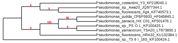
[30] | Dereeper A, Guignon V, Blanc G, et al. (2008) Phylogeny.fr: robust phylogenetic analysis for the non-specialist. Nucleic Acids Res 36: W465–W469. |
[31] |
Dereeper A, Audic S, Claverie JM, et al. (2010) BLAST-EXPLORER helps you building datasets for phylogenetic analysis. BMC Evol Biol 10: 8. doi: 10.1186/1471-2148-10-8
![]() |
[32] | Turner RJ, Weiner JH, Taylor DE (1998) Selenium metabolism in Escherichia coli. Biometals 11: 223–227. |
[33] |
Heider J, Bröck A (1993) Selenium metabolism in microorganisms. Adv Microb Physiol 35: 71–109. doi: 10.1016/S0065-2911(08)60097-1
![]() |
[34] | Macy JM, Rech S, Auling G, et al. (1993) Thauera selenatis gen. nov., sp. nov., a member of the beta subclass of Proteobacteria with a novel type of anaerobic respiration. Int J Syst Evol Micr 43: 135–142. |
[35] |
Gharieb MM, Gadd GM (2004) The kinetics of 75[Se]-selenite uptake by Saccharomyces cerevisiae and the vacuolization response to high concentrations. Mycol Res 108: 1415–1422. doi: 10.1017/S0953756204001418
![]() |
[36] | Kari C, Nagy Z, Kovácz P, et al. (1971) Mechanism of the growth inhibitory effect of cysteine on Escherichia coli. J Gen Microbiol 68: 349–356. |
[37] |
Harrison G, Curle C, Laishley EJ (1984) Purification and characterization of an inducible dissimilatory type sulfite reductase from Clostridium pasteurianum. Arch Microbiol 138: 72–78. doi: 10.1007/BF00425411
![]() |
[38] | DeMoll-Decker H, Macy JM (1993) The periplasmic nitrite reductase of Thauera selenatis may catalyze the reduction of selenite to elemental selenium. Arch Microbiol 160: 241–247. |
[39] |
Hudman JF, Glenn AR (1984) Selenite uptake and incorporation by Selenomonas ruminantium. Arch Microbiol 140: 252–256. doi: 10.1007/BF00454937
![]() |
[40] |
Brown TA, Shrift A (1980) Assimilation of selenate and selenite by Salmonella typhimurium. Can J Microbiol 26: 671–675. doi: 10.1139/m80-117
![]() |
[41] |
Smith FW, Hawkesford MJ, Prosser IM, et al. (1995) Isolation of cDNA from Saccharomyces cerevisiae that encodes a high-affinity sulfate transporter at the plasma membrane. Mol Gen Genet 247: 709–715. doi: 10.1007/BF00290402
![]() |
[42] |
Guzzo J, Dubow MS (2000) A novel selenite- and tellurite-inducible gene in Escherichia coli. Appl Environ Microb 66: 4972–4978. doi: 10.1128/AEM.66.11.4972-4978.2000
![]() |
[43] |
Yamada A, Miyashita M, Inoue K, et al. (1997) Extracellular reduction of selenite by a novel marine photosynthetic bacterium. Appl Microbiol Biotechnol 48: 367–372. doi: 10.1007/s002530051064
![]() |
[44] |
Bryant RD, Laishley EJ (1988) Evidence for two transporters of sulfur and selenium oxyanions in Clostridium pasteurianum. Can J Microbiol 34: 700–703. doi: 10.1139/m88-118
![]() |
[45] |
Hartke A, Bouche S, Laplace JM, et al. (1995) UV-inducible proteins and UV-induced cross-protection against acid, ethanol, H2O2 or heat treatments in Lactococcus lactis subsp. lactis. Arch Microbiol 163: 329–336. doi: 10.1007/BF00404205
![]() |
[46] |
Nepple BB, Bachofen R (1997) Induction of stress proteins in the phototrophic bacterium Rhodobacter sphaeroides. FEMS Microbiol Lett 153: 173–180. doi: 10.1111/j.1574-6968.1997.tb10479.x
![]() |
[47] | Keto-Timonen R, Hietala N, Palonen E, et al. (2016) Cold shock proteins: A minireview with special emphasis on Csp-family of enteropathogenic Yersinia. Front Microbiol 7. |
[48] |
Van FV, Chasteen TG, Pickering IJ, et al. (2000) Fate of selenate and selenite metabolized by Rhodobacter sphaeroides. Appl Environ Microb 66: 4849–4853. doi: 10.1128/AEM.66.11.4849-4853.2000
![]() |
1. | Merja Lusa, Jenna Knuutinen, Marcus Lindgren, Juhani Virkanen, Malin Bomberg, Microbial communities in a former pilot-scale uranium mine in Eastern Finland – Association with radium immobilization, 2019, 686, 00489697, 619, 10.1016/j.scitotenv.2019.05.432 | |
2. | Merja Lusa, Hanna Help, Ari-Pekka Honkanen, Jenna Knuutinen, Joni Parkkonen, Dominika Kalasová, Malin Bomberg, The reduction of selenium(IV) by boreal Pseudomonas sp. strain T5-6-I – Effects on selenium(IV) uptake in Brassica oleracea, 2019, 177, 00139351, 108642, 10.1016/j.envres.2019.108642 | |
3. | Ari-Pekka Honkanen, Sami Ollikkala, Taru Ahopelto, Antti-Jussi Kallio, Merja Blomberg, Simo Huotari, Johann-type laboratory-scale x-ray absorption spectrometer with versatile detection modes, 2019, 90, 0034-6748, 033107, 10.1063/1.5084049 | |
4. | Merja Lusa, Malin Bomberg, Microbial Community Composition Correlates with Metal Sorption in an Ombrotrophic Boreal Bog: Implications for Radionuclide Retention, 2021, 5, 2571-8789, 19, 10.3390/soilsystems5010019 | |
5. | Eva Garcia-Lopez, Ana Moreno, Miguel Bartolomé, Maria Leunda, Carlos Sancho, Cristina Cid, Glacial Ice Age Shapes Microbiome Composition in a Receding Southern European Glacier, 2021, 12, 1664-302X, 10.3389/fmicb.2021.714537 | |
6. | Roberto Avendaño, Said Muñoz‐Montero, Diego Rojas‐Gätjens, Paola Fuentes‐Schweizer, Sofía Vieto, Rafael Montenegro, Manuel Salvador, Rufus Frew, Juhyun Kim, Max Chavarría, Jose I. Jiménez, Production of selenium nanoparticles occurs through an interconnected pathway of sulphur metabolism and oxidative stress response in Pseudomonas putida KT2440 , 2023, 1751-7915, 10.1111/1751-7915.14215 | |
7. | Adriana Riveros, Benhur K. Asefaw, Qingshi Wang, Tahir Maqbool, Youneng Tang, Daqian Jiang, Selenium treatment via integrating flow electrode capacitive deionization (FCDI) and bio-electrochemical systems (BES), 2024, 00431354, 122844, 10.1016/j.watres.2024.122844 |
Macronutrient solutions | Selenite solutions | Selenate solutions | |||
E1 | 6 mM NO2– | E5 | 6 mM SeO32– | E10 | 6 mM SeO42– |
E2 | 6 mM NO3– | E6 | 3 mM SeO32– + 3 mM NO2– | E11 | 3 mM SeO42– + 3 mM NO2– |
E3 | 6 mM SO32– | E7 | 3 mM SeO32– + 3 mM NO3– | E12 | 3 mM SeO42– + 3 mM NO3– |
E4 | 6 mM SO42– | E8 | 3 mM SeO32– + 3 mM SO32– | E13 | 3 mM SeO42– + 3 mM SO32– |
E9 | 3 mM SeO32– + 3 mM SO42– | E14 | 3 mM SeO42– + 3 mM SO42– |