Citation: Arsenio M. Fialho, Nuno Bernardes, Ananda M Chakrabarty. Exploring the anticancer potential of the bacterial protein azurin[J]. AIMS Microbiology, 2016, 2(3): 292-303. doi: 10.3934/microbiol.2016.3.292
[1] | Jinhu Xu, Yicang Zhou . Bifurcation analysis of HIV-1 infection model with cell-to-cell transmission and immune response delay. Mathematical Biosciences and Engineering, 2016, 13(2): 343-367. doi: 10.3934/mbe.2015006 |
[2] | Shunyi Li . Hopf bifurcation, stability switches and chaos in a prey-predator system with three stage structure and two time delays. Mathematical Biosciences and Engineering, 2019, 16(6): 6934-6961. doi: 10.3934/mbe.2019348 |
[3] | Huan Kong, Guohong Zhang, Kaifa Wang . Stability and Hopf bifurcation in a virus model with self-proliferation and delayed activation of immune cells. Mathematical Biosciences and Engineering, 2020, 17(5): 4384-4405. doi: 10.3934/mbe.2020242 |
[4] | Yuan Ma, Yunxian Dai . Stability and Hopf bifurcation analysis of a fractional-order ring-hub structure neural network with delays under parameters delay feedback control. Mathematical Biosciences and Engineering, 2023, 20(11): 20093-20115. doi: 10.3934/mbe.2023890 |
[5] | Bruno Buonomo, Marianna Cerasuolo . The effect of time delay in plant--pathogen interactions with host demography. Mathematical Biosciences and Engineering, 2015, 12(3): 473-490. doi: 10.3934/mbe.2015.12.473 |
[6] | Hongfan Lu, Yuting Ding, Silin Gong, Shishi Wang . Mathematical modeling and dynamic analysis of SIQR model with delay for pandemic COVID-19. Mathematical Biosciences and Engineering, 2021, 18(4): 3197-3214. doi: 10.3934/mbe.2021159 |
[7] | Shishi Wang, Yuting Ding, Hongfan Lu, Silin Gong . Stability and bifurcation analysis of SIQR for the COVID-19 epidemic model with time delay. Mathematical Biosciences and Engineering, 2021, 18(5): 5505-5524. doi: 10.3934/mbe.2021278 |
[8] | Sarita Bugalia, Jai Prakash Tripathi, Hao Wang . Mathematical modeling of intervention and low medical resource availability with delays: Applications to COVID-19 outbreaks in Spain and Italy. Mathematical Biosciences and Engineering, 2021, 18(5): 5865-5920. doi: 10.3934/mbe.2021295 |
[9] | J. Leonel Rocha, Sandra M. Aleixo . An extension of Gompertzian growth dynamics: Weibull and Fréchet models. Mathematical Biosciences and Engineering, 2013, 10(2): 379-398. doi: 10.3934/mbe.2013.10.379 |
[10] | Xiaomeng Ma, Zhanbing Bai, Sujing Sun . Stability and bifurcation control for a fractional-order chemostat model with time delays and incommensurate orders. Mathematical Biosciences and Engineering, 2023, 20(1): 437-455. doi: 10.3934/mbe.2023020 |
For nonconducting solids, Smoluchowski [1] derived equations that are valid for any shape of a particle or pores inside a solid, provided the (local) curvature radius, aparticle, largely exceeds the Debye length, λD = κ−1, where k is derived from the Poisson-Boltzmann theory in the Debye-Hückel approximation [2]. For 1:1 NaCl electrolyte with bulk concentration cNaCl at T = 25 ℃ the Debye length equals to:
λD≅0.304/√cNaCl(in nm) | (1) |
where cNaCl is the concentration of the salt, in mol/dm3
The simplification that the surface charge is uniformly smeared along the surface renders the governing equations one-dimensional and, thereby, greatly facilitates their solution. The classical Smoluchowski equation was derived under two important approximations [1]: (i) the diffuse layer is rather thin as compare to the capillary radius, acapillary, that is, kacapillary » 1 and (ii) ζ potential of the surface is low, that is, the electrical energy is small as compared to thermal energy |ζze| < kBT, where z is the valence of ions (assuming symmetric electrolyte, that is, z+ = z- = z), e is the electron charge, kB is the Boltzmann constant, and T is absolute temperature. For ambient temperature of 25 ℃ in water it translates to |ζ| < 25.7 mV for the 1:1 electrolyte. Theoretical treatments are available for calculation of streaming current in the case of cylindrical capillary [3] and for the case of a close packing of spheres [4,5].
Borghi et al. [6] observed a remarkable reduction by several pH units of the isoelectric point (IEP) on rough nanostructured surfaces, with respect to flat crystalline rutile TiO2. In order to explain the observed behavior of IEP, Borghi et al. [6] considered the roughness-induced self-overlap of the electrical double layers as a potential source of deviation from the trend expected for flat surfaces. We recently reported an increase by about three pH units in the values of point of zero charge (PZC) of AlOOH-coated siliceous particles seen in Figure 1 with nanoscale roughness [7] as compare to smooth [8] and AlOOH-coated silica surfaces with (unspecified) nanoscale roughness [9].
Numerical models for pressure driven flow with an arbitrary but periodic patch-wise heterogeneous surface pattern over a flat heterogeneous region perpendicular to the flow axis have been developed for 2D [10,11] and for 3D [12,13,14] flows. The models were based on a simultaneous solution to the Nernst-Planck, Poisson-Boltzmann, and Navier-Stokes equations to determine the local ionic concentration, velocity vectors and electrostatic potential contours in the double layer distribution. The presence of a heterogeneous patch is shown to induce flow in all three coordinate directions, including a circulation pattern perpendicular to the main flow axis [10,11,12,13,14]. The normal velocities in the 2D case [10] increase linearly with pressure drop and increase with the square of the difference in magnitude between the surface charge densities of two distinctly charged patches. These velocities vary with the fifth power of the Debye length, i.e., λ5 [10]. The difference in magnitude between the charge densities, |σ1| and |σ2|, generates the normal velocities in 2D case and the subsequent electric field distortion. However, more important than just the values of |σ1| and |σ2|, is the difference in their magnitudes, Ds, defined as
Δσ=‖σ1‖−‖σ2‖ | (2) |
Colloid stability is often predicted by the Derjaguin-Landau-Verwey-Overbeek (DLVO) theory[15,16], which was developed for smooth, homogeneous particles with ideal geometries and with no DL overlap. Despite the success of DLVO theory, numerous investigators have modified the DLVO theory to include factors not accounted for in the DLVO model since the real colloidal systems generally contain some degree of nonuniformity, in the form of surface roughness and/or chemical heterogeneities [17,18,19,20,21,22]. The general impact of roughness is to amplify the long range behavior of noncontact DLVO forces with the approximate amplification factor of exponential force between a rough and smooth surfaces of exp (σroughness2/λDebye2), where σroughness is the mean roughness [20]. Significant work has been focused on extending the precepts of the traditional DLVO model to accommodate these non-DLVO forces (energies). The effect of hemispherical asperities on the long range van der Waals and electrostatic interactions between a rough surface and smooth colloidal particle can further contribute to the distortion [19]. A review paper [22] discussed many of the interactions that play a role in environmental systems and are not commonly included in the traditional DLVO model: e.g., hydrogen bonding and the hydrophobic effect, hydration pressure, non-charge transfer Lewis acid base interactions, and steric interactions.
The purpose of this paper is to investigate the effect of nanoscale surface roughness on filtration of micron and sub-micron particles from aqueous solution by AlOOH-coated siliceous particles associated with pressure-driven flow through micrometer size pores of non-woven filter media and aluminized diatomaceous earth (DE) particles packed into a short column.
Experimental data suggest that the generation of flow velocities in all three coordinate directions, including a circulation pattern perpendicular to the main flow axis results in exceptionally high microbes and ions removal efficiencies from aqueous suspension by the alumina's surfaces with combined microscale and nanoscale structures.
The TEM images were recorded on the JEOL JEM2010 TEM at 200 kV accelerating voltage. No coating was applied. The SEM images were recorded on the JEOL JSM6335F SEM at 5 kV. Sample preparation for a nonconductive materials seen in Figure 1 was by applying a gold-palladium alloy by sputtering in vacuum to a thickness of a few nanometers ( < 5 nm). A coating of this thickness would not be visible in images at this magnification. Figure 1a and 1b shows a TEM and SEM images of the alumina nanofibers, 2 nm in diameter and approximately 0.25 μm long that are electroadhesively grafted to a microglass fiber at 40 weight percent (wt%) AlOOH loadings bonded to a 0.6 mm diameter glass microfiber. The alumina nanofibers appear as a fuzzy layer on the microglass core. The active component has been classified as quantum wire [7].
The active component in the filter media of Ref. [7] is a nanolayer in the form of monocrystalline oxide/hydroxide (γ-AlOOH) with a thickness of approximately 1.2 nm electroadhesively deposited onto siliceous support material, thereby forming a highly positive composite. The active component has been classified as quantum dots rather than 2D continuous nanocoating [7].
For the purpose of this study the alumina nanofiber content bonded to 0.6 μm glass microfiber scaffolding seen in Figure 1a and 1b were optimized on 20, 40 and 60 wt% of alumina loadings following a procedure described in Example 4 of Ref. [23]. No other fibers or binders were added to the mulch.
The filter media is manufactured and sold as non-woven media by Ahlstrom Filtration LLC under the tradename DisruptorTM. Argonide Corporation manufactures the filter cartridges from Disruptor media. Polymeric fibers, primarily polyester are added for formability and pleatability. The alumina nanofibers completely occupy the available surface of the 0.6 μm glass microfiber scaffolding. The pore size of the media can be varied from about 1 to 30 μm, although a 1.0 ± 0.3 μm pore size was selected for purifying drinking water from sub-micron particles (see Table 1 below) and a 4 ± 2 μm pore size was selected as a low pressure drop prefilter to the drinking water filter element (see Table 1 below). The filter media's thicknesses are about 0.8 mm and 1.2 mm thick for drinking water filter and prefilter respectively, resulting in approximately 800 and 300 pores correspondently that a particle must transit before exiting as filtrate. The media have a rapid dynamic response for adsorbing bacteria, virus and nucleic acids from biological solutions. Typically 5 LRV (logarithm reduction value (Eq. (3)) of bacteria and 4 LRV of viruses are retained by a pleated layer at flow velocity 0.7 mm/s at a ΔP of 2-6 kPa for prefilter and 15 kPa for drinking water filter media where:
Media | Grade | Thickness (mm) | Mean particle sizea (μm) | Bubble point (kPa) | Mean flow pore diameter, dmean flow (μm) | Largest pore diameter, dlargest (μm) |
Non-woven a | Prefilter media c | 1.2 | ~1 a | 9.8 ± 0.8 e | 2.3 ± 0.3 | 29 ± 2 e |
Drinking water filter media d | 0.8 | ~1 a | 20 ± 2 e | 0.7 ± 0.3 | 14 ± 1 e | |
Aluminized DE b | Fine grade aluminized DE | 10 | 3.0 ± 0.5 | |||
Medium grade aluminized DE | 18 | 5.5 ± 1.5 | ||||
Coarse grade aluminized DE | 80 | 24.0 ± 3.5 | ||||
Notes: a Figure 1a and 1b; b Figure 1c and 1d; c Disruptor grade 5292; d Disruptor grade 5283; e Average value for 10 cartridges. |
LRV=−log10(Cfinal/Cinitial) | (3) |
where Cinitial and Cfinal are the initial and final concentrations of particles.
The precoats were formed using either a Nalgene filter holders with receiver (available from Cole-Palmer, Cat # S-06730-53) or a 25 mm diameter syringe filter holder (available from VWR, Cat # 28144-109). The precoats have surface area of ~10 cm2 and ~3.7 cm2, respectively, and are ~3 mm thick.
Monodispersed polystyrene latex microspheres (beads) coated with sulfate groups with a mean diameter of 30 ± 5 nm were obtained from Duke Scientific Corporation. According to the manufacturer, the spheres have a net negative charge in aqueous solution. Particles were used as supplied by the manufacturer and re-suspended in DI RO water whose conductivity is less than 0.1 μS/cm. The charge is provided by a mixture of sulfate and hydroxyl groups on the latex surface with zeta potential of approximately z ≅-82 mV [24].
Suspensions of MS2 and fr bacteriophages with titers of ~1012 particles/mL were prepared as described in Ref. [25], stored at 4 ℃, and then diluted in deionized reverse osmosis (DI RO) water with conductivity less than 0.1 μS/cm. MS2 (ATCC 15597-B1) and fr (ATCC 15767-B1) bacteriophages are icosahedral in shape with a diameter of 26-27 nm [26] and 19 nm [27], respectively. The isoelectric point (IEP) of fr bacteriophage is 8.9-9.0 [27]. In this study, MS2 bacteriophage was chosen because it is often used as a surrogate for human enteric viruses and fr bacteriophage was chosen because it is positive in aqueous suspensions at pH range of 3-9. The zeta potential of MS2 (ζ ≅-(33-38) mV [28]).
Stocks of E. coli (ATCC 15597), Raoultella terrigena (ATCC 33257) and B. diminuta (ATCC 19146) with titers of ~109 particles/mL were prepared and stored at 4 ℃, and then diluted in DI RO water with conductivity less than 0.1 μS/cm. E. coli bacterium has a cell IEP of 3.5 and zeta potential of ζ ≅-35 mV. Zeta potential is fairly insensitive within a pH range of 6 to 9 [29]. However, it should be noted that the zeta-potential is physically meaningful only for the so-called hard colloids. Instead, the bioparticles are models of soft colloids, i.e., colloids where charges are three-dimensionally distributed [30]. For example, the IEP value of MS2 bacteriophage in different environments based on averaging of 10 measurements shows a mean IEP value of 3.5 ± 0.6 and with the discrepancy in IEP (ΔIEP) of 1.8 [31]. Two recent studies by Duek et al. [26] and Chrysikopoulos et al. [28] determined the alteration of the IEP by a change in water chemistry (e.g., ionic strength or ionic composition) with IEP of the former case in the range from less than 2 at ionic strengths of 0.0 and 2.6 mM to IEP of 4.4 in the latter case that was measured in double distilled water. Consequently, zeta-potential and IEP parameters should be interpreted very carefully for viruses with their adhesion capacity onto charged abiotic surfaces [30].
In the streaming potential process, electrolyte solution is forced through the filtration media pores by applying a pressure drop, ΔP. Because the walls of the channel are charged, the fluid adjacent to the surface bears an equal but opposite charge density, and the hydrodynamic flow carries a convection countercurrent. The net current through the cross-sectional area of the pore must be zero so that a voltage differential, Δϕ, is generated.
Zeta potential is an important and reliable indicator of the surface charge of filters. It can be deduced from experimental data, e.g., from measuring the streaming potential. It has been long recognized [32,33,34] that in the plug flow and in the networks of fibrous materials situations, the apparent zeta potential (ζapparent) is a function of bulk conductivity of filtered liquid (KL) while the true zeta potential (ζtrue) is a function of bulk conductivity of filtered liquid (KL) as well as surface conductance (Ks). The true zeta potential and specific surface conductance can be evaluated by plotting of reciprocal of the apparent zeta potential as a function of the resistivity of water. The apparent zeta potential (ζapparent) was calculated using the weighted least-squared procedure [35] for at least three different conductivities of water adjusted with NaCl
Estreaming=ε0εζapparentΔpμKL | (4) |
where μ is the dynamic viscosity of the electrolyte solution (for water at 25 ℃ is equals to ~890.02 μPa·s [36]) and Δp is the differential pressure across a packed bed or non-woven media.
The true zeta potential (ζtrue) has contributions from both, the surface (Kσ) and bulk (KL) conductions (see equation 4.3.41 of Ref. [37]):
Estreaming=ε0εζtrueΔpμ(KL+2Kσ/asurface) | (5) |
After re-arraignment we obtain:
1/ζapparent=1/ζtrue+2Kσ/ζtrueasurfaceR | (6) |
where R is the resistivity of the water (in Ω·m) being filtered (by definition R = 1/KL). Another way to determine the true zeta potential would be to perform additional electrical resistance measurements at high ionic strength[38].
The streaming potential is measured by means of a pair of Ag/AgCl electrodes located on both sides of two parts of a channel (ID = 19 mm) separated by a septum (three layers of two dimensional filter media having an irregular pore pattern and an irregular pore size of 0.1-0.2 mm. The aluminized DE powder was packed inside the tube on the septum with depth of 3.5-4.0 cm). A solution of a known conductivity, as measured by Oakton ECTestr Pure, is placed in a pressurized, aluminized DE-packed column and pressure in the range from 7 to 70 kPa is applied to the vessel and therefore the pressure is transferred to the filter media. The streaming potential is found by applying at a given time an over-pressure (ΔP, bar) to the vessel and measuring the resulting potential difference (Δφ ≡ Estreaming, V) on both sides of the filter bed. The Estreaming and ΔP values are measured at ± 0.1 mV and ± 700 Pa, respectively. The bulk conductivity of the solutions ranged from 2.9 μS/cm for 0.03 mmol/dm3 NaCl to 12.96 mS/cm for 0.12 mol/dm3 NaCl solutions in deionized reverse osmosis (DI RO) water with initial conductivity less than 0.1 μS/cm. The pH values were adjusted with 1 mol/dm3 NaOH or with 1 mol/dm3 HCl.
A thoroughly wetted and capped from both ends filter cartridge is immersed into a tank filled with water and air is exerted into the capped cavity. The upstream air pressure is then slowly increased, eventually resulting in the selective intrusion of air through the largest pores and formation of an air bubbles stream [39]. The pressure is recorded at the point where the first stream of bubbles is seen in the effluent. The bubble point values were determined for both, the prefilter and drinking water filter cartridges (available from Argonide Corporation, USA) used in present work. The largest pore diameter, dlargest, was determined assuming that the surface tension of the water within the media at T = 25 oC is γ = 71.97 mN/m [36] and boehmite surface is hydrophilic with the contact angle θ = 0o.
dlargest=4γcosθ/DPbubble point | (7) |
The mean flow pore diameter, d mean flow, was determined as described in Ref. [40,41]:
dmeanflow=√32B/ε | (8) |
where permeability B = vμz/ΔP; v is the flow velocity through the media; μ is the viscosity; z is the thickness of the media; ΔP is the pressure drop across the media, and ε is the porosity.
Table 1 shows values of the mean flow and largest flow pore diameter of the prefilter and drinking water cartridges as well as the mean flow pore diameter of aluminized DE powders packed in empty refillable canisters (available from Watts Filtration, Cat # RC-C-975).
The filter's active component is a nanoalumina fiber with an external surface area of ~500 m2/g (see Figure 1a and 1b). It has been identified as crystalline boehmite (AlOOH) using x-ray diffraction [7]. Boehmite nanofibers create high zeta potential (ζtrue ≥ 46 mV) in aqueous solutions in the pH range of 3-8. The point of zero charge (PZC) of alumina non-woven media was determined to be 11.6 ± 0.2 [7].
Figure 2 shows breakthrough curves of 30 nm size latex spheres (beads) by a 0.8 mm thick layer of nanoalumina filter of at a flow velocity of 0.17 mm/s. The sorption capacity is approximately proportional to the nanoalumina fiber loading.
Table 2 shows correlation of zeta potential with bacteriophages, and bacteria removal efficiencies by one and two layer of 35 wt% nanoalumina/microglass media at flow velocity of 0.7 mm/s.
pH | z potential (mV) | fr removal efficiency, LRV b | MS2 removal efficiency, LRV b | PRD-1 removal efficiency, LRV b | E. coli removal efficiency, LRV b | B. diminuta removal efficiency, LRV b | |
1 layer c | 2 layers c | 2 layers c | 2 layers c | 2 layers c | 2 layers c | ||
3 | 46 ± 7 a | 3.0 ± 0.3 | 5.0 ± 0.3 | ||||
5 | 46 ± 7 a | > 6.2 | > 6.2 | > 5.0 | |||
7 | 47 ± 9 a | > 6.2 | > 6.2 | > 6.0 | > 7.0 | > 7.5 | > 7.6 |
8 | 46 ± 6 a | > 6.1 | > 6.1 | > 5.0 | 6.3 | > 6.2 | |
9 | 26 ± 4 a | > 6.0 | > 6.0 | > 5.0 | |||
10 | 23 ± 5 a | 4.2 ± 0.3 | > 6.0 | ||||
11 | 22 ± 5 a | 0.2 ± 0.2 | 1.5 ± 0.7 | ||||
Notes: a Ref. [7]; b Logarithm reduction value; c By 1-or 2-layers of drinking water filter media (Table 1) at flow velocity of 0.7 mm/s. |
Initial removal efficiencies are high for both bacteriophages and bacteria. Most bacteria and most other contaminants encountered in nature are negatively charged in water with notable exception of the fr bacteriophage that is positive at pH less than about 8.9-9.0 [27]. Yet, the initial removal efficiency for the fr bacteriophage at pH = 7 is high as well (see Table 2), i.e., > 99.9999% with a double layer of the media at a flow velocity 0.7 mm/s.
Table 3 shows removal efficiencies of MS2 bacteriophage and RT bacteria at neutral pH = 7 by a single layer of non-woven prefilter media in a form of pleated cartridge with surface area of 0.3 m2 as a function of flow velocity and electrolyte ionic strength.
Electrolyte ionic strength, NaCl (mM) | λD a (nm) | Flow velocity (mm/s) | Contact time (s) | Brownian displacemen b of RT (mm) | Brownian displacemen b of MS2 (mm) | MS2 removal efficiency, LRV c | RT removal efficiency, LRV c |
0.002(1) d | 215 | 0.05 | 22.5 | 4.7 | 28 | > 6.4 | 6.4 ± 0.7 |
0.2 | 5.6 | 2.4 | 14 | > 6.4 | 6.2 ± 1.0 | ||
0.8 | 1.4 | 1.2 | 7.0 | > 6.4 | 5.8 ± 0.4 | ||
3.4 | 0.35 | 0.6 | 3.5 | > 6.4 | 6.0 ± 0.5 | ||
0.05(1) d | 43 | 0.05 | 22.5 | 4.7 | 28 | > 5.7 | 5.9 ± 0.2 |
0.2 | 5.6 | 2.4 | 14 | > 5.7 | 5.8 ± 0.2 | ||
0.8 | 1.4 | 1.2 | 7.0 | > 5.7 | 5.6 ± 0.2 | ||
3.4 | 0.35 | 0.6 | 3.5 | > 5.7 | 5.2 ± 0.6 | ||
0.35(4) d | 16 | 0.05 | 22.5 | 4.7 | 28 | > 5.2 | 5.8 ± 0.2 |
0.2 | 5.6 | 2.4 | 14 | > 5.2 | 5.3 ± 0.2 | ||
0.8 | 1.4 | 1.2 | 7.0 | > 5.2 | 5.2 ± 0.4 | ||
3.4 | 0.35 | 0.6 | 3.5 | > 5.2 | 5.0 ± 0.8 | ||
1.0(1) d | 9.6 | 0.05 | 22.5 | 4.7 | 28 | > 6.6 | 5.7 ± 0.4 |
0.2 | 5.6 | 2.4 | 14 | > 6.6 | 5.2 ± 0.5 | ||
0.8 | 1.4 | 1.2 | 7.0 | 5.6 ± 0.2 | 4.8 ± 0.5 | ||
3.4 | 0.35 | 0.6 | 3.5 | 5.1 ± 0.2 | 4.4 ± 0.5 | ||
3.0(3) d | 5.5 | 0.05 | 22.5 | 4.7 | 28 | > 5.8 | 4.9 ± 0.3 |
0.2 | 5.6 | 2.4 | 14 | > 5.8 | 4.6 ± 0.3 | ||
0.8 | 1.4 | 1.2 | 7.0 | > 5.8 | 4.2 ± 0.7 | ||
3.4 | 0.35 | 0.6 | 3.5 | 5.2 ± 0.6 | 4.3 ± 0.9 | ||
10(1) d | 3.0 | 0.05 | 22.5 | 4.7 | 28 | > 5.9 | 4.2 ± 0.2 |
0.2 | 5.6 | 2.4 | 14 | > 5.9 | 4.1 ± 0.2 | ||
0.8 | 1.4 | 1.2 | 7.0 | > 5.9 | 3.7 ± 0.3 | ||
3.4 | 0.35 | 0.6 | 3.5 | 4.7 ± 0.3 | 3.7 ± 0.4 | ||
40(4) d | 1.5 | 0.05 | 22.5 | 4.7 | 28 | 4.8 ± 0.2 | 3.9 ± 0.2 |
0.2 | 5.6 | 2.4 | 14 | 5.1 ± 0.3 | 3.7 ± 0.4 | ||
0.8 | 1.4 | 1.2 | 7.0 | 4.7 ± 0.2 | 3.1 ± 0.4 | ||
3.4 | 0.35 | 0.6 | 3.5 | 3.1 ± 0.2 | 2.8 ± 0.5 | ||
100(10) d | 0.96 | 0.05 | 22.5 | 4.7 | 28 | > 4.5 | 4.0 ± 0.5 |
0.2 | 5.6 | 2.4 | 14 | > 4.5 | 3.7 ± 0.4 | ||
0.8 | 1.4 | 1.2 | 7.0 | > 4.5 | 3.1 ± 0.4 | ||
3.4 | 0.35 | 0.6 | 3.5 | 3.5 ± 0.3 | 2.8 ± 0.4 | ||
250(25) d | 0.61 | 0.05 | 22.5 | 4.7 | 28 | 4.2 ± 1.0 | 3.1 ± 0.2 |
0.2 | 5.6 | 2.4 | 14 | 3.7 ± 0.3 | 2.7 ± 0.2 | ||
0.8 | 1.4 | 1.2 | 7.0 | 3.0 ± 0.2 | 2.5 ± 0.2 | ||
3.4 | 0.35 | 0.6 | 3.5 | 1.2 ± 0.2 | 1.8 ± 0.2 | ||
510(51) d | 0.43 | 0.05 | 22.5 | 4.7 | 28 | 2.3 ± 0.6 | 2.4 ± 0.6 |
0.2 | 5.6 | 2.4 | 14 | 1.8 ± 0.2 | 2.2 ± 0.4 | ||
0.8 | 1.4 | 1.2 | 7.0 | 1.8 ± 0.4 | 1.9 ± 0.2 | ||
3.4 | 0.35 | 0.6 | 3.5 | 1.0 ± 0.5 | 1.5 ± 0.4 | ||
3000(300) d | 0.18 | 0.05 | 22.5 | 4.7 | 28 | 2.1 ± 0.3 | 2.0 ± 0.3 |
0.2 | 5.6 | 2.4 | 14 | 1.6 ± 0.3 | 1.6 ± 0.2 | ||
0.8 | 1.4 | 1.2 | 7.0 | 1.3 ± 0.3 | 1.4 ± 0.2 | ||
3.4 | 0.35 | 0.6 | 3.5 | 1.3 ± 0.5 | 1.0 ± 0.2 | ||
Notes: a Calculated with the use of equation (1); b Calculated with the use of equation (9); c Logarithm reduction value; d Error limits in parentheses are accuracy in the units of the last digit reported. |
The electrolyte ionic strength was adjusted with NaCl salt (purity > 99.0%) that was added to distilled water (DW) with conductivity less than 0.1 mS/cm. An aliquot of 15-20 mL of MS2 suspension was added whose input concentration was ~5.0 × 10⁹ particles/mL with a conductivity of ~28 μS/cm together with an aliquot of 12 mL of washed in DW water RT bacteria whose input concentration was ~5.0 × 10⁹ particles/mL to 50 L of the DW water. After passing 5 liters of the suspension at a specified flow rate, a 250 mL aliquot was collected. The sample was serially diluted by a factor of 10 and analyzed as plaque forming units (PFU) for counting MS2 bacteriophages with the use of the double-layer method [25,42]. Briefly, dilutions of each sample were mixed with host cells, plated on nutrient agar, and incubated overnight. The resulting plaque counts were converted to PFU/mL. The RT bacteria was enumerated with the use of a membrane method [43] for counting coliform forming units (CFU).
Results of Table 3 indicate that the MS2 and RT removal efficiencies do not depend within 2s accuracy on the flow velocity in the range from 0.05 to 3.4 mm/s and at contact times from 0.35 to 22.5 seconds for electrolyte's ionic strength less than 1 mmol. Application of the Stokes-Einstein equation [44]:
⟨x⟩2Brownian/2t=kBT/6πμa | (9) |
to a spherical particle suspended in water (μ = 890.02 μPa·s at 25 ℃ [36]) produces a root-mean-square (rms) displacements for MS2 virus and RT bacteria (see Table 3) that are much lesser than the radius of the largest pore of alargest = 15 ± 1 mm (see Table 1). Yet, the removal efficiencies at ionic strengths less or equal to 0.35 mM is greater than 99.999%.
Such a behavior can be described based on the characteristic effect of the local EDL field distortion by generating of circulating velocity pattern normal to the flow direction.
Aluminum oxide-hydroxide nanolayer (AlOOH) with estimated thickness of 1.2 nm (see Eqs. (13)-(17) of Ref. [7]) was deposited onto siliceous DE support material as described in Ref. [45]. Figure 3 indicates that aluminized DE18 powders with average particle size of 18 mm are highly positive (ζ > +50 mV) at aluminum oxide/hydroxide solids loading greater than ~20 wt% as compared to the highly negative character of the bare DE18 support (ζ~-70 mV). Alumina's nanolayer surface can be further functionalized by adding other sub-micron or nano-size particles to target a specific contaminant. For example, aluminized DE coated with nano-size iron monohydrate (FeOOH) yields an arsenic sorbent that shows high sorption capacity (see Examples 15 and 16 of Ref. [45]).
Similar behavior to that described in Section 3.1.3 was observed while adsorbing soluble 59Co isotope in short packed columns of aluminized DE60 (see Table 4) with average particle size of 48 mm and mean flow pore size of dmean flow = 14.5 ± 2.5 mm (see Table 1) where the adsorption efficiency does not depend on flow velocity in the range from 3.3 to 10 mm/s and at contact times from 5 to 60 seconds. Similarly to the microbial removal efficiencies such a behavior can be described based on the characteristic effect of local EDL field distortion by generating of circulating velocity pattern normal to the flow direction.
Bed volume (dm3) | pH | Column depth (mm) | Flow velocity (mm/s) | Contact time (s) | Input 59Co concentration (ppb) | Output 59Co concentration (ppb) | Removal efficiency (%) |
0.16 | 10 | 50 | 10 | 5.0 | 635 | < 5 | > 99.2 |
0.32 | 10 | 100 | 6 | 17 | 635 | < 5 | > 99.2 |
0.64 | 10 | 200 | 3.3 | 60 | 635 | < 5 | > 99.2 |
0.16 | 8 | 50 | 10 | 5.0 | 656 | < 5 | > 99.2 |
0.32 | 8 | 100 | 6 | 17 | 656 | < 5 | > 99.2 |
0.64 | 8 | 200 | 3.3 | 60 | 656 | < 5 | > 99.2 |
The incorporation of a nonuniform surface charge in the streaming potential process beyond the one-dimensional analysis and by not restricting the solution to low charges was first undertaken in Refs. [10,11]. Particularly, it was shown how the nonuniform surface charge will affect the value of the zeta potential by over 30% higher than expected value due to a severe distortion of Poisson-Boltzmann EDL field especially at the ionic strengths less than 1 mM. Numerical model as well as a first order perturbation theory model for pressure driven flow through a slit with an arbitrary but periodic patchwise heterogeneous surface pattern has been developed for a 2D case [10,11]. The impact of bipolar surface charge distributions on pressure-driven transport has been investigated theoretically in Ref. [46] for systems with weakly charged nanopores with radius smaller than the Debye screening length.
The surfaces of DE80 and aluminized DE80 have similar but opposite values of ζ potentials. In such a case, there is an excess of positive ions over the DE surface but negative ions dominate over the aluminized DE surface to balance out the positive surface charge. The difference in the surface potential results in an electrostatic potential gradient within the double layer region near the transition zone between the two regions. The presence of this potential gradient results in a body force on the microbes and/or ions in the double layer; however, in this case an opposite body force is applied to the negative ions over the positively charged particle. As a result, the net body force over the region is zero and the circulation velocity perpendicular to the main flow becomes negligible. Table 5 provides experimental evidence of such behavior when mixing together the DE80 and aluminized DE80 powders, the resulting mix formed in a 3 mm precoat is losing properties of the aluminized DE tested in the same configuration. This is consistent with the case when one of the neighboring particles has opposite sign of the surface charge, i.e., -|σ2| for DE, than other neighboring particles, i.e., +|σ1| for aluminized DE (see Eq. (2)).
Furthermore, Table 5 provides an estimate of number of layers formed by aluminized DE particles arranged a group of four neighboring particles in the plane perpendicular to the flow in a 3 mm thick precoat as a function of weight percent of DE in the aluminized DE/DE mix. The estimate shows some correlation between a number of layers formed by four neighboring aluminized DE particles and the LRV values (see Eq. (3)) of MS2 virus and RT bacteria.
Percent of DE in aluminized DE/DE precoat | Number of layers formed by aluminized DE c | MS2 removal efficiency a, LRV b | RT removal efficiency a, LRV b |
0 | 40 | 6.1 ± 0.1 | > 7.7 |
10 | 38 | 5.1 ± 0.2 | > 7.7 |
25 | 16 | 3.6 ± 0.4 | 3.9 ± 0.3 |
50 | 7 | 2.6 ± 0.9 | 3.0 ± 0.3 |
75 | 2 | 1.0 ± 0.2 | 2.2 ± 0.1 |
100 | 0 | 0.5 ± 0.1 | 0.7 ± 0.2 |
Notes: a At flow velocity of 0.67 mm/s; b Logarithm reduction value; c Arranged in a group of four neighboring aluminized DE particles in the plane perpendicular to the flow. |
Figure 4 compares values of true zeta potential, ζtrue, and surface conductance, Kσ, of aluminized DE powder as well as non-woven drinking water filter media. In both cases, the Kσ values are greater by two order of magnitude at pH3 and pH12 as compare to the values at neutral pH in the range from pH = 6 to pH = 8. In addition, the Kσ values of aluminized DE are greater than these of non-woven filter media by a factor of 4.6 ± 3.0 over the pH range of 3-12.
This paper demonstrates how the positive charge properties of alumina's surfaces with combined microscale and nanoscale structures dispersed in aqueous solution can be used as a means of collecting submicron and micron particles such as viruses, bacteriophages, and bacteria. Because the nonuniformity in charge in the direction of the applied pressure drop, circulation velocities are generated that represents a symptom of electric field distortion. The larger the electric field distortion, the greater the deviation is for the zeta potential from its expected value for a smooth surface at a given pH. Experimental data suggest that the circulation velocities increase with applied pressure drop.
The most remarkable result of our studies is the observation of the shift of the point of zero charge (PZC) of quantum dot (aluminized DE, Figure 1c and 1d) and quantum wire (Figure 1a) assembled nanostructures in the form of monocrystalline oxide/hydroxide by more than three pH units towards more basic character with respect to reference g-AlOOH crystalline surfaces.
We thank Dr. R. Ristau of IMS, University of Connecticut for providing us with TEM and SEM images of alumina nanofibers bonded to glass and microfibers (Figures 1a and 1b) and Dr. A. Slesarev of Rice University for providing us with SEM, and TEM images of aluminized DE powders (Figures 1c and 1d).
No potential conflict of interest was reported by the authors.
[1] | De Rienzo F, Gabdoulline RR, Menziani MC, et al. (2000) Blue copper proteins: a comparative analysis of their molecular interaction properties. Protein Sci 98: 1439–1454. |
[2] |
De Rienzo F, Gabdoulline RR, Wade RC, et al. (2004) Computational approaches to structural and functional analysis of plastocyanin and other blue copper proteins. Cell Mol Life Sci 61: 1123–1142. doi: 10.1007/s00018-004-3181-5
![]() |
[3] |
Fialho AM, Stevens FJ, Das Gupta TK, et al. (2007) Beyond host-pathogen interactions: microbial defense strategy in the host environment. Curr Opin Biotechnol 18: 279–286. doi: 10.1016/j.copbio.2007.04.001
![]() |
[4] |
Stevens FJ (2008) Homology versus analogy: possible evolutionary relationship of immunoglobulins, cupredoxins, and Cu,Zn-superoxide dismutase. J Mol Recognit 21: 20–29. doi: 10.1002/jmr.861
![]() |
[5] |
Warren JJ, Lancaster KM, Richards JH, et al. (2012) Inner- and outer-sphere metal coordination in blue copper proteins. J Inorg Biochem 115: 119–126. doi: 10.1016/j.jinorgbio.2012.05.002
![]() |
[6] |
Yanagisawa S, Banfield MJ, Dennison C (2006) The role of hydrogen bonding at the active site of a cupredoxin: the Phe114Pro azurin variant. Biochemistry 45: 8812–8822. doi: 10.1021/bi0606851
![]() |
[7] | Cannon JG (1989) Conserved lipoproteins of pathogenic neisseria species bearing the H.8 epitope: lipid-modified azurin and H.8 outer membrane protein. Clin Microbiol Rev 2: S1–S4. |
[8] |
Hashimoto W, Ochiai A, Hong CS, et al. (2015) Structural studies on Laz, a promiscuous anticancer Neisserial protein. Bioengineered 6: 141–148. doi: 10.1080/21655979.2015.1022303
![]() |
[9] |
Chaudhari A, Fialho AM, Ratner D, et al. (2006) Azurin, Plasmodium falciparum and HIV/AIDS: inhibition of parasitic and viral growth by azurin. Cell Cycle 5: 1642–1648. doi: 10.4161/cc.5.15.2992
![]() |
[10] |
Cruz-Gallardo, Díaz-Moreno, Díaz-Quintana A, et al. (2013) Antimalarial activity of cupredoxins: the interaction of Plasmodium merozoite surface protein 119 (MSP119) and rusticyanin. J Biol Chem 288: 20896–20907. doi: 10.1074/jbc.M113.460162
![]() |
[11] |
Naguleswaran A, Fialho AM, Chaudhari A, et al. (2008) Azurin-like protein blocks invasion of Toxoplasma gondii through potential interactions with parasite surface antigen SAG1. Antimicrob Agents Ch 52: 402–408. doi: 10.1128/AAC.01005-07
![]() |
[12] |
Škrlec K, Štrukelj B, Berlec A (2015) Non-immunoglobulin scaffolds: a focus on their targets. Trends Biotechnol 33: 408–418. doi: 10.1016/j.tibtech.2015.03.012
![]() |
[13] |
Yamada T, Hiraoka Y, Ikehata M, et al. (2004) Apoptosis or growth arrest: modulation of tumor suppressor p53’s specificity by bacterial redox protein azurin. Proc Natl Acad Sci USA 101: 4770–4775. doi: 10.1073/pnas.0400899101
![]() |
[14] |
Punj V, Bhattacharyya S, Saint-dic D, et al. (2004) Bacterial cupredoxin azurin as an inducer of apoptosis and regression in human breast cancer. Oncogene 23: 2367–2378. doi: 10.1038/sj.onc.1207376
![]() |
[15] |
Bernardes N, Chakrabarty AM, Fialho AM (2013) Engineering of bacterial strains and their products for cancer therapy. Appl Microbiol Biotechnol 97: 5189–5199. doi: 10.1007/s00253-013-4926-6
![]() |
[16] |
Apiyo D, Wittung-Stafshede P (2005) Unique complex between bacterial azurin and tumor-suppressor protein p53. Biochem Biophys Res Commun 332: 965–968. doi: 10.1016/j.bbrc.2005.05.038
![]() |
[17] | Goto M, Yamada T, Kimbara K, et al. (2002) Induction of apoptosis in macrophages by Pseudomonas aeruginosa azurin: tumour-suppressor protein p53 and reactive oxygen species, but not redox activity, as critical elements in cytotoxicity. Mol Microbiol 47: 549–559. |
[18] |
Yamada T, Fialho AM, Punj V, et al. (2005) Internalization of bacterial redox protein azurin in mammalian cells: entry domain and specificity. Cell Microbiol 7: 1418–1431. doi: 10.1111/j.1462-5822.2005.00567.x
![]() |
[19] | Hong CS, Yamada T, Hashimoto W, et al. (2006) Disrupting the entry barrier and attacking brain tumors: The role of the Neisseria H.8 epitope and the Laz protein. Cell Cycle 5: 1633–1641. |
[20] |
Chaudhari A, Mahfouz M, Fialho AM, et al. (2007) Cupredoxin-cancer interrelationship: azurin binding with EphB2, interference in EphB2 tyrosine phosphorylation and inhibition of cancer growth. Biochemistry 46: 1799–1810. doi: 10.1021/bi061661x
![]() |
[21] |
Mehta RR, Yamada T, Taylor BN, et al. (2011) A cell penetrating peptide derived from azurin inhibits angiogenesis and tumor growth by inhibiting phosphorylation of VEGFR-2, FAK and Akt. Angiogenesis 14: 355–369. doi: 10.1007/s10456-011-9220-6
![]() |
[22] |
Mehta RR, Hawthorne M, Peng X, et al. (2010) A 28-amino-acid peptide fragment of the cupredoxin azurin prevents carcinogen-induced mouse mammary lesions. Cancer Prev Res 3: 1351–1360. doi: 10.1158/1940-6207.CAPR-10-0024
![]() |
[23] |
Warso MA, Richards JM, Mehta D, et al. (2013) A first-in-class, first-in-human, phase I trial of p28, a non-HDM2-mediated peptide inhibitor of p53 ubiquitination in patients with advanced solid tumours. Br J Cancer 108: 1061–1070. doi: 10.1038/bjc.2013.74
![]() |
[24] | Lulla RR, Goldman S, Yamada T, et al. (2016) Phase 1 trial of p28 (NSC745104), a non-HDM2-mediated peptide inhibitor of p53 ubiquitination in pediatric patients with recurrent or progressive central nervous system tumors: a pediatric brain tumor consortium study. Neuro Oncol pii: now047. |
[25] |
Taylor BN, Mehta RR, Yamada T, et al. (2009) Noncationic peptides obtained from azurin preferentially enter cancer cells. Cancer Res 69: 537–546. doi: 10.1158/0008-5472.CAN-08-2932
![]() |
[26] |
Jia L, Gorman GS, Coward LU, et al. (2011) Preclinical pharmacokinetics, metabolism, and toxicity of azurin-p28 (NSC745104) a peptide inhibitor of p53 ubiquitination. Cancer Chemother Pharmacol 68: 513–524. doi: 10.1007/s00280-010-1518-3
![]() |
[27] |
Fialho AM, Bernardes N, Chakrabarty AM (2012) Recent patents on live bacteria and their products as potential anticancer agents. Recent Pat Anticancer Drug Discov 7: 31–55. doi: 10.2174/157489212798357949
![]() |
[28] |
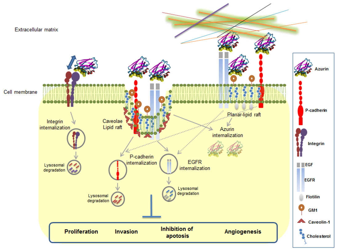
Bernardes N, Abreu S, Carvalho FA, et al. (2016) Modulation of membrane properties of lung cancer cells by azurin enhances the sensitivity to EGFR-targeted therapy and decreased β1 integrin-mediated adhesion. Cell Cycle 15: 1415–1424. doi: 10.1080/15384101.2016.1172147
![]() |
[29] | Yamada T, Das Gupta TK, Beattie CW (2016) p28-mediated activation of p53 in G2-M phase of the cell cycle enhances the efficacy of DNA damaging and antimitotic chemotherapy. Cancer Res 76: 2354–2365. |
[30] |
Zaborina O, Dhiman N, Ling Chen M, et al. (2000) Secreted products of a nonmucoid Pseudomonas aeruginosa strain induce two modes of macrophage killing: external-ATP-dependent, P2Z-receptor-mediated necrosis and ATP-independent, caspase-mediated apoptosis. Microbiology 146: 2521–2530. doi: 10.1099/00221287-146-10-2521
![]() |
[31] |
Bernardes N., Ribeiro AS., Abreu S, et al. (2013) The bacterial protein azurin impairs invasion and FAK/Src signaling in P-cadherin-overexpressing breast cancer cell models. PLoS One 8: e69023. doi: 10.1371/journal.pone.0069023
![]() |
[32] |
Ribeiro AS, Albergaria A, Sousa B, et al. (2010) Extracellular cleavage and shedding of P-cadherin: a mechanism underlying the invasive behavior of breast cancer cells. Oncogene 29: 392–402. doi: 10.1038/onc.2009.338
![]() |
[33] |
Bernardes N, Ribeiro AS, Abreu S, et al. (2014) High-throughput molecular profiling of a P-cadherin overexpressing breast cancer model reveals new targets for the anti-cancer bacterial protein azurin. Int J Biochem Cell Biol 50: 1–9. doi: 10.1016/j.biocel.2014.01.023
![]() |
[34] |
Mollinedo F, de la Iglesia-Vicente J, Gajate C, et al. (2010) Lipid raft-targeted therapy in multiple myeloma. Oncogene 29: 3748–3757. doi: 10.1038/onc.2010.131
![]() |
[35] | Coppari E, Yamada T, Bizzarri AR, et al. (2014) A nanotechnological molecular-modeling, and immunological approach to study the interaction of the anti-tumorigenic peptide p28 with the p53 family of proteins. Intl J Nanomedicine 9: 1799–1813. |
[36] | Yamada T, Das Gupta TK, Beattie CW (2013) P28, an anionic cell-penetrating peptide, increases the activity of wild type and mutated p53 without altering its conformation. Mol. Pharm 10: 3375–3383. |
[37] | Cho I, Blaser MJ (2012) The human microbiome: at the interface of health and disease. Nat Rev Genet 13: 260–270. |
[38] |
Shaikh F, Abhinand P, Ragunath P (2012) Identification & characterization of Lactobacillus salavarius bacteriocins and its relevance in cancer therapeutics. Bioinformation 8: 589–594. doi: 10.6026/97320630008589
![]() |
[39] | Nguyen C, Nguyen VD (2016) Discovery of azurin-like anticancer bacteriocins from human gut microbiome through homology modeling and molecular docking against the tumor suppressor p53. Biomed Res Int 2016: 8490482. |
[40] | Guex N, Peitsch MC (1997) SWISS-MODEL and the Swiss-PdbViewer: An environment for comparative protein modeling. Electrophoresis 18: 2714–2723. |
[41] |
Gibrat JF, Madej T, Bryant SH (1996) Surprising similarities in structure comparison. Curr Opin Struct Biol 6: 377–385. doi: 10.1016/S0959-440X(96)80058-3
![]() |
[42] | Paz I, Kligun E, Bengad B, et al. (2016) BindUP: a web server for non-homology-based prediction of DNA and RNA binding proteins. Nucleic Acids Res pii: gkw454. |
[43] |
Jo S, Kim T, Iyer VG, et al. (2008) CHARMM-GUI: A web-based graphical user interface for CHARMM. J Comput Chem 29: 1859–1865. doi: 10.1002/jcc.20945
![]() |
1. | A. S. Lozhkomoev, M. Shakirov, 2019, 2167, 0094-243X, 020200, 10.1063/1.5132067 | |
2. | Leonid A. Kaledin, Fred Tepper, Yuly Vesga, Tatiana G. Kaledin, The effect of the surface roughness and ageing characteristics of boehmite on the removal of biological particles from aqueous solution, 2019, 6, 2372-0484, 498, 10.3934/matersci.2019.4.498 | |
3. | Leonid A. Kaledin, Fred Tepper, Yuly Vesga, Tatiana G. Kaledin, Boehmite and Akaganeite 1D and 2D Mesostructures: Synthesis, Growth Mechanism, Ageing Characteristics and Surface Nanoscale Roughness Effect on Water Purification, 2019, 2019, 1687-4110, 1, 10.1155/2019/9516156 |
Media | Grade | Thickness (mm) | Mean particle sizea (μm) | Bubble point (kPa) | Mean flow pore diameter, dmean flow (μm) | Largest pore diameter, dlargest (μm) |
Non-woven a | Prefilter media c | 1.2 | ~1 a | 9.8 ± 0.8 e | 2.3 ± 0.3 | 29 ± 2 e |
Drinking water filter media d | 0.8 | ~1 a | 20 ± 2 e | 0.7 ± 0.3 | 14 ± 1 e | |
Aluminized DE b | Fine grade aluminized DE | 10 | 3.0 ± 0.5 | |||
Medium grade aluminized DE | 18 | 5.5 ± 1.5 | ||||
Coarse grade aluminized DE | 80 | 24.0 ± 3.5 | ||||
Notes: a Figure 1a and 1b; b Figure 1c and 1d; c Disruptor grade 5292; d Disruptor grade 5283; e Average value for 10 cartridges. |
pH | z potential (mV) | fr removal efficiency, LRV b | MS2 removal efficiency, LRV b | PRD-1 removal efficiency, LRV b | E. coli removal efficiency, LRV b | B. diminuta removal efficiency, LRV b | |
1 layer c | 2 layers c | 2 layers c | 2 layers c | 2 layers c | 2 layers c | ||
3 | 46 ± 7 a | 3.0 ± 0.3 | 5.0 ± 0.3 | ||||
5 | 46 ± 7 a | > 6.2 | > 6.2 | > 5.0 | |||
7 | 47 ± 9 a | > 6.2 | > 6.2 | > 6.0 | > 7.0 | > 7.5 | > 7.6 |
8 | 46 ± 6 a | > 6.1 | > 6.1 | > 5.0 | 6.3 | > 6.2 | |
9 | 26 ± 4 a | > 6.0 | > 6.0 | > 5.0 | |||
10 | 23 ± 5 a | 4.2 ± 0.3 | > 6.0 | ||||
11 | 22 ± 5 a | 0.2 ± 0.2 | 1.5 ± 0.7 | ||||
Notes: a Ref. [7]; b Logarithm reduction value; c By 1-or 2-layers of drinking water filter media (Table 1) at flow velocity of 0.7 mm/s. |
Electrolyte ionic strength, NaCl (mM) | λD a (nm) | Flow velocity (mm/s) | Contact time (s) | Brownian displacemen b of RT (mm) | Brownian displacemen b of MS2 (mm) | MS2 removal efficiency, LRV c | RT removal efficiency, LRV c |
0.002(1) d | 215 | 0.05 | 22.5 | 4.7 | 28 | > 6.4 | 6.4 ± 0.7 |
0.2 | 5.6 | 2.4 | 14 | > 6.4 | 6.2 ± 1.0 | ||
0.8 | 1.4 | 1.2 | 7.0 | > 6.4 | 5.8 ± 0.4 | ||
3.4 | 0.35 | 0.6 | 3.5 | > 6.4 | 6.0 ± 0.5 | ||
0.05(1) d | 43 | 0.05 | 22.5 | 4.7 | 28 | > 5.7 | 5.9 ± 0.2 |
0.2 | 5.6 | 2.4 | 14 | > 5.7 | 5.8 ± 0.2 | ||
0.8 | 1.4 | 1.2 | 7.0 | > 5.7 | 5.6 ± 0.2 | ||
3.4 | 0.35 | 0.6 | 3.5 | > 5.7 | 5.2 ± 0.6 | ||
0.35(4) d | 16 | 0.05 | 22.5 | 4.7 | 28 | > 5.2 | 5.8 ± 0.2 |
0.2 | 5.6 | 2.4 | 14 | > 5.2 | 5.3 ± 0.2 | ||
0.8 | 1.4 | 1.2 | 7.0 | > 5.2 | 5.2 ± 0.4 | ||
3.4 | 0.35 | 0.6 | 3.5 | > 5.2 | 5.0 ± 0.8 | ||
1.0(1) d | 9.6 | 0.05 | 22.5 | 4.7 | 28 | > 6.6 | 5.7 ± 0.4 |
0.2 | 5.6 | 2.4 | 14 | > 6.6 | 5.2 ± 0.5 | ||
0.8 | 1.4 | 1.2 | 7.0 | 5.6 ± 0.2 | 4.8 ± 0.5 | ||
3.4 | 0.35 | 0.6 | 3.5 | 5.1 ± 0.2 | 4.4 ± 0.5 | ||
3.0(3) d | 5.5 | 0.05 | 22.5 | 4.7 | 28 | > 5.8 | 4.9 ± 0.3 |
0.2 | 5.6 | 2.4 | 14 | > 5.8 | 4.6 ± 0.3 | ||
0.8 | 1.4 | 1.2 | 7.0 | > 5.8 | 4.2 ± 0.7 | ||
3.4 | 0.35 | 0.6 | 3.5 | 5.2 ± 0.6 | 4.3 ± 0.9 | ||
10(1) d | 3.0 | 0.05 | 22.5 | 4.7 | 28 | > 5.9 | 4.2 ± 0.2 |
0.2 | 5.6 | 2.4 | 14 | > 5.9 | 4.1 ± 0.2 | ||
0.8 | 1.4 | 1.2 | 7.0 | > 5.9 | 3.7 ± 0.3 | ||
3.4 | 0.35 | 0.6 | 3.5 | 4.7 ± 0.3 | 3.7 ± 0.4 | ||
40(4) d | 1.5 | 0.05 | 22.5 | 4.7 | 28 | 4.8 ± 0.2 | 3.9 ± 0.2 |
0.2 | 5.6 | 2.4 | 14 | 5.1 ± 0.3 | 3.7 ± 0.4 | ||
0.8 | 1.4 | 1.2 | 7.0 | 4.7 ± 0.2 | 3.1 ± 0.4 | ||
3.4 | 0.35 | 0.6 | 3.5 | 3.1 ± 0.2 | 2.8 ± 0.5 | ||
100(10) d | 0.96 | 0.05 | 22.5 | 4.7 | 28 | > 4.5 | 4.0 ± 0.5 |
0.2 | 5.6 | 2.4 | 14 | > 4.5 | 3.7 ± 0.4 | ||
0.8 | 1.4 | 1.2 | 7.0 | > 4.5 | 3.1 ± 0.4 | ||
3.4 | 0.35 | 0.6 | 3.5 | 3.5 ± 0.3 | 2.8 ± 0.4 | ||
250(25) d | 0.61 | 0.05 | 22.5 | 4.7 | 28 | 4.2 ± 1.0 | 3.1 ± 0.2 |
0.2 | 5.6 | 2.4 | 14 | 3.7 ± 0.3 | 2.7 ± 0.2 | ||
0.8 | 1.4 | 1.2 | 7.0 | 3.0 ± 0.2 | 2.5 ± 0.2 | ||
3.4 | 0.35 | 0.6 | 3.5 | 1.2 ± 0.2 | 1.8 ± 0.2 | ||
510(51) d | 0.43 | 0.05 | 22.5 | 4.7 | 28 | 2.3 ± 0.6 | 2.4 ± 0.6 |
0.2 | 5.6 | 2.4 | 14 | 1.8 ± 0.2 | 2.2 ± 0.4 | ||
0.8 | 1.4 | 1.2 | 7.0 | 1.8 ± 0.4 | 1.9 ± 0.2 | ||
3.4 | 0.35 | 0.6 | 3.5 | 1.0 ± 0.5 | 1.5 ± 0.4 | ||
3000(300) d | 0.18 | 0.05 | 22.5 | 4.7 | 28 | 2.1 ± 0.3 | 2.0 ± 0.3 |
0.2 | 5.6 | 2.4 | 14 | 1.6 ± 0.3 | 1.6 ± 0.2 | ||
0.8 | 1.4 | 1.2 | 7.0 | 1.3 ± 0.3 | 1.4 ± 0.2 | ||
3.4 | 0.35 | 0.6 | 3.5 | 1.3 ± 0.5 | 1.0 ± 0.2 | ||
Notes: a Calculated with the use of equation (1); b Calculated with the use of equation (9); c Logarithm reduction value; d Error limits in parentheses are accuracy in the units of the last digit reported. |
Bed volume (dm3) | pH | Column depth (mm) | Flow velocity (mm/s) | Contact time (s) | Input 59Co concentration (ppb) | Output 59Co concentration (ppb) | Removal efficiency (%) |
0.16 | 10 | 50 | 10 | 5.0 | 635 | < 5 | > 99.2 |
0.32 | 10 | 100 | 6 | 17 | 635 | < 5 | > 99.2 |
0.64 | 10 | 200 | 3.3 | 60 | 635 | < 5 | > 99.2 |
0.16 | 8 | 50 | 10 | 5.0 | 656 | < 5 | > 99.2 |
0.32 | 8 | 100 | 6 | 17 | 656 | < 5 | > 99.2 |
0.64 | 8 | 200 | 3.3 | 60 | 656 | < 5 | > 99.2 |
Percent of DE in aluminized DE/DE precoat | Number of layers formed by aluminized DE c | MS2 removal efficiency a, LRV b | RT removal efficiency a, LRV b |
0 | 40 | 6.1 ± 0.1 | > 7.7 |
10 | 38 | 5.1 ± 0.2 | > 7.7 |
25 | 16 | 3.6 ± 0.4 | 3.9 ± 0.3 |
50 | 7 | 2.6 ± 0.9 | 3.0 ± 0.3 |
75 | 2 | 1.0 ± 0.2 | 2.2 ± 0.1 |
100 | 0 | 0.5 ± 0.1 | 0.7 ± 0.2 |
Notes: a At flow velocity of 0.67 mm/s; b Logarithm reduction value; c Arranged in a group of four neighboring aluminized DE particles in the plane perpendicular to the flow. |