Citation: Andrea Bianchi, Chiara Lanzuolo. Into the chromatin world: Role of nuclear architecture in epigenome regulation[J]. AIMS Biophysics, 2015, 2(4): 585-612. doi: 10.3934/biophy.2015.4.585
[1] | Théo Lebeaupin, Hafida Sellou, Gyula Timinszky, Sébastien Huet . Chromatin dynamics at DNA breaks: what, how and why?. AIMS Biophysics, 2015, 2(4): 458-475. doi: 10.3934/biophy.2015.4.458 |
[2] | Bertrand R. Caré, Pierre-Emmanuel Emeriau, Ruggero Cortini, Jean-Marc Victor . Chromatin epigenomic domain folding: size matters. AIMS Biophysics, 2015, 2(4): 517-530. doi: 10.3934/biophy.2015.4.517 |
[3] | Larisa I. Fedoreyeva, Boris F. Vanyushin, Ekaterina N. Baranova . Peptide AEDL alters chromatin conformation via histone binding. AIMS Biophysics, 2020, 7(1): 1-16. doi: 10.3934/biophy.2020001 |
[4] | E.N. Baranova, R.M. Sarimov, A.A. Gulevich . Stress induced «railway for pre-ribosome export» structure as a new model for studying eukaryote ribosome biogenesis. AIMS Biophysics, 2019, 6(2): 47-67. doi: 10.3934/biophy.2019.2.47 |
[5] | Vladimir B. Teif, Andrey G. Cherstvy . Chromatin and epigenetics: current biophysical views. AIMS Biophysics, 2016, 3(1): 88-98. doi: 10.3934/biophy.2016.1.88 |
[6] | Claudia Tanja Mierke . Physical role of nuclear and cytoskeletal confinements in cell migration mode selection and switching. AIMS Biophysics, 2017, 4(4): 615-658. doi: 10.3934/biophy.2017.4.615 |
[7] | Michael J. Hamilton, Matthew D. Young, Silvia Sauer, Ernest Martinez . The interplay of long non-coding RNAs and MYC in cancer. AIMS Biophysics, 2015, 2(4): 794-809. doi: 10.3934/biophy.2015.4.794 |
[8] | Raghvendra P Singh, Guillaume Brysbaert, Marc F Lensink, Fabrizio Cleri, Ralf Blossey . Kinetic proofreading of chromatin remodeling: from gene activation to gene repression and back. AIMS Biophysics, 2015, 2(4): 398-411. doi: 10.3934/biophy.2015.4.398 |
[9] | Larisa I. Fedoreyeva, Tatiana A. Dilovarova, Boris F. Vanyushin, Inna A. Chaban, Neonila V. Kononenko . Regulation of gene expression in Nicotiana tabacum seedlings by the MKASAA peptide through DNA methylation via the RdDM pathway. AIMS Biophysics, 2022, 9(2): 113-129. doi: 10.3934/biophy.2022011 |
[10] | Audrey Lejart, Gilles Salbert, Sébastien Huet . Cytosine hydroxymethylation by TET enzymes: From the control of gene expression to the regulation of DNA repair mechanisms, and back. AIMS Biophysics, 2018, 5(3): 182-193. doi: 10.3934/biophy.2018.3.182 |
Abbreviations
HP1 | Heterochromatin Protein 1 |
PcG proteins | Polycomb group of proteins |
PRC1 | Polycomb Repressive Complex 1 |
PRC2 | Polycomb Repressive Complex 2 |
Pax7 | Paired box protein Pax-7 |
3C | Chromosome Conformantion Capture |
4C | Chromosome Conformantion Capture on Chip |
Hi-C | High resolution Capture |
PRE | Polycomb Response elements |
TAD | Topological Associated Domain |
HOXD | Homeobox protein Hox D |
MET-2 | Histone-lysine N-methyltransferase met-2 |
SET-25 | SET-25 |
NB | Nuclear Body |
FRAP | Fluorescence Recovery After Photobleaching |
HOX | Homeobox protein Hox |
ESC | Embryonic Stem Cell |
LAD | Lamina Associated Domain |
ASC | Adipose Stem Cell |
Klf4 | Kruppel-like factor 4 |
Oct4 | Octamer-binding transcription factor 4 |
NPC | Neuronal Precursor Cell |
HGPS | Hutchinson-Gilford Progeria Syndrome |
NPC | Nuclear Pore Complex |
AF | Atrial Fibrillation |
Nup | Nucleoporin |
NAD | Nucleolar Associated Domain |
LIN28A | Protein lin-28 homolog A |
BER | Base Excision Repair |
NER | Nucleotide Excision Repair |
MMR | Mismatch Repair |
DSB | Double Strand Break |
NHEJ | Non Homologous End Joining |
HR | Homologous Recombination |
Ku70 (XRCC6) | X-ray repair cross-complementing protein 6 |
Ku80 (XRCC5) | X-ray repair cross-complementing protein 5 |
DNA-PKcs | DNA-dependent Protein Kinase |
ATM | Ataxia Telangiectasia Mutated |
ATR | Ataxia Telangiectasia and Rad3-related protein |
XRCC4 | X-ray repair cross-complementing protein 4 |
RPA | Replication Protein A |
Rad 51 | DNA repair protein RAD51 homolog 1 |
Rad 52 | DNA repair protein RAD52 homolog |
DDR | DNA Damage Response |
53BP1 | Tumor suppressor p53-binding protein 1 |
MRE11 | Meiotic Recombination 11 |
NSB1 | Nijmegen breakage syndrome protein 1 |
ROS | Reactive Oxygen Species |
NADPH | Nicotinamide Adenine Dinucleotide Phosphate |
Rb | Retinoblastoma protein |
p53 | Cellular tumor antigen p53 |
NF-kB | Nuclear factor NF-kappa-B |
FoxO | Forkhead box protein O |
HIV-1 | Human Immunodeficiency Virus 1 |
In eukaryotic cells, DNA is packaged inside the nucleus through its ordered and repetitive aggregation with histones and is bound by several proteins and RNA molecules: the overall complex of nucleic acids and their associated proteins is known as “chromatin”. The first level of chromatin organization is the nucleosome: four histone proteins; H2A, H2B, H3, H4 surrounded by DNA [1]. Each nucleosome is separated from the next one by a DNA linker, variable in length, which is associated to histone H1 and or its variants [2,3]. These DNA-histones complexes fold in 30 nm fibers. This process is driven in part by the histone H1, which regulates the intranuclear electrostatic balance and by nucleosome repeats which ensure the stabilization of chromatin fibers [4,5,6]. In interphase the chromatin shows different levels of compaction, clearly visible under electron microscopy, that allowed scientists to classify it into euchromatin and heterochromatin, observed for the first time by Emil Heizt (1928) [7].The more decondensed euchromatin is generally enriched with expressed genes (reviewed in [8,9]). In fact, being more accessible, euchromatin favours the binding of transcription factors and RNA polymerase [10] and is correlated to high transcriptional activity [11]. On the other hand, heterochromatin is more condensed, gene-poor and is further divided into “facultative” and “constitutive”. The constitutive heterochromatin includes pericentric and telomeric regions [12,13,14], both critically important for the higher order structure folding and the maintenance of intact chromosomes [8]. The facultative heterochromatin is a repressive environment able to rapidly shift between activated and repressed state through the crosstalk with epigenetic regulators that lead to a chromatin reorganization [8].
Histones are characterized by amino and carboxy-protruding tails that are the target of several modifications, critically important for regulating the balance between activated or repressed genes. These modifications mainly include acetylation, methylation, phosphorylation, ubiquitination and sumoylation (reviewed in [15]). Multiple histone modifications can also coexist on the same tail, dictating specific biological readouts [16]. Euchromatin and heterochromatin are distinguishable biochemically by different covalent histone modifications [17] (Figure 1). Euchromatin is enriched with H3 and H4 acetylation and H3K4 methylation [18,19] while facultative heterochromatin is enriched with hypoacetylated histones, H4K20me1, H3K9me2, H3K27me3, H2AK119ub1 (reviewed in [20]). On the other hand, constitutive heterochromatin is marked with H3K9me3 catalyzed by Suv39H [21], which in turn stimulates the recruitment of Heterochromatin Protein 1(HP1) [22], so contributing to form a closed chromatin structure normally associated with a repressive transcriptional state. However, despite this classification, histone marks should not be considered exclusively restricted to specific chromatin compartments because they were also found in other chromatin states. For instance, although H3K9me3 and HP1 are considered key features of heterochromatin, they have also been found in euchromatic region in flies [23,24] and in mammals [25], thus also suggesting their role in reinforcing gene repression in euchromatin.
At the chromatin level, specific epigenetic factors are able to post-translationally modify the histone proteins. Then, specific set of histone modifications generate a chromatin environment that is recognized by specific binding proteins; these, in turn, influence gene expression and other chromatin functions and represent an additional layer of gene regulation complexity. One of the most studied families of epigenetic regulators that include both histone modifiers and chromatin binding proteins are the Polycomb group of proteins (PcG proteins) which act as a repressor of gene expression (reviewed in [26]). PcG proteins aggregate into different complexes. The most studied are Polycomb Repressive Complex 1 and 2 (PRC1 and PRC2) that deposit H2AK119ub and H3K27me3 marks, respectively. The catalytic subunit responsible for the histone signature placement are E3 ubiquitin-protein ligase RING1 (Ring 1A/B) for PRC1 and Enhancer of zeste 2 (Ezh2) for PRC2 (reviewed in [27]). A classical, hierarchical model suggests that the PRC2-dependent H3K27me3 signature recruits the PRC1 complex which, through the ubiquitination of H2AK119, inhibits RNA polymerase II-dependent transcriptional elongation [28,29,30]. In recent years, several studies have challenged this model (reviewed in [31]) by showingindependent functions and recruitment mechanisms for PRC1 and PRC2. Thus, it has been demonstrated that PRC1 and PRC2 complexes share only a subset of binding sites [28,32,33] and that there are several PRC1 complexes containing distinct subunits that differ in genomic binding [34]. Moreover, PRC1 can recruit PRC2 [32,35,36] while recruitment of PRC1 and PRC2 can also be mediated by noncoding RNA (reviewed in [37]), specific factors or structures [38,39,40,41] and by their own histone marks [42,43]. These findings suggest that PRC recruitment relies on several alternative mechanisms and that the combination of mechanisms tethering PcG to chromatin is probably locus-specific. The plasticity of the facultative heterochromatin to switch on or off the gene expression through histone modifications is extremely important for lineage commitment, development and cell differentiation, when a proper timing of gene expression is needed [28,44,45]. Most of differentiation genes in mammals are under the control of Polycomb group of proteins [46]. Upon differentiation stimuli, PcG proteins leave lineage specific muscle promoters and bind genes important for stemness maintenance [46]. This dynamics has been extensively shown in muscle differentiation: in muscle stem cells (satellite cells) muscle specific genes are maintained repressed by PcG proteins; at the onset of muscle differentiation PcG proteins are displaced from muscle gene and are relocalized at stemness genes, such as Pax7 (marker of satellite cells), ensuring the correct timing of the muscle differentiation [47,48,49,50,51].
In addition to histone modifications, the chromatin can fold in specific higher order structures that favour the activation or the repression of genes (reviewed in [52]). In particular, structure-mediated gene expression control regards genes that are regulated by a large set of enhancers, insulators or repressors, which are often located at a considerable distance from the target gene. Thus, the establishment of topologically distinct chromatin domains could play a fundamental role in the modulation of gene expression (Figure 1). PcG proteins also exert gene repression by mediating chromatin higher order structures (reviewed in [26,53]). The development of Chromosome Conformation Capture (3C) technology [54] and its derivative technologies (reviewed in [55]), such as Chromosome Conformation Capture on chip (4C) [56] and High resolution Capture (Hi-C) [57] shed light on the chromatin contacts occurring in the nucleus allowing the high-through-put mapping of the genome conformation. The use of these technologies has allowed important advances in understanding PcG functions, demonstrating that the coordinated action of PcG proteins is required to mediate the formation of multi-looped structures where all the major PcG targets are gathered together, by cis and trans interactions [58,59,60,61]. This PcG-mediated DNA conformation is cell cycle regulated and fundamental for the maintenance of gene repression [58,59,62,63]. Notably, although PcG recruitment is not conserved from fly to mammals, being mediated in Drosophila by specific DNA sequences called Polycomb Response Elements (PRE) [64], the higher order structures of PcG-regulated genes are similarly organized in multi-loops among different species, thus suggesting a functional role for DNA higher order structures in the control of PcG mediated transcriptional repression.
At the genome wide level, the combination of all DNA-DNA interactions determines the formation of specific macro-domains of active and inactive chromatin, named Topological Associated Domains (TADs) observed in human, in mouse and in Drosophila melanogaster genomes [65,66,67] (Figure 1). TADs are characterized by a central region of high chromatin interactions where reside the tissue-specific genes, surrounded by less-interacting boundary regions enriched of housekeeping genes [52]. Although TADs share a common organization in different cell-types and are partially conserved from human to mouse, suggesting a very stable conformation, they can be subjected to important structural rearrangements. During the mouse hind limb development, the HOXD genes are expressed following specific timing from early genes to later genes; this transition is guided by the reorganization of TADs which allow the switch of HOXD locus interactions from one compartment to another one [68]. This dynamics is required for the formation of an intermediate area that will develop into the wrist.
Another level of chromatin organization is represented by the chromosome position. Several studies have shown that chromosomes are not randomly assembled and packaged in the nucleus, but occupy specific regions called chromosome territories (reviewed in [69]) (Figure 1). The positioning of these “territories” is cell specific and conserved between human and other primates, suggesting a functional role of specific chromosome organization inside the nucleus [52,70,71]. In fact, chromosome territories have an internal structure thought to modulate the accessibility of proteins and consequently regulates the gene expression through its remodeling (reviewed in [72,73]). In particular, it has been shown that within the territories the gene-rich regions are separated from heterochromatin and generally are positioned at the periphery of chromosome territories while gene-poor regions are at the interior of chromosome territories [74,75]. How chromosome territories form and occupy specific regions inside the nucleus is still unclear, but researchers have suggested two different hypothesis. The first suggests that preferential positioning of chromosome territories inside the nucleus is self-organized and guided in part by the interchromatin compartment, which creates channels for the interconnection between the chromosome territories and in part by the content of gene-rich or gene-poor areas of each specific chromosome [72,76,77]. According to this hypothesis chromosome folding within territories should allow that the gene-rich zones (euchromatin) will assume a position compatible with expression, whereas not commonly expressed genes, tissue specific or developmental regulated genes (heterochromatin) will be buried inside the chromosome territories and expressed only after a conformational change [72]. However, some exceptions to this role have been described, showing ubiquitously and tissue-specific genes located within the chromosome territories after their activation, suggesting that the inner region of chromosome territories is not an insurmountable barrier to transcription machinery and transcription factors [74,78]. The second emerging hypothesis supports the involvement of nuclear proteins and nuclear envelope in the chromosome positioning (reviewed in ([76,79]). This hypothesis does not conflict with the first one and could also explain how the chromosome can be oriented in the nuclear space. Thus, even though some progress has been made on the functional role of chromosomal territories in controlling gene expression, there is still a long way to go before discovering the mechanism behind their positioning and assembling inside the nucleus.
Inside the nucleus the coexistence of different environments ensure an additional level of gene transcription regulation (reviewed in [52]). Indeed, chromatin is non-homogenously distributed inside the nucleus, as shown by electron microscopy. In particular, most of the darkly stained chromatin (heterochromatin) is found around the nucleoli or at the nuclear periphery interrupted with euchromatin at nuclear pores (Figure 1). One of the most studied microenvironments is the nuclear periphery directly linked to a repressive transcriptional state (reviewed in [80,81]). Indeed, artificial tethering of a reporter gene to the nuclear periphery favours the establishment of silent chromatin, by mediating the access to local high concentration of repressors and chromatin modifiers, as observed in yeast and human cells (reviewed in [80] and [82,83]). Although endogenous molecular mechanisms driving the periphery localization are still under investigation, a genetic screen in worms revealed that histone H3 lysine 9 (H3K9) methylation marks target repetitive heterochromatin to the nuclear envelope and that the combined elimination of the genes encoding two histone H3K9 methyl transferase, MET-2 and SET-25, determines the loss of peripheral localization [84]. Interestingly, loss of SET-25 alone de-represses the transgene array, but does not lead to array delocalization, suggesting that H3K9me1 or me2, catalyzed by SET-25, are sufficient to anchor chromatin to the nuclear envelope and that further modification, generating H3K9me3, is necessary for transcriptional repression [84]. In line with and further corroborating these observations, in budding yeast, several highly regulated classes of genes were described as translocating to the nuclear periphery concurrent with their activation (reviewed in [85]). Interestingly, it has been shown that when an inducible active gene is repressed again, its persistent presence at the nuclear periphery allows a rapid reactivation upon stimuli. This process has been described as “transcriptional memory, ” since it allows the cell to “remember” recently activated loci (reviewed in [85]), so suggesting that the nuclear periphery environment can establish an epigenetic memory. Thus, in the nuclear periphery there is an alternation on permissive and non-permissive environment that can influence a given gene’s transcriptional state.
Within the nuclear space the non-uniform spatial distribution of macromolecules gives rise to a formation of a variety of functionally distinct nuclear bodies (NBs), highly dynamic structures formed by protein-protein interactions and having a large impact on gene regulation (reviewed in [86,87]). NBs are nuclear niches, in which machineries responsible for specific nuclear functions, such as transcription (transcriptional factories), replication (replication foci) or repression (repressive bodies) are situated. The presence of NBs implies a non-diffusive transport of factors involved in chromatin regulation. This compartmentalization of nuclear processes avoids the stochastic non-directional transport of nuclear regulators and speeds up the dynamics in the nucleus [87]. The most prominent NB in the nucleus is the nucleolus, responsible of ribosomal RNA transcription (Figure 1). Interestingly the nucleolus has a specific structural organization in three distinct regions that mirror nucleolar functions (reviewed in [86]). An example of epigenetic regulators forming aggregates in the nucleus are again the PcG proteins, forming structures called PcG bodies, often localized close to pericentric heterochromatin [88] (Figure 2). The formation of PcG bodies is mediated by intrinsic [89]
and extrinsic [90] protein-protein interactions. Studies on the kinetics of PcG proteins exchange with the body have revealed that these protein aggregates are very stable and that distinct PcG factors exhibit large variability in their mobilities [91]. In terms of kinetics, three distinguishable components form the foci: fast, slow and an immobile on the time scales of measurements [91]. The slow binding component amplitudes directly correlate with the intensities and sizes of PcG foci, this suggesting that the PcG bodies contain multiple PcG targets. On the other hand, FRAP analysis has revealed that the size and the kinetics inside the foci is mostly slower than PcG complexes localized outside the foci [89,91], suggesting a coordinated overlap between genome higher order structures and PcG proteic compartments. In fact, previous studies revealed that Drosophila Hox clusters co-localize in PcG bodies in the tissue where they are repressed, this nuclear localization being required for efficient silencing [58]. In line with these findings, PcG bodies are clearly distinguishable at the end of early embryogenesis; they progressively increase in size and number during stages 5-11 of late embryogenesis when the transcriptional programs need to be maintained through cell division [92]. Similarly, the mobility of PcG bodies increases at the onset of ESC (Embryonic Stem Cell) differentiation and decreases as differentiation progresses [93]. Further studies of the composition of different PcG foci compartments could shed light on the coordinated functions of PcG complexes.
In spite of all classifications of epigenetic signatures, the epigenome is highly dynamic and can rapidly respond to the environmental stimuli. The occasional presence of repressive marks on the heterochromatin, as in the case of H3K9me3 and HP1 [11], the heterochromatin’s ability to switch from a repressive to an active state (or vice-versa) and recent findings showing that epigenetic repressors can be bound to active transcribed chromatin [94,95] all underline the thin line separating euchromatin from heterochromatin. All these evidence also demonstrate that chromatin cannot be considered a static entity rigorously classified in an active or repressive state, but should be considered dynamic, and that this plasticity is the key for precise gene expression.
The nuclear lamina is a proteinaceous layer tightly associated with the inner nuclear membrane. The main components of the nuclear lamina are the lamin proteins [96], classified as intermediate filament V and further divided in vertebrates into A-type lamins (lamin A and lamin C) and the B-type lamins (lamin B1 and lamin B2) [97,98]. Whereas B-type lamins are ubiquitously expressed and firmly anchored at the nuclear membrane, A-type lamins are mostly absent from early embryos [99,100], developmentally regulated, expressed in lineage-committed progenitor cells and differentiated cells [101,102]. Moreover, A-type lamins are present in both peri-nuclear and nucleoplasmatic compartments [103]. A and B-type lamins, form distinct homopolimers that interweave each other at the inner membrane to create an interconnected plot [104]. One of the lamina’s roles is to maintain nuclear shape and integrity. Indeed, knocking down lamin B1 results in an enlargement of Lamin A and Lamin B2 network and in the formation of nuclear blebs. These alterations are accompanied by a perturbation of epigenetic signatures, as a reduction of H3K9me2 and an increase of H3 acetylation, suggesting that the impairment of the nuclear envelope affects the proper organization of chromatin [104]. Other cellular functions are attributed to the lamins such as transcription, cell cycle regulation, development and differentiation (reviewed in [105]).
The importance of lamins and nuclear lamina as epigenetic regulators is reinforced by the fact that the nuclear lamina interacts with the genome at specific DNA sequences called Lamina Associated Domains (LADs) (Figure 1). These domains show variable lengths from 0.1 to 10 Mb and generally create an environment that maintains genes repressed [106,107] and marked by H3K9me2, H3K9me3 or H3K27me3 [108,109,110,111]. Interestingly, the borders of LADs include promoters that often contain transcription-associated histone modification indicating the transcriptional start site of the activated genes [112]. This characteristic may suggest that LADs define genomic regions in which genes are repressed, neighboured by activated genes. Some LADs are cell-specific, suggesting a possible role in cell identity and differentiation [106,113]. Recently, it has been shown in worms that in a lamin-defective background muscle promoters do not re-localize from the nuclear periphery to a more internal location in differentiating muscle cells [114]; this further indicates that the genome architecture dynamics mediated by Lamin A are necessary for a correct differentiation. This was confirmed by studies in mammals, showing that during the differentiation of adipose stem cells (ASCs) into adipocytes, genes that regulate a specific cell lineage, are released from the lamina while genes that maintain the cell undifferentiated are retained near the nuclear lamina [110]. Similarly, during the differentiation of Embryonic stem cells (ESCs) into neural precursor cells (NPCs), the so named “stemness genes” such as Nanog, Klf4 and Oct4, exhibit significantly increased interactions with the nuclear lamina in NPCs compared to ESCs [115]. In parallel, other LADs, seem to be constitutively associated to the lamina and conserved between species, suggesting a role in the organization of chromosome architecture [106,107]. One possibility is that the presence of these constitutive LADs could help the correct DNA organization (and gene expression) in daughter cells after mitosis, acting as a “bookmarks” of the genome architecture [116]. However, in contrast with this hypothesis, recent evidence has shown that LADs-nuclear lamina interactions are not rigid and fixed domains, but on the contrary, are dynamic and organized in a random manner after cell division; this stochastic organization is related to H3K9me2 and guided in part by Lamin A [108,117]. Moreover, in spite of all the efforts until now to characterize the LADs and the intriguing hypothesis concerning the role of LADs during genome organization, a recent work has questioned some aspects of LADs, asserting that nuclear lamins are not essential for the formation of LADs [118]. Thus it is clear that the research is still far from clearly defining the role of these domains in genome organization and regulation of gene expression.
A-type Lamin proteins, being situated in a strategic position at the interface between the nuclear membrane and chromatin, cover an important role also in mechanotrasduction, the process by which the external mechanical stimuli are converted into biochemical signals inside cells (reviewed in [119]). An intriguing hypothesis suggests that mechanical signals from the extracellular environment can be transmitted physically by the contractile cytoskeleton to the nucleus through the nuclear membrane and the nuclear lamina [120] and then translated into gene expression response through Lamin-mediated distortion of chromatin higher order structure and changes in transcription factor accessibility [121]. Recent findings support this hypothesis, showing that stress, matrix stiffness and the environment act on gene expression by perturbing the Lamin A expression and consequently the lamin A:B ratio. The altered Lamin A content, in turn, influences specific cell lineage determination and differentiation [122].
Mutations in lamin proteins can cause a wide set of diseases, named laminopathies, affecting specific tissues or acting upon multiple types of tissue creating overlapping or systemic phenotypes. Only few mutations in Lamin B have been described as being compatible with life [123,124] so confirming its fundamental role in sustaining the nuclear structure [125,126]. On the other hand, pathology with mutations related to Lamin A are more frequent and give rise to muscular dystrophy, lipodystrophy, neuropathy, cardiomyopathy and HGPS syndrome (Hutchinson-Gilford progeria syndrome) (reviewed in [126]). Many of the laminopathies are characterized by nuclear abnormalities [127,128]. Thus it is reasonable to assume that in these diseases, mutations in lamins or alteration of lamin A:B ratio act on gene expression in different ways: on the one hand by increasing nuclear stress sensitive and improper signalling with an aberrant mechanotrasduction [129] and on the other hand by altering the chromatin organization and modulating gene expression epigenetically. Both these aspects could be involved in the pathogenesis and disease progression [122,129]. Hutchinson-Gilford Progeria Syndrome (HGPS) is one of the most known and studied diseases belonging to laminopathies and is characterized by a premature aging. In 90% of cases, the disease is caused by a point mutation 1824C > T that activates a cryptic site of splicing creating an immature form of Lamin A/C (called progerin) that accumulates at the nuclear membrane, giving rise to abnormal nuclear shapes (reviewed in [130]). Several epigenetic marks have been found altered in HGPS, such as a reduction of H3K27me3, H3K9me3 and lamina-heterochromatin associations [131,132]. Although it is not clear if these alterations precede the senescent phenotype, it has been proposed that epigenetic dysfunctions and aberrant chromatin higher order structures could play a role in HGPS pathogenesis and progression [131]. This suggests a direct interplay between the alteration of the nuclear membrane and epigenome misregulation. Further confirming the idea that Lamin A/C plays a key role in epigenome regulation, studies on laminopathies have revealed that distinct mutations in Lamin A gene can give rise to the same clinical condition while a single mutation can result in different phenotypes and disease severity [133,134,135], suggesting an involvement of the individual epigenetic background to lamin dependent diseases.In line with this hypothesis, recently we found that Lamin A/C maintains transcriptional repression at muscle-specific genes by sustaining the intranuclear positioning of PcG bodies and the activity of PcG proteins [136]. Lamin A/C depletion does indeed lead to an earlier muscle differentiation onset due to a premature PcG displacement from their targets.
The nuclear pore complexes (NPCs) are multi-component protein complexes that form selectively permeable channels through the nuclear envelope, mediating nucleo-cytoplasmic transport of molecules between the nucleus and the cytoplasm (reviewed in [137]). The NPC is composed of multiple copies of nucleoporins. The overall structure of the NPC is highly conserved and displays nucleoporins organized into an eightfold symmetrical ring structures lining the circumference of the pore where they interact with the membrane. Various studies have implicated the NPCs as the binding platform for several highly transcribed housekeeping genes and genes strongly induced by changes in environmental conditions [137] (Figure 1). The presence of several expressed loci in the close proximity of the nuclear pore inspired the gene gating hypothesis, in which active genes are found near nuclear pore complexes to facilitate efficient export of mRNAs from the nucleus [138,139,140]. 3C analysis confirmed this hypothesis showing that some inducible genes are arranged in chromatin loops the ends of which are anchored at the NPC [141]. However, NPCs have also been associated with transcriptional repression [142,143,144,145], suggesting distinctive roles for individual components of the NPC in facilitating both gene activation and repression through its crosstalk with the epigenome. The NPC control of gene transcription is further confirmed by their central role during cell differentiation [38,146,147,148,149,150,151]. Intriguingly, NPC have been also found to be involved in the maintenance of the pluripotency via controlling levels of the pluripotency factors in the nucleus [152,153], underlying functional roles of the NPC in regulating developmental states and transitions. As described above for lamins, also mutations in key components of the NPC have been described to be associated with specific diseases. Thus, inherited cases of a cardiac disorder, the atrial fibrillation (AF), have been mapped to a missense mutation in the human nucleoporin Nup155 [154]. Studies in mice and human have shown that the impairment of Nup155 nuclear localization affects mRNA and protein transport leading to cardiovascular disease. Other studies have shown that mutations in the nucleoporin Nup62 are responsible for the familial form of infantile bilateral striatal necrosis, determining the degeneration of the basal ganglia in humans [155].
Although heterochromatic satellite regions were described near the nucleolus in the 1930s the role of the nucleolus as chromatin organizer is a recent hypothesis. Two independent high-throughput sequencing studies have isolated and sequenced the genomic DNA associated with purified nucleoli [156,157], finding the nucleolar-associated domains (NADs) (Figure 1). These genomic regions are gene poor and enriched for satellite DNA repeats. Interestingly, NAD peaks overlap with previously published LADs [156] and live imaging-based experiments have revealed an exchange between nuclear lamin and nucleolus border after mitosis. These data were confirmed by an independent study done by tracking LADs during the cell cycle [108]. Intriguingly, while Lamin B tends to interact only with LADs at the nuclear periphery, the subfraction of Lamin A that localize in the nucleoplasm preferentially accumulates around the nucleoli [117]. This suggests an interplay between the nucleolus and the nuclear membrane. Taken together these findings suggest that the nucleolus, as well as the nuclear envelope, could be involved in various nuclear functions, including gene transcription and the maintenance of chromatin structure. In line with this hypothesis, two seminal papers have provided insights into independent nucleolar mechanisms regulating pluripotency in embryonic stem cells (ESCs) [158,159]. In the first paper the authors found that the long noncoding RNA, pRNA, determines the progressive condensation of euchromatin into heterochromatin at nucleolus reducing epigenetic plasticity during differentiation [159]. In the second work authors showed that the methylation of the key pluripotency factor LIN28A leads to nucleolar localization and sequestration, preventing LIN28A processing and maintaining stemness programs [158]. These studies support the view that nuclear components are an important scaffold for spatial genome organization, which bears direct relevance to the establishment of cell type specific gene expression programs. Further studies are necessary to elucidate how the nuclear structures work together in a coordinated manner.
In recent years, what is emerging is that, besides the plasticity of the chromatin fundamental for fine-regulated process, the nuclear architecture can also influence important cellular processes and is a hallmark of the healthy cell. Indeed an increasing number of tissue-specific pathologies in humans and tissue-specific phenotypes in model organisms have been described for mutations in a variety of the nuclear architecture components (reviewed in [160]) (Table 1).
NUCLEAR ARCHITECTURE COMPONENTS | MUTATIONS | ASSOCIATED DISEASE | REFERENCE |
Lamin A/C | 1824 C Cryptic splice variant at LMNA gene < T | Hutchinson-Gilford Progeria Syndrome (HGPS) | [130] |
Q6X, R25G/P, R50S/P, R133P, S143F, H222P, R249Q, Y267C, M371K, R377H/L, R453W, W498R, R527P/H, L530P, R644C | Emery Dreifuss Muscular dystrophy (EDMD) | [134] | |
S143P, E161K, R190W/Q, N195K, R377H/L, R644C | Dilated Cardiomyopathy (DCM) | [134] | |
R482W/Q, K486N, R60G, G62G, R133L | Familial Partial Lipodystrophy 2 (FPLD2) | [134] | |
Lamin B1 | Partial duplication of LMNB1 gene | Leukodystrophy (ADLD) | [123] |
Lamin B2 | R215Q, A407T | Acquired Partial Lipodystrophy (APL) | [124] |
Nup155 | R391H | Atrial fibrillation (AF) | [154] |
Nup62 | Q391P | Infatile bilateral striatal necrosis | [155] |
During cancerogenesis the nuclear shape is used as parameter to identify the grade of malignancy (reviewed in [161,162]); in spite of all molecular mechanisms found to be involved in cancer, cyto-pathologists still make their official diagnosis by basing it on nuclear morphological features. Thus, it is possible that a number of different mechanisms involved in cancer progression finally determine the same nuclear abnormalities which in turn have functional consequence. The other view is that the nuclear architecture can drive the chromosomal instability and the aberrant gene expression underlying cancer pathogenesis and progression. In line with this hypothesis, proteomics screening methods for the identification of diagnostic and prognostic biomarkers in prostate cancer showed an increased expression of Lamin A in higher risk tumours and indicated a link to the progression of the disease and tumor aggressiveness [163]. Moreover, expression of Lamin A in pre-metastatic colon adenocarcinoma cell line determines an increase of spindle-like morphology and motility, two parameters normally associated with metastatic behaviour [164]. Live-imaging studies on a prostatic cancer cell line have confirmed these results, showing unexpected dynamics of the nuclear envelope, with cells showing irregular nuclei and cells showing dynamic interphase deflections of the lamina [165,166]. Intriguingly, these nuclear alterations were not dependent on actin microfilaments or microtubules. Thus the authors suggest that an intranuclear force, possibly chromatin-based, actively deforms the interphase nuclear envelope in a subset of chromosomally unstable cells. On the other hand, it is now well known that cancer cells are strongly influenced by their biomechanical environment (reviewed in [167]). Correspondingly, the nuclear envelope, in particular Lamin A, can influence nuclear mechanical properties [168,169], being at the crossroad between nucleoskeleton and cytoskeleton. Taken together such evidence leads to the hypothesis that the nuclear envelope can directly mediate the crosstalk between the tumor environment and the epigenome. Further studies will elucidate if the unique microenvironment of a tumor can influence the epigenome through remodeling of the nuclear architecture and if is possible to counteract tumor progression by reverting nuclear structure abnormalities.
When DNA damage happens, distinct pathways are activated to repair depending on the type of damage [170]. DNA damage involving only one strand is repaired through Base Excision Repair (BER) or Nucleotide excision repair (NER), using undamaged strand as a template for repair [170]. Alternatively, cells activate Mismatch Repair (MMR) that repairs mismatch, deletion or insertion which all occur during recombination or replication [170]. The DNA could also be subjected to the formation of adducts such as alkylation that is toxic for the cell. These lesions are repaired “directly” through DNA repair by reversal of DNA damage [171]. The double strand break (DSB), in mammalian cells are repaired with non-homologous end-joining (NHEJ) and homologous recombination (HR) (reviewed in [172]) (Figure 3). NHEJ doesnot require a homologous template and performing the religation of the DNA ends can be error prone, making errors caused by the end processing [172]. Indeed, during NHEJ, the ends of damaged DNA are recognized and bound by the heterodimer Ku70/Ku80, which in turn recruits DNA-PKcs, stimulating its kinase activity. DNA-PKcs autophosporylates, so phosphorylating histone H2AX(γH2AX). This can be also phosphorylated directly by ATM and or ATR [173], two key sensors for DNA damage, which can activate the checkpoint of DSB repair in distinct manner and timing [174]. These events stimulate the recruitment of other downstream components of the NHEJ pathway including DNA ligase IV that religate the ends with the help of XRCC4 [175]. On the other hand, the homologous recombination (HR) is more accurate than NHEJ and requires a sister chromatid used as a template [170,172] (Figure 3). During homologous recombination, one filament of DNA is processed by several proteins to produce a 3’ overhang that is recognized and bound by RPA. Subsequently, Rad51 and Rad52 bind the single strand of DNA and search the homologous sequence. Then, the DNA/protein complex invades the duplex template and starts the branch migration, forming a displacement loop (D-loop) between the invading 3' overhang strand and the homologous chromosome. A DNA polymerase extends the 3’ end of the invading strand; this changes the D-loop to a Holliday junction. Finally a DNA-Ligase and Resolvase complete the process [176].
Emerging findings have revealed that the DNA repair is another cellular process that can be influenced by the nuclear position. Tethering a DNA sequence containing a double strand break (DSB) at nuclear lamina determines a delay in DNA Damage Response (DDR) and a repression of the homologous recombination (HR) repair pathway, whereas the non-homologous end-joining (NHEJ) is not influenced [177]. On the other hand, the forced localization of the same DNA sequence at nuclear pores did not give the same results, confirming again the difference between the two peripheral compartments. At the genome wide level, gene-rich regions and euchromatin marked by H3K36me3 are preferentially repaired by HR [177,178] while, when damaged, the heterochromatin associated with LADs, characterized by enrichment in A/T repeats undergoes the NHEJ rather than HR [107]. This choice probably is to avoid the genomic instability caused by the recombination of A/T repeats mediated by HR. These observations suggest that the grade of organization, and the state of chromatin in term of condensation and transcribed gene content, could partly regulate the choice of repair pathway after DSB. Parallel studies in yeast have confirmed these findings, further showing that different perinuclear anchorage sites define distinct outcomes on repair, arguing that the spatial segregation of damage at the nuclear envelope participates selectively in the choice of survival pathways [179]. The mobility of the chromatin after the DSB could be an important determinant of different DNA repair choice (reviewed in [180]). In fact through single-particle tracking it has been shown that DSBs move within a larger radius than does its undamaged counterpart [181]. This depends on chromatin remodelers [181,182] and proteins involved in HR pathway [181,183]. However, the chromatin conformational changes and epigenetic signatures involved in these processes need to be elucidated. Intriguingly, Lamin A seems to limit the DSB movement in mammalian cells [184]. This confirms that nuclear lamina, closely associated to heterochromatin, favours the NHEJ and further suggesting that Lamin A and the nuclear lamina can influence the DNA repair choice through regulation of chromatin dynamics and structure. Recently, a role for chromatin structure in DSB repair has been described also at the nucleolus level [185]. When DSB occurs at ribosomal gene (rDNA) buried inside nucleolus a nucleolar re-organization was observed, this allowing the damaged DNA to be relocated at the nucleolar periphery. Reorganization renders rDNA accessible to repair factors normally excluded from nucleoli. Importantly, this chromatin re-organization is thought to predominantly allow the HR repair rather than NHEJ. The exact factors driving this choice remain unknown. One possibility is that the suppression of NHEJ in nucleolar damage is dependent on the acetylation of histone H4 on K16 which inhibits the recognition of H4K20me2 by 53BP1, an important protein that promotes NHEJ [186,187]. These findings further support the view that the “chromatin remodelers” acting on histone modifications and chromatin organization are able to dictate the DNA repair choice, enlarging the roles of chromatin modifiers also in this field [188]. On the other hand, the nuclear components, such as lamins or nucleoporins, probably through their influence on the epigenome, can also determine the DNA repair choice. In line with this idea, Hutchinson-Gilford Progeria Syndrome (HGPS) patient-derived cells with nuclear shape abnormalities as described above, show DNA repair defects. In particular, HGPS fibroblasts accumulate DNA damage, showing increased levels of the marker of DNA damage such as the phosphorylated histone variant γH2AX and an impaired recruitment on DNA damage of 53BP1, Rad51 [172,173,174], MRE11 and phospho NBS1 [189], key components of HR repair pathway (Figure 4). These findings suggest that both the HR and the NHEJ pathway could be affected in HGPS. It has been proposed that the accumulation of DNA damage in progeria could depend on the accumulation of reactive oxygen species (ROS), produced by mitochondria and NADPH oxidases. This leads to oxidative stress possibly due to chromatin alterations [190]. In line with this hypothesis it has been shown that while DNA damage induced by the etoposide could be repaired in progeroid fibroblasts, ROS-induced DSB are unrepairable; this suggests that the accumulation of DNA damage could be mainly due to higher ROS levels in progeroid fibroblasts [191]. In response to oxidative stress, the gene expression is physiologically modulated in order to activate various redox-transcription factors, such as Rb, p53, NF-kB and FoxO (reviewed in [192]), fundamental for the resistance to oxidative stress. Accumulation of farnesylated prelamin A in progeric laminopathies results in increased production of ROS, leading respectively to senescence and apoptosis and to a reduction of antioxidant enzymes [193,194,195](Figure 4); this indicates that, under normal condition, lamins could act as recruiters and/or stabilizers also for antioxidant enzyme [190]. In line with this hypothesis, in Atypical Werner Syndrome, another diseases associated with Lamin A/C mutations [196,197] the level of the antioxidant enzymes is lower than normal [198]. Such evidence underlines the role of lamins as a shield against the ROS and shows that aberrant lamins lead to a lowering of this defence so adding another possible cause to the aging phenotype of laminopathies.
As summarized in this review, the organization of the nucleus and the compartmentalization of chromatin are fundamental for gene expression regulation and, as recent studies show, can both influence specific cellular processes such as DNA repair. Recently, DNA integration has been indicated as another mechanism that could be dependent on the nuclear architecture. Previous studies on Human immunodeficiency virus 1 (HIV-1) have already suggested a preference for an integration within actively expressed chromosomal locations [199]. However it was unclear whether transcription directly or indirectly helps integration (reviewed in [200]). Recently, it has been shown that HIV-1 preferentially integrates in genes localized at the periphery, close to the nuclear pores and characterized by active transcription chromatin marks before viral infection [201]. After integration, the viral DNA needs the activity of the nucleoporin Nup 153 for transcriptional activation while, for replication purposes, it needs the activity of Tpr, also located at the nuclear basket site [201,202]. Overall such evidence promotes a new concept supporting the role of nuclear architecture in HIV integration. Thus it is tempting to speculate that other cellular processes (such as DNA replication, cellular migration or the stress response) can also be dependent on the nuclear architecture and on its influence on epigenome regulation.
We are grateful to Prof. Martin Bennett for “proofreading work” of the manuscript. This work was supported by grants from the Italian Ministry of Research and University (Futuro in Ricerca RBFR106S1Z_001) and the flagship CNR project (Epigen). Authors declare that there are no conflicts of interest.
All authors declare no conflicts of interest in this paper.
[1] | Luger K, Mader AW, Richmond RK, et al. (1997) Crystal structure of the nucleosome core particle at 2.8 A resolution. Nature 389: 251-260. |
[2] |
Travers A (1999) The location of the linker histone on the nucleosome. Trends Biochem Sci 24: 4-7. doi: 10.1016/S0968-0004(98)01339-5
![]() |
[3] | Goytisolo FA, Gerchman SE, Yu X, et al. (1996) Identification of two DNA-binding sites on the globular domain of histone H5. Embo J 15: 3421-3429. |
[4] |
Thoma F, Koller T, Klug A (1979) Involvement of histone H1 in the organization of the nucleosome and of the salt-dependent superstructures of chromatin. J Cell Biol 83: 403-427. doi: 10.1083/jcb.83.2.403
![]() |
[5] |
Woodcock CL, Skoultchi AI, Fan Y (2006) Role of linker histone in chromatin structure and function: H1 stoichiometry and nucleosome repeat length. Chromosome Res 14: 17-25. doi: 10.1007/s10577-005-1024-3
![]() |
[6] |
Fan Y, Nikitina T, Morin-Kensicki EM, et al. (2003) H1 linker histones are essential for mouse development and affect nucleosome spacing in vivo. Mol Cell Biol 23: 4559-4572. doi: 10.1128/MCB.23.13.4559-4572.2003
![]() |
[7] | Zacharias H (1995) Emil Heitz (1892-1965): chloroplasts, heterochromatin, and polytene chromosomes. Genetics 141: 7-14. |
[8] |
Huisinga KL, Brower-Toland B, Elgin SC (2006) The contradictory definitions of heterochromatin: transcription and silencing. Chromosoma 115: 110-122. doi: 10.1007/s00412-006-0052-x
![]() |
[9] |
Gilbert N, Boyle S, Fiegler H, et al. (2004) Chromatin architecture of the human genome: gene-rich domains are enriched in open chromatin fibers. Cell 118: 555-566. doi: 10.1016/j.cell.2004.08.011
![]() |
[10] |
Gorisch SM, Wachsmuth M, Toth KF, et al. (2005) Histone acetylation increases chromatin accessibility. J Cell Sci 118: 5825-5834. doi: 10.1242/jcs.02689
![]() |
[11] |
Vakoc CR, Mandat SA, Olenchock BA, et al. (2005) Histone H3 lysine 9 methylation and HP1gamma are associated with transcription elongation through mammalian chromatin. Mol Cell 19: 381-391. doi: 10.1016/j.molcel.2005.06.011
![]() |
[12] |
Gilbert N, Allan J (2001) Distinctive higher-order chromatin structure at mammalian centromeres. Proc Natl Acad Sci U S A 98: 11949-11954. doi: 10.1073/pnas.211322798
![]() |
[13] |
Blasco MA (2007) The epigenetic regulation of mammalian telomeres. Nat Rev Genet 8: 299-309. doi: 10.1038/nrg2047
![]() |
[14] | Slotkin RK, Martienssen R (2007) Transposable elements and the epigenetic regulation of the genome. Nat Rev Genet 8: 272-285. |
[15] |
Kouzarides T (2007) Chromatin modifications and their function. Cell 128: 693-705. doi: 10.1016/j.cell.2007.02.005
![]() |
[16] |
Bernstein BE, Kamal M, Lindblad-Toh K, et al. (2005) Genomic maps and comparative analysis of histone modifications in human and mouse. Cell 120: 169-181. doi: 10.1016/j.cell.2005.01.001
![]() |
[17] | Allshire RC, Ekwall K (2015) Epigenetic Regulation of Chromatin States in Schizosaccharomyces pombe. Cold Spring Harb Perspect Biol 7. |
[18] |
Nakamura T, Mori T, Tada S, et al. (2002) ALL-1 is a histone methyltransferase that assembles a supercomplex of proteins involved in transcriptional regulation. Mol Cell 10: 1119-1128. doi: 10.1016/S1097-2765(02)00740-2
![]() |
[19] |
Milne TA, Briggs SD, Brock HW, et al. (2002) MLL targets SET domain methyltransferase activity to Hox gene promoters. Mol Cell 10: 1107-1117. doi: 10.1016/S1097-2765(02)00741-4
![]() |
[20] |
Trojer P, Reinberg D (2007) Facultative heterochromatin: is there a distinctive molecular signature? Mol Cell 28: 1-13. doi: 10.1016/j.molcel.2007.09.011
![]() |
[21] |
Rea S, Eisenhaber F, O'Carroll D, et al. (2000) Regulation of chromatin structure by site-specific histone H3 methyltransferases. Nature 406: 593-599. doi: 10.1038/35020506
![]() |
[22] |
Lachner M, O'Carroll D, Rea S, et al. (2001) Methylation of histone H3 lysine 9 creates a binding site for HP1 proteins. Nature 410: 116-120. doi: 10.1038/35065132
![]() |
[23] |
Cowell IG, Aucott R, Mahadevaiah SK, et al. (2002) Heterochromatin, HP1 and methylation at lysine 9 of histone H3 in animals. Chromosoma 111: 22-36. doi: 10.1007/s00412-002-0182-8
![]() |
[24] |
Fanti L, Berloco M, Piacentini L, et al. (2003) Chromosomal distribution of heterochromatin protein 1 (HP1) in Drosophila: a cytological map of euchromatic HP1 binding sites. Genetica 117: 135-147. doi: 10.1023/A:1022971407290
![]() |
[25] |
Nielsen SJ, Schneider R, Bauer UM, et al. (2001) Rb targets histone H3 methylation and HP1 to promoters. Nature 412: 561-565. doi: 10.1038/35087620
![]() |
[26] | Lanzuolo C, Orlando V (2012) Memories from the Polycomb Group Proteins. Annu Rev Genet. |
[27] |
Simon JA, Kingston RE (2009) Mechanisms of polycomb gene silencing: knowns and unknowns. Nat Rev Mol Cell Biol 10: 697-708. doi: 10.1038/nrn2731
![]() |
[28] |
Boyer LA, Plath K, Zeitlinger J, et al. (2006) Polycomb complexes repress developmental regulators in murine embryonic stem cells. Nature 441: 349-353. doi: 10.1038/nature04733
![]() |
[29] |
Sauvageau M, Sauvageau G (2010) Polycomb group proteins: multi-faceted regulators of somatic stem cells and cancer. Cell Stem Cell 7: 299-313. doi: 10.1016/j.stem.2010.08.002
![]() |
[30] |
Zhou W, Zhu P, Wang J, et al. (2008) Histone H2A monoubiquitination represses transcription by inhibiting RNA polymerase II transcriptional elongation. Mol Cell 29: 69-80. doi: 10.1016/j.molcel.2007.11.002
![]() |
[31] | van Kruijsbergen I, Hontelez S, Veenstra GJ (2015) Recruiting polycomb to chromatin. Int J Biochem Cell Biol. |
[32] |
Blackledge NP, Farcas AM, Kondo T, et al. (2014) Variant PRC1 complex-dependent H2A ubiquitylation drives PRC2 recruitment and polycomb domain formation. Cell 157: 1445-1459. doi: 10.1016/j.cell.2014.05.004
![]() |
[33] |
Ku M, Koche RP, Rheinbay E, et al. (2008) Genomewide analysis of PRC1 and PRC2 occupancy identifies two classes of bivalent domains. PLoS Genet 4: e1000242. doi: 10.1371/journal.pgen.1000242
![]() |
[34] |
Gao Z, Zhang J, Bonasio R, et al. (2012) PCGF Homologs, CBX Proteins, and RYBP Define Functionally Distinct PRC1 Family Complexes. Mol Cell 45: 344-356. doi: 10.1016/j.molcel.2012.01.002
![]() |
[35] |
Cooper S, Dienstbier M, Hassan R, et al. (2014) Targeting polycomb to pericentric heterochromatin in embryonic stem cells reveals a role for H2AK119u1 in PRC2 recruitment. Cell Rep 7: 1456-1470. doi: 10.1016/j.celrep.2014.04.012
![]() |
[36] |
Kalb R, Latwiel S, Baymaz HI, et al. (2014) Histone H2A monoubiquitination promotes histone H3 methylation in Polycomb repression. Nat Struct Mol Biol 21: 569-571. doi: 10.1038/nsmb.2833
![]() |
[37] |
Nazer E, Lei EP (2014) Modulation of chromatin modifying complexes by noncoding RNAs in trans. Curr Opin Genet Dev 25: 68-73. doi: 10.1016/j.gde.2013.11.019
![]() |
[38] |
Jacinto FV, Benner C, Hetzer MW (2015) The nucleoporin Nup153 regulates embryonic stem cell pluripotency through gene silencing. Genes Dev 29: 1224-1238. doi: 10.1101/gad.260919.115
![]() |
[39] |
Mozzetta C, Pontis J, Fritsch L, et al. (2014) The histone H3 lysine 9 methyltransferases G9a and GLP regulate polycomb repressive complex 2-mediated gene silencing. Mol Cell 53: 277-289. doi: 10.1016/j.molcel.2013.12.005
![]() |
[40] |
Sarma K, Cifuentes-Rojas C, Ergun A, et al. (2014) ATRX directs binding of PRC2 to Xist RNA and Polycomb targets. Cell 159: 869-883. doi: 10.1016/j.cell.2014.10.019
![]() |
[41] |
Wu S, Shi Y, Mulligan P, et al. (2007) A YY1-INO80 complex regulates genomic stability through homologous recombination-based repair. Nat Struct Mol Biol 14: 1165-1172. doi: 10.1038/nsmb1332
![]() |
[42] |
Margueron R, Justin N, Ohno K, et al. (2009) Role of the polycomb protein EED in the propagation of repressive histone marks. Nature 461: 762-767. doi: 10.1038/nature08398
![]() |
[43] |
Xu C, Bian C, Yang W, et al. (2010) Binding of different histone marks differentially regulates the activity and specificity of polycomb repressive complex 2 (PRC2). Proc Natl Acad Sci U S A 107: 19266-19271. doi: 10.1073/pnas.1008937107
![]() |
[44] | Posfai E, Kunzmann R, Brochard V, et al. (2012) Polycomb function during oogenesis is required for mouse embryonic development. Genes Dev. |
[45] |
Surface LE, Thornton SR, Boyer LA (2010) Polycomb group proteins set the stage for early lineage commitment. Cell Stem Cell 7: 288-298. doi: 10.1016/j.stem.2010.08.004
![]() |
[46] |
Bracken AP, Dietrich N, Pasini D, et al. (2006) Genome-wide mapping of Polycomb target genes unravels their roles in cell fate transitions. Genes Dev 20: 1123-1136. doi: 10.1101/gad.381706
![]() |
[47] |
Asp P, Blum R, Vethantham V, et al. (2011) Genome-wide remodeling of the epigenetic landscape during myogenic differentiation. Proc Natl Acad Sci U S A 108: E149-158. doi: 10.1073/pnas.1102223108
![]() |
[48] |
Caretti G, Di Padova M, Micales B, et al. (2004) The Polycomb Ezh2 methyltransferase regulates muscle gene expression and skeletal muscle differentiation. Genes Dev 18: 2627-2638. doi: 10.1101/gad.1241904
![]() |
[49] |
Juan AH, Derfoul A, Feng X, et al. (2011) Polycomb EZH2 controls self-renewal and safeguards the transcriptional identity of skeletal muscle stem cells. Genes Dev 25: 789-794. doi: 10.1101/gad.2027911
![]() |
[50] |
Juan AH, Kumar RM, Marx JG, et al. (2009) Mir-214-dependent regulation of the polycomb protein Ezh2 in skeletal muscle and embryonic stem cells. Mol Cell 36: 61-74. doi: 10.1016/j.molcel.2009.08.008
![]() |
[51] |
Palacios D, Mozzetta C, Consalvi S, et al. (2010) TNF/p38alpha/polycomb signaling to Pax7 locus in satellite cells links inflammation to the epigenetic control of muscle regeneration. Cell Stem Cell 7: 455-469. doi: 10.1016/j.stem.2010.08.013
![]() |
[52] | Pombo A, Dillon N (2015) Three-dimensional genome architecture: players and mechanisms. Nat Rev Mol Cell Biol 16: 245-257. |
[53] |
Bantignies F, Cavalli G (2011) Polycomb group proteins: repression in 3D. Trends Genet 27: 454-464. doi: 10.1016/j.tig.2011.06.008
![]() |
[54] |
Dekker J, Rippe K, Dekker M, et al. (2002) Capturing chromosome conformation. Science 295: 1306-1311. doi: 10.1126/science.1067799
![]() |
[55] |
de Wit E, de Laat W (2012) A decade of 3C technologies: insights into nuclear organization. Genes Dev 26: 11-24. doi: 10.1101/gad.179804.111
![]() |
[56] |
Simonis M, Klous P, Splinter E, et al. (2006) Nuclear organization of active and inactive chromatin domains uncovered by chromosome conformation capture-on-chip (4C). Nat Genet 38: 1348-1354. doi: 10.1038/ng1896
![]() |
[57] |
Lieberman-Aiden E, van Berkum NL, Williams L, et al. (2009) Comprehensive mapping of long-range interactions reveals folding principles of the human genome. Science 326: 289-293. doi: 10.1126/science.1181369
![]() |
[58] |
Bantignies F, Roure V, Comet I, et al. (2011) Polycomb-Dependent Regulatory Contacts between Distant Hox Loci in Drosophila. Cell 144: 214-26. doi: 10.1016/j.cell.2010.12.026
![]() |
[59] |
Lanzuolo C, Roure V, Dekker J, et al. (2007) Polycomb response elements mediate the formation of chromosome higher-order structures in the bithorax complex. Nat Cell Biol 9: 1167-1174. doi: 10.1038/ncb1637
![]() |
[60] |
Lo Sardo F, Lanzuolo C, Comoglio F, et al. (2013) PcG-Mediated Higher-Order Chromatin Structures Modulate Replication Programs at the Drosophila BX-C. PLoS Genet 9: e1003283. doi: 10.1371/journal.pgen.1003283
![]() |
[61] |
Tolhuis B, Blom M, Kerkhoven RM, et al. (2011) Interactions among Polycomb domains are guided by chromosome architecture. PLoS Genet 7: e1001343. doi: 10.1371/journal.pgen.1001343
![]() |
[62] |
Lanzuolo C, Lo Sardo F, Diamantini A, et al. (2011) PcG Complexes Set the Stage for Epigenetic Inheritance of Gene Silencing in Early S Phase before Replication. PLoS Genet 7: e1002370. doi: 10.1371/journal.pgen.1002370
![]() |
[63] |
Schoenfelder S, Sugar R, Dimond A, et al. (2015) Polycomb repressive complex PRC1 spatially constrains the mouse embryonic stem cell genome. Nat Genet 47: 1179-1186. doi: 10.1038/ng.3393
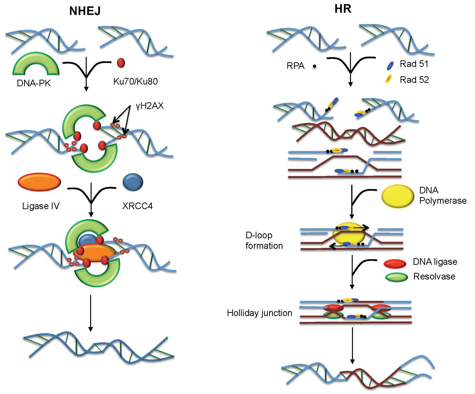
![]() |
[64] |
Ringrose L, Paro R (2007) Polycomb/Trithorax response elements and epigenetic memory of cell identity. Development 134: 223-232. doi: 10.1242/dev.02723
![]() |
[65] |
Dekker J, Marti-Renom MA, Mirny LA (2013) Exploring the three-dimensional organization of genomes: interpreting chromatin interaction data. Nat Rev Genet 14: 390-403. doi: 10.1038/nrg3454
![]() |
[66] |
Dixon JR, Selvaraj S, Yue F, et al. (2012) Topological domains in mammalian genomes identified by analysis of chromatin interactions. Nature 485: 376-380. doi: 10.1038/nature11082
![]() |
[67] |
Hou C, Li L, Qin ZS, et al. (2012) Gene density, transcription, and insulators contribute to the partition of the Drosophila genome into physical domains. Mol Cell 48: 471-484. doi: 10.1016/j.molcel.2012.08.031
![]() |
[68] |
Andrey G, Montavon T, Mascrez B, et al. (2013) A switch between topological domains underlies HoxD genes collinearity in mouse limbs. Science 340: 1234167. doi: 10.1126/science.1234167
![]() |
[69] |
Cremer T, Cremer M, Dietzel S, et al. (2006) Chromosome territories--a functional nuclear landscape. Curr Opin Cell Biol 18: 307-316. doi: 10.1016/j.ceb.2006.04.007
![]() |
[70] |
Parada LA, McQueen PG, Misteli T (2004) Tissue-specific spatial organization of genomes. Genome Biol 5: R44. doi: 10.1186/gb-2004-5-7-r44
![]() |
[71] |
Tanabe H, Muller S, Neusser M, et al. (2002) Evolutionary conservation of chromosome territory arrangements in cell nuclei from higher primates. Proc Natl Acad Sci U S A 99: 4424-4429. doi: 10.1073/pnas.072618599
![]() |
[72] |
Meaburn KJ, Misteli T (2007) Cell biology: chromosome territories. Nature 445: 379-781. doi: 10.1038/445379a
![]() |
[73] | Cremer T, Kreth G, Koester H, et al. (2000) Chromosome territories, interchromatin domain compartment, and nuclear matrix: an integrated view of the functional nuclear architecture. Crit Rev Eukaryot Gene Expr 10: 179-212. |
[74] |
Schneider R, Grosschedl R (2007) Dynamics and interplay of nuclear architecture, genome organization, and gene expression. Genes Dev 21: 3027-3043. doi: 10.1101/gad.1604607
![]() |
[75] |
Cremer M, von Hase J, Volm T, et al. (2001) Non-random radial higher-order chromatin arrangements in nuclei of diploid human cells. Chromosome Res 9: 541-567. doi: 10.1023/A:1012495201697
![]() |
[76] |
Rajapakse I, Groudine M (2011) On emerging nuclear order. J Cell Biol 192: 711-721. doi: 10.1083/jcb.201010129
![]() |
[77] |
Albiez H, Cremer M, Tiberi C, et al. (2006) Chromatin domains and the interchromatin compartment form structurally defined and functionally interacting nuclear networks. Chromosome Res 14: 707-733. doi: 10.1007/s10577-006-1086-x
![]() |
[78] |
Mahy NL, Perry PE, Gilchrist S, et al. (2002) Spatial organization of active and inactive genes and noncoding DNA within chromosome territories. J Cell Biol 157: 579-589. doi: 10.1083/jcb.200111071
![]() |
[79] |
Branco MR, Pombo A (2006) Intermingling of chromosome territories in interphase suggests role in translocations and transcription-dependent associations. PLoS Biol 4: e138. doi: 10.1371/journal.pbio.0040138
![]() |
[80] |
Meister P, Taddei A (2013) Building silent compartments at the nuclear periphery: a recurrent theme. Curr Opin Genet Dev 23: 96-103. doi: 10.1016/j.gde.2012.12.001
![]() |
[81] |
Deniaud E, Bickmore WA (2009) Transcription and the nuclear periphery: edge of darkness? Curr Opin Genet Dev 19: 187-191. doi: 10.1016/j.gde.2009.01.005
![]() |
[82] |
Andrulis ED, Neiman AM, Zappulla DC, et al. (1998) Perinuclear localization of chromatin facilitates transcriptional silencing. Nature 394: 592-595. doi: 10.1038/29100
![]() |
[83] |
Finlan LE, Sproul D, Thomson I, et al. (2008) Recruitment to the nuclear periphery can alter expression of genes in human cells. PLoS Genet 4: e1000039. doi: 10.1371/journal.pgen.1000039
![]() |
[84] |
Towbin BD, Gonzalez-Aguilera C, Sack R, et al. (2012) Step-wise methylation of histone H3K9 positions heterochromatin at the nuclear periphery. Cell 150: 934-947. doi: 10.1016/j.cell.2012.06.051
![]() |
[85] |
Steglich B, Sazer S, Ekwall K (2013) Transcriptional regulation at the yeast nuclear envelope. Nucleus 4: 379-389. doi: 10.4161/nucl.26394
![]() |
[86] |
Dundr M (2012) Nuclear bodies: multifunctional companions of the genome. Curr Opin Cell Biol 24: 415-422. doi: 10.1016/j.ceb.2012.03.010
![]() |
[87] |
Zhu L, Brangwynne CP (2015) Nuclear bodies: the emerging biophysics of nucleoplasmic phases. Curr Opin Cell Biol 34: 23-30. doi: 10.1016/j.ceb.2015.04.003
![]() |
[88] |
Cmarko D, Verschure PJ, Otte AP, et al. (2003) Polycomb group gene silencing proteins are concentrated in the perichromatin compartment of the mammalian nucleus. J Cell Sci 116: 335-343. doi: 10.1242/jcs.00225
![]() |
[89] |
Isono K, Endo TA, Ku M, et al. (2013) SAM domain polymerization links subnuclear clustering of PRC1 to gene silencing. Dev Cell 26: 565-577. doi: 10.1016/j.devcel.2013.08.016
![]() |
[90] |
Gonzalez I, Mateos-Langerak J, Thomas A, et al. (2014) Identification of regulators of the three-dimensional polycomb organization by a microscopy-based genome-wide RNAi screen. Mol Cell 54: 485-499. doi: 10.1016/j.molcel.2014.03.004
![]() |
[91] |
Vandenbunder B, Fourre N, Leray A, et al. (2014) PRC1 components exhibit different binding kinetics in Polycomb bodies. Biol Cell 106: 111-125. doi: 10.1111/boc.201300077
![]() |
[92] |
Cheutin T, Cavalli G (2012) Progressive Polycomb Assembly on H3K27me3 Compartments Generates Polycomb Bodies with Developmentally Regulated Motion. PLoS Genet 8: e1002465. doi: 10.1371/journal.pgen.1002465
![]() |
[93] |
Ren X, Vincenz C, Kerppola TK (2008) Changes in the distributions and dynamics of polycomb repressive complexes during embryonic stem cell differentiation. Mol Cell Biol 28: 2884-2895. doi: 10.1128/MCB.00949-07
![]() |
[94] |
Gao Z, Lee P, Stafford JM, et al. (2014) An AUTS2-Polycomb complex activates gene expression in the CNS. Nature 516: 349-354. doi: 10.1038/nature13921
![]() |
[95] | Mousavi K, Zare H, Wang AH, et al. (2011) Polycomb Protein Ezh1 Promotes RNA Polymerase II Elongation. Mol Cell. |
[96] |
Goldman RD, Gruenbaum Y, Moir RD, et al. (2002) Nuclear lamins: building blocks of nuclear architecture. Genes Dev 16: 533-547. doi: 10.1101/gad.960502
![]() |
[97] |
Goldman AE, Maul G, Steinert PM, et al. (1986) Keratin-like proteins that coisolate with intermediate filaments of BHK-21 cells are nuclear lamins. Proc Natl Acad Sci U S A 83: 3839-3843. doi: 10.1073/pnas.83.11.3839
![]() |
[98] |
McKeon FD, Kirschner MW, Caput D (1986) Homologies in both primary and secondary structure between nuclear envelope and intermediate filament proteins. Nature 319: 463-468. doi: 10.1038/319463a0
![]() |
[99] | Rober RA, Sauter H, Weber K, et al. (1990) Cells of the cellular immune and hemopoietic system of the mouse lack lamins A/C: distinction versus other somatic cells. J Cell Sci 95 ( Pt 4): 587-598. |
[100] |
Solovei I, Wang AS, Thanisch K, et al. (2013) LBR and lamin A/C sequentially tether peripheral heterochromatin and inversely regulate differentiation. Cell 152: 584-598. doi: 10.1016/j.cell.2013.01.009
![]() |
[101] | Guilly MN, Bensussan A, Bourge JF, et al. (1987) A human T lymphoblastic cell line lacks lamins A and C. Embo J 6: 3795-3799. |
[102] |
Stewart C, Burke B (1987) Teratocarcinoma stem cells and early mouse embryos contain only a single major lamin polypeptide closely resembling lamin B. Cell 51: 383-392. doi: 10.1016/0092-8674(87)90634-9
![]() |
[103] |
Kolb T, Maass K, Hergt M, et al. (2011) Lamin A and lamin C form homodimers and coexist in higher complex forms both in the nucleoplasmic fraction and in the lamina of cultured human cells. Nucleus 2: 425-433. doi: 10.4161/nucl.2.5.17765
![]() |
[104] |
Shimi T, Pfleghaar K, Kojima S, et al. (2008) The A- and B-type nuclear lamin networks: microdomains involved in chromatin organization and transcription. Genes Dev 22: 3409-3421. doi: 10.1101/gad.1735208
![]() |
[105] |
Dubinska-Magiera M, Zaremba-Czogalla M, Rzepecki R (2013) Muscle development, regeneration and laminopathies: how lamins or lamina-associated proteins can contribute to muscle development, regeneration and disease. Cell Mol Life Sci 70: 2713-2741. doi: 10.1007/s00018-012-1190-3
![]() |
[106] |
Collas P, Lund EG, Oldenburg AR (2014) Closing the (nuclear) envelope on the genome: How nuclear lamins interact with promoters and modulate gene expression. Bioessays 36: 75-83. doi: 10.1002/bies.201300138
![]() |
[107] |
Meuleman W, Peric-Hupkes D, Kind J, et al. (2013) Constitutive nuclear lamina-genome interactions are highly conserved and associated with A/T-rich sequence. Genome Res 23: 270-280. doi: 10.1101/gr.141028.112
![]() |
[108] |
Kind J, Pagie L, Ortabozkoyun H, et al. (2013) Single-cell dynamics of genome-nuclear lamina interactions. Cell 153: 178-192. doi: 10.1016/j.cell.2013.02.028
![]() |
[109] |
Kind J, van Steensel B (2010) Genome-nuclear lamina interactions and gene regulation. Curr Opin Cell Biol 22: 320-325. doi: 10.1016/j.ceb.2010.04.002
![]() |
[110] | Lund E, Oldenburg A, Delbarre E, et al. (2013) Lamin A/C-promoter interactions specify chromatin state-dependent transcription outcomes. Genome Res. |
[111] |
Pickersgill H, Kalverda B, de Wit E, et al. (2006) Characterization of the Drosophila melanogaster genome at the nuclear lamina. Nat Genet 38: 1005-1014. doi: 10.1038/ng1852
![]() |
[112] |
Guelen L, Pagie L, Brasset E, et al. (2008) Domain organization of human chromosomes revealed by mapping of nuclear lamina interactions. Nature 453: 948-951. doi: 10.1038/nature06947
![]() |
[113] |
Kind J, Pagie L, de Vries SS, et al. (2015) Genome-wide Maps of Nuclear Lamina Interactions in Single Human Cells. Cell 163: 134-147. doi: 10.1016/j.cell.2015.08.040
![]() |
[114] | Mattout A, Pike BL, Towbin BD, et al. (2011) An EDMD mutation in C. elegans lamin blocks muscle-specific gene relocation and compromises muscle integrity. Curr Biol 21: 1603-1614. |
[115] |
Peric-Hupkes D, Meuleman W, Pagie L, et al. (2010) Molecular maps of the reorganization of genome-nuclear lamina interactions during differentiation. Mol Cell 38: 603-613. doi: 10.1016/j.molcel.2010.03.016
![]() |
[116] |
Zaidi SK, Young DW, Montecino MA, et al. (2010) Mitotic bookmarking of genes: a novel dimension to epigenetic control. Nat Rev Genet 11: 583-589. doi: 10.1038/nrg2827
![]() |
[117] |
Kind J, van Steensel B (2014) Stochastic genome-nuclear lamina interactions: Modulating roles of Lamin A and BAF. Nucleus 5: 124-130. doi: 10.4161/nucl.28825
![]() |
[118] |
Amendola M, van Steensel B (2015) Nuclear lamins are not required for lamina-associated domain organization in mouse embryonic stem cells. EMBO Rep 16: 610-617. doi: 10.15252/embr.201439789
![]() |
[119] |
Osmanagic-Myers S, Dechat T, Foisner R (2015) Lamins at the crossroads of mechanosignaling. Genes Dev 29: 225-237. doi: 10.1101/gad.255968.114
![]() |
[120] |
Haque F, Lloyd DJ, Smallwood DT, et al. (2006) SUN1 interacts with nuclear lamin A and cytoplasmic nesprins to provide a physical connection between the nuclear lamina and the cytoskeleton. Mol Cell Biol 26: 3738-3751. doi: 10.1128/MCB.26.10.3738-3751.2006
![]() |
[121] | Gruenbaum Y, Foisner R (2015) Lamins: Nuclear Intermediate Filament Proteins with Fundamental Functions in Nuclear Mechanics and Genome Regulation. Annu Rev Biochem. |
[122] |
Swift J, Ivanovska IL, Buxboim A, et al. (2013) Nuclear lamin-A scales with tissue stiffness and enhances matrix-directed differentiation. Science 341: 1240104. doi: 10.1126/science.1240104
![]() |
[123] |
Padiath QS, Saigoh K, Schiffmann R, et al. (2006) Lamin B1 duplications cause autosomal dominant leukodystrophy. Nat Genet 38: 1114-1123. doi: 10.1038/ng1872
![]() |
[124] |
Hegele RA, Cao H, Liu DM, et al. (2006) Sequencing of the reannotated LMNB2 gene reveals novel mutations in patients with acquired partial lipodystrophy. Am J Hum Genet 79: 383-389. doi: 10.1086/505885
![]() |
[125] | Harborth J, Elbashir SM, Bechert K, et al. (2001) Identification of essential genes in cultured mammalian cells using small interfering RNAs. J Cell Sci 114: 4557-4565. |
[126] | Zaremba-Czogalla M, Dubinska-Magiera M, Rzepecki R (2011) Laminopathies: the molecular background of the disease and the prospects for its treatment. Cell Mol Biol Lett 16: 114-148. |
[127] |
Broers JL, Peeters EA, Kuijpers HJ, et al. (2004) Decreased mechanical stiffness in LMNA-/- cells is caused by defective nucleo-cytoskeletal integrity: implications for the development of laminopathies. Hum Mol Genet 13: 2567-2580. doi: 10.1093/hmg/ddh295
![]() |
[128] |
Sullivan T, Escalante-Alcalde D, Bhatt H, et al. (1999) Loss of A-type lamin expression compromises nuclear envelope integrity leading to muscular dystrophy. J Cell Biol 147: 913-920. doi: 10.1083/jcb.147.5.913
![]() |
[129] |
Hernandez L, Roux KJ, Wong ES, et al. (2010) Functional coupling between the extracellular matrix and nuclear lamina by Wnt signaling in progeria. Dev Cell 19: 413-425. doi: 10.1016/j.devcel.2010.08.013
![]() |
[130] | Sinha JK, Ghosh S, Raghunath M (2014) Progeria: a rare genetic premature ageing disorder. Indian J Med Res 139: 667-674. |
[131] |
McCord RP, Nazario-Toole A, Zhang H, et al. (2013) Correlated alterations in genome organization, histone methylation, and DNA-lamin A/C interactions in Hutchinson-Gilford progeria syndrome. Genome Res 23: 260-269. doi: 10.1101/gr.138032.112
![]() |
[132] |
Shumaker DK, Dechat T, Kohlmaier A, et al. (2006) Mutant nuclear lamin A leads to progressive alterations of epigenetic control in premature aging. Proc Natl Acad Sci U S A 103: 8703-8708. doi: 10.1073/pnas.0602569103
![]() |
[133] |
Landires I, Pascale JM, Motta J (2007) The position of the mutation within the LMNA gene determines the type and extent of tissue involvement in laminopathies. Clin Genet 71: 592-593; author reply 594-596. doi: 10.1111/j.1399-0004.2007.00772.x
![]() |
[134] |
Scharner J, Gnocchi VF, Ellis JA, et al. (2010) Genotype-phenotype correlations in laminopathies: how does fate translate? Biochem Soc Trans 38: 257-262. doi: 10.1042/BST0380257
![]() |
[135] |
Sewry CA, Brown SC, Mercuri E, et al. (2001) Skeletal muscle pathology in autosomal dominant Emery-Dreifuss muscular dystrophy with lamin A/C mutations. Neuropathol Appl Neurobiol 27: 281-290. doi: 10.1046/j.0305-1846.2001.00323.x
![]() |
[136] | Cesarini E, Mozzetta C, Marullo F, et al. (2015) Lamin A/C sustains PcG proteins architecture maintaining transcriptional repression at target genes. J Cell Biol. |
[137] |
Ptak C, Aitchison JD, Wozniak RW (2014) The multifunctional nuclear pore complex: a platform for controlling gene expression. Curr Opin Cell Biol 28: 46-53. doi: 10.1016/j.ceb.2014.02.001
![]() |
[138] |
Blobel G (1985) Gene gating: a hypothesis. Proc Natl Acad Sci U S A 82: 8527-8529. doi: 10.1073/pnas.82.24.8527
![]() |
[139] |
Cabal GG, Genovesio A, Rodriguez-Navarro S, et al. (2006) SAGA interacting factors confine sub-diffusion of transcribed genes to the nuclear envelope. Nature 441: 770-773. doi: 10.1038/nature04752
![]() |
[140] |
Taddei A, Van Houwe G, Hediger F, et al. (2006) Nuclear pore association confers optimal expression levels for an inducible yeast gene. Nature 441: 774-778. doi: 10.1038/nature04845
![]() |
[141] |
Tan-Wong SM, Wijayatilake HD, Proudfoot NJ (2009) Gene loops function to maintain transcriptional memory through interaction with the nuclear pore complex. Genes Dev 23: 2610-2624. doi: 10.1101/gad.1823209
![]() |
[142] |
Green EM, Jiang Y, Joyner R, et al. (2012) A negative feedback loop at the nuclear periphery regulates GAL gene expression. Mol Biol Cell 23: 1367-1375. doi: 10.1091/mbc.E11-06-0547
![]() |
[143] | Yoshida T, Shimada K, Oma Y, et al. (2010) Actin-related protein Arp6 influences H2A.Z-dependent and -independent gene expression and links ribosomal protein genes to nuclear pores. PLoS Genet 6: e1000910. |
[144] |
Van de Vosse DW, Wan Y, Lapetina DL, et al. (2013) A role for the nucleoporin Nup170p in chromatin structure and gene silencing. Cell 152: 969-983. doi: 10.1016/j.cell.2013.01.049
![]() |
[145] |
Galy V, Olivo-Marin JC, Scherthan H, et al. (2000) Nuclear pore complexes in the organization of silent telomeric chromatin. Nature 403: 108-112. doi: 10.1038/47528
![]() |
[146] |
Buchwalter AL, Liang Y, Hetzer MW (2014) Nup50 is required for cell differentiation and exhibits transcription-dependent dynamics. Mol Biol Cell 25: 2472-2484. doi: 10.1091/mbc.E14-04-0865
![]() |
[147] |
Gomez-Cavazos JS, Hetzer MW (2015) The nucleoporin gp210/Nup210 controls muscle differentiation by regulating nuclear envelope/ER homeostasis. J Cell Biol 208: 671-681. doi: 10.1083/jcb.201410047
![]() |
[148] |
Iwamoto M, Koujin T, Osakada H, et al. (2015) Biased assembly of the nuclear pore complex is required for somatic and germline nuclear differentiation in Tetrahymena. J Cell Sci 128: 1812-1823. doi: 10.1242/jcs.167353
![]() |
[149] |
Liang Y, Franks TM, Marchetto MC, et al. (2013) Dynamic association of NUP98 with the human genome. PLoS Genet 9: e1003308. doi: 10.1371/journal.pgen.1003308
![]() |
[150] |
Lupu F, Alves A, Anderson K, et al. (2008) Nuclear pore composition regulates neural stem/progenitor cell differentiation in the mouse embryo. Dev Cell 14: 831-842. doi: 10.1016/j.devcel.2008.03.011
![]() |
[151] |
D'Angelo MA, Gomez-Cavazos JS, Mei A, et al. (2012) A change in nuclear pore complex composition regulates cell differentiation. Dev Cell 22: 446-458. doi: 10.1016/j.devcel.2011.11.021
![]() |
[152] |
Lin Y, Yang Y, Li W, et al. (2012) Reciprocal regulation of Akt and Oct4 promotes the self-renewal and survival of embryonal carcinoma cells. Mol Cell 48: 627-640. doi: 10.1016/j.molcel.2012.08.030
![]() |
[153] | Yang J, Cai N, Yi F, et al. (2014) Gating pluripotency via nuclear pores. Trends Mol Med 20: 1-7. |
[154] |
Zhang X, Chen S, Yoo S, et al. (2008) Mutation in nuclear pore component NUP155 leads to atrial fibrillation and early sudden cardiac death. Cell 135: 1017-1027. doi: 10.1016/j.cell.2008.10.022
![]() |
[155] |
Basel-Vanagaite L, Muncher L, Straussberg R, et al. (2006) Mutated nup62 causes autosomal recessive infantile bilateral striatal necrosis. Ann Neurol 60: 214-222. doi: 10.1002/ana.20902
![]() |
[156] |
van Koningsbruggen S, Gierlinski M, Schofield P, et al. (2010) High-resolution whole-genome sequencing reveals that specific chromatin domains from most human chromosomes associate with nucleoli. Mol Biol Cell 21: 3735-3748. doi: 10.1091/mbc.E10-06-0508
![]() |
[157] |
Nemeth A, Conesa A, Santoyo-Lopez J, et al. (2010) Initial genomics of the human nucleolus. PLoS Genet 6: e1000889. doi: 10.1371/journal.pgen.1000889
![]() |
[158] |
Kim SK, Lee H, Han K, et al. (2014) SET7/9 methylation of the pluripotency factor LIN28A is a nucleolar localization mechanism that blocks let-7 biogenesis in human ESCs. Cell Stem Cell 15: 735-749. doi: 10.1016/j.stem.2014.10.016
![]() |
[159] |
Savic N, Bar D, Leone S, et al. (2014) lncRNA maturation to initiate heterochromatin formation in the nucleolus is required for exit from pluripotency in ESCs. Cell Stem Cell 15: 720-734. doi: 10.1016/j.stem.2014.10.005
![]() |
[160] | Talamas JA, Capelson M (2015) Nuclear envelope and genome interactions in cell fate. Front Genet 6: 95. |
[161] |
Fischer AH (2014) The diagnostic pathology of the nuclear envelope in human cancers. Adv Exp Med Biol 773: 49-75. doi: 10.1007/978-1-4899-8032-8_3
![]() |
[162] |
de Las Heras JI, Schirmer EC (2014) The nuclear envelope and cancer: a diagnostic perspective and historical overview. Adv Exp Med Biol 773: 5-26. doi: 10.1007/978-1-4899-8032-8_1
![]() |
[163] |
Skvortsov S, Schafer G, Stasyk T, et al. (2011) Proteomics profiling of microdissected low- and high-grade prostate tumors identifies Lamin A as a discriminatory biomarker. J Proteome Res 10: 259-268. doi: 10.1021/pr100921j
![]() |
[164] |
Willis ND, Cox TR, Rahman-Casans SF, et al. (2008) Lamin A/C is a risk biomarker in colorectal cancer. PLoS One 3: e2988. doi: 10.1371/journal.pone.0002988
![]() |
[165] |
Vargas JD, Hatch EM, Anderson DJ, et al. (2012) Transient nuclear envelope rupturing during interphase in human cancer cells. Nucleus 3: 88-100. doi: 10.4161/nucl.18954
![]() |
[166] |
Fischer AH, Bardarov S, Jr., Jiang Z (2004) Molecular aspects of diagnostic nucleolar and nuclear envelope changes in prostate cancer. J Cell Biochem 91: 170-184. doi: 10.1002/jcb.10735
![]() |
[167] |
Suresh S (2007) Biomechanics and biophysics of cancer cells. Acta Biomater 3: 413-438. doi: 10.1016/j.actbio.2007.04.002
![]() |
[168] |
Lammerding J, Fong LG, Ji JY, et al. (2006) Lamins A and C but not lamin B1 regulate nuclear mechanics. J Biol Chem 281: 25768-25780. doi: 10.1074/jbc.M513511200
![]() |
[169] | Chow KH, Factor RE, Ullman KS (2012) The nuclear envelope environment and its cancer connections. Nat Rev Cancer 12: 196-209. |
[170] |
Mjelle R, Hegre SA, Aas PA, et al. (2015) Cell cycle regulation of human DNA repair and chromatin remodeling genes. DNA Repair (Amst) 30: 53-67. doi: 10.1016/j.dnarep.2015.03.007
![]() |
[171] |
Yi C, He C (2013) DNA repair by reversal of DNA damage. Cold Spring Harb Perspect Biol 5: a012575. doi: 10.1101/cshperspect.a012575
![]() |
[172] | Davis AJ, Chen DJ (2013) DNA double strand break repair via non-homologous end-joining. Transl Cancer Res 2: 130-143. |
[173] |
Shroff R, Arbel-Eden A, Pilch D, et al. (2004) Distribution and dynamics of chromatin modification induced by a defined DNA double-strand break. Curr Biol 14: 1703-1711. doi: 10.1016/j.cub.2004.09.047
![]() |
[174] |
Jazayeri A, Falck J, Lukas C, et al. (2006) ATM- and cell cycle-dependent regulation of ATR in response to DNA double-strand breaks. Nat Cell Biol 8: 37-45. doi: 10.1038/ncb1337
![]() |
[175] |
Radhakrishnan SK, Jette N, Lees-Miller SP (2014) Non-homologous end joining: emerging themes and unanswered questions. DNA Repair (Amst) 17: 2-8. doi: 10.1016/j.dnarep.2014.01.009
![]() |
[176] |
Shrivastav M, De Haro LP, Nickoloff JA (2008) Regulation of DNA double-strand break repair pathway choice. Cell Res 18: 134-147. doi: 10.1038/cr.2007.111
![]() |
[177] |
Lemaitre C, Grabarz A, Tsouroula K, et al. (2014) Nuclear position dictates DNA repair pathway choice. Genes Dev 28: 2450-2463. doi: 10.1101/gad.248369.114
![]() |
[178] |
Aymard F, Bugler B, Schmidt CK, et al. (2014) Transcriptionally active chromatin recruits homologous recombination at DNA double-strand breaks. Nat Struct Mol Biol 21: 366-374. doi: 10.1038/nsmb.2796
![]() |
[179] |
Horigome C, Oma Y, Konishi T, et al. (2014) SWR1 and INO80 chromatin remodelers contribute to DNA double-strand break perinuclear anchorage site choice. Mol Cell 55: 626-639. doi: 10.1016/j.molcel.2014.06.027
![]() |
[180] |
Dion V, Gasser SM (2013) Chromatin movement in the maintenance of genome stability. Cell 152: 1355-1364. doi: 10.1016/j.cell.2013.02.010
![]() |
[181] |
Dion V, Kalck V, Horigome C, et al. (2012) Increased mobility of double-strand breaks requires Mec1, Rad9 and the homologous recombination machinery. Nat Cell Biol 14: 502-509. doi: 10.1038/ncb2465
![]() |
[182] |
Seeber A, Dion V, Gasser SM (2013) Checkpoint kinases and the INO80 nucleosome remodeling complex enhance global chromatin mobility in response to DNA damage. Genes Dev 27: 1999-2008. doi: 10.1101/gad.222992.113
![]() |
[183] |
Neumann FR, Dion V, Gehlen LR, et al. (2012) Targeted INO80 enhances subnuclear chromatin movement and ectopic homologous recombination. Genes Dev 26: 369-383. doi: 10.1101/gad.176156.111
![]() |
[184] |
Mahen R, Hattori H, Lee M, et al. (2013) A-type lamins maintain the positional stability of DNA damage repair foci in mammalian nuclei. PLoS One 8: e61893. doi: 10.1371/journal.pone.0061893
![]() |
[185] |
van Sluis M, McStay B (2015) A localized nucleolar DNA damage response facilitates recruitment of the homology-directed repair machinery independent of cell cycle stage. Genes Dev 29: 1151-1163. doi: 10.1101/gad.260703.115
![]() |
[186] |
Tang J, Cho NW, Cui G, et al. (2013) Acetylation limits 53BP1 association with damaged chromatin to promote homologous recombination. Nat Struct Mol Biol 20: 317-325. doi: 10.1038/nsmb.2499
![]() |
[187] |
Botuyan MV, Lee J, Ward IM, et al. (2006) Structural basis for the methylation state-specific recognition of histone H4-K20 by 53BP1 and Crb2 in DNA repair. Cell 127: 1361-1373. doi: 10.1016/j.cell.2006.10.043
![]() |
[188] | Misteli T, Soutoglou E (2009) The emerging role of nuclear architecture in DNA repair and genome maintenance. Nat Rev Mol Cell Biol 10: 243-254. |
[189] |
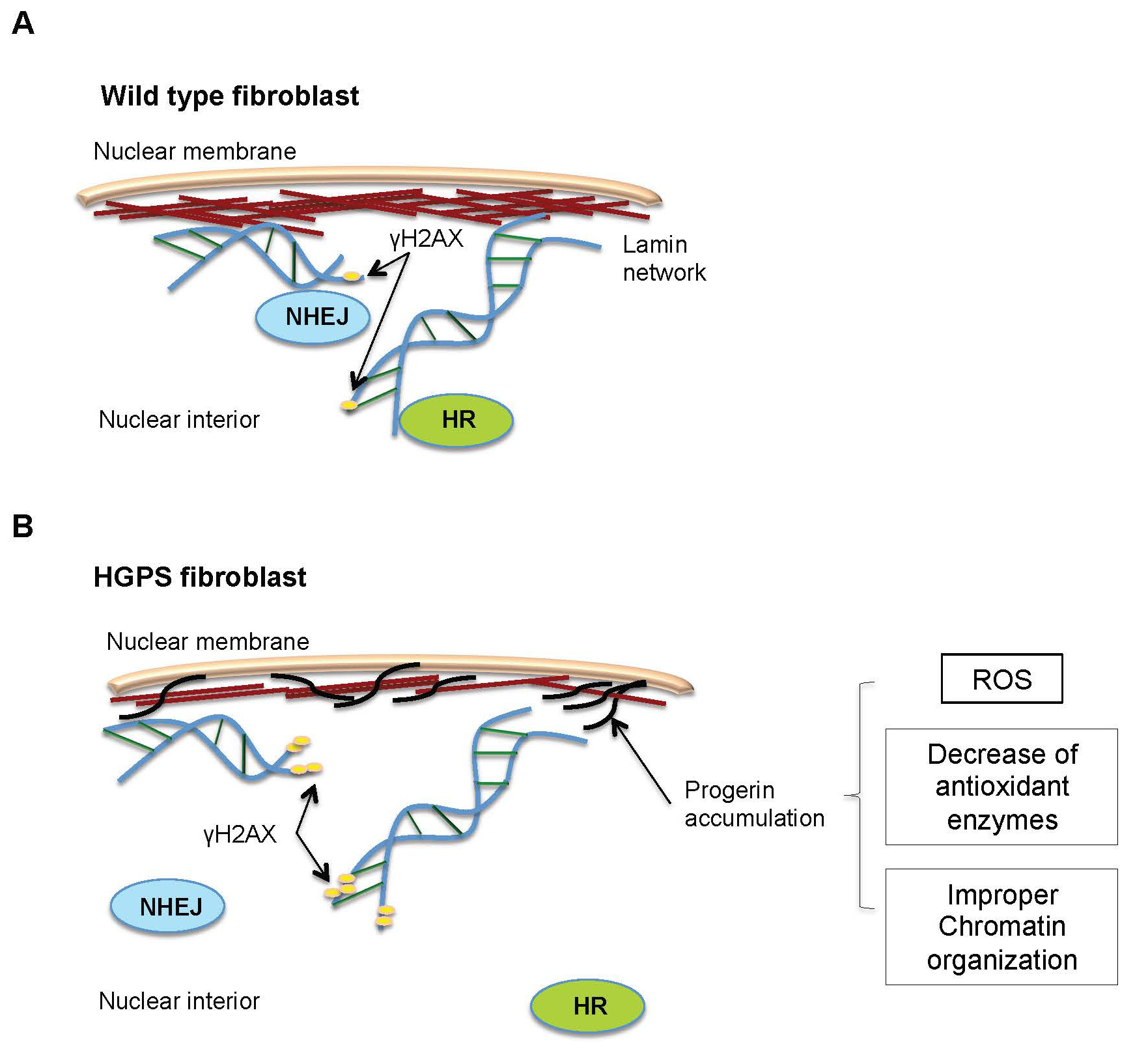
Constantinescu D, Csoka AB, Navara CS, et al. (2010) Defective DSB repair correlates with abnormal nuclear morphology and is improved with FTI treatment in Hutchinson-Gilford progeria syndrome fibroblasts. Exp Cell Res 316: 2747-2759. doi: 10.1016/j.yexcr.2010.05.015
![]() |
[190] |
Lattanzi G, Marmiroli S, Facchini A, et al. (2012) Nuclear damages and oxidative stress: new perspectives for laminopathies. Eur J Histochem 56: e45. doi: 10.4081/ejh.2012.e45
![]() |
[191] |
Richards SA, Muter J, Ritchie P, et al. (2011) The accumulation of un-repairable DNA damage in laminopathy progeria fibroblasts is caused by ROS generation and is prevented by treatment with N-acetyl cysteine. Hum Mol Genet 20: 3997-4004. doi: 10.1093/hmg/ddr327
![]() |
[192] |
Allen RG, Tresini M (2000) Oxidative stress and gene regulation. Free Radic Biol Med 28: 463-499. doi: 10.1016/S0891-5849(99)00242-7
![]() |
[193] |
Sieprath T, Corne T, Nooteboom M, et al. (2015) Sustained accumulation of prelamin A and depletion of lamin A/C both cause oxidative stress and mitochondrial dysfunction but induce different cell fates. Nucleus 6: 236-246. doi: 10.1080/19491034.2015.1050568
![]() |
[194] |
Pekovic V, Gibbs-Seymour I, Markiewicz E, et al. (2011) Conserved cysteine residues in the mammalian lamin A tail are essential for cellular responses to ROS generation. Aging Cell 10: 1067–1079. doi: 10.1111/j.1474-9726.2011.00750.x
![]() |
[195] |
Viteri G, Chung YW, Stadtman ER (2010) Effect of progerin on the accumulation of oxidized proteins in fibroblasts from Hutchinson Gilford progeria patients. Mech Ageing Dev 131: 2–8. doi: 10.1016/j.mad.2009.11.006
![]() |
[196] |
Csoka AB, Cao H, Sammak PJ, et al. (2004) Novel lamin A/C gene (LMNA) mutations in atypical progeroid syndromes. J Med Genet 41: 304–308. doi: 10.1136/jmg.2003.015651
![]() |
[197] |
Doubaj Y, De Sandre-Giovannoli A, Vera EV, et al. (2012) An inherited LMNA gene mutation in atypical Progeria syndrome. Am J Med Genet A 158A: 2881–2887. doi: 10.1002/ajmg.a.35557
![]() |
[198] | Seco-Cervera M, Spis M, Garcia-Gimenez JL, et al. (2014) Oxidative stress and antioxidant response in fibroblasts from Werner and atypical Werner syndromes. Aging (Albany NY) 6: 231–245. |
[199] |
Schroder AR, Shinn P, Chen H, et al. (2002) HIV-1 integration in the human genome favors active genes and local hotspots. Cell 110: 521–529. doi: 10.1016/S0092-8674(02)00864-4
![]() |
[200] | Wong RW, Mamede JI, Hope TJ (2015) The Impact of Nucleoporin Mediated Chromatin localization and Nuclear Architecture on HIV Integration Site Selection. J Virol. |
[201] |
Marini B, Kertesz-Farkas A, Ali H, et al. (2015) Nuclear architecture dictates HIV-1 integration site selection. Nature 521: 227–231.202. Lelek M, Casartelli N, Pellin D, et al. (2015) Chromatin organization at the nuclear pore favours HIV replication. Nat Commun 6: 6483. doi: 10.1038/nature14226
![]() |
1. | Vladimir B. Teif, Andrey G. Cherstvy, Chromatin and epigenetics: current biophysical views, 2016, 3, 2377-9098, 88, 10.3934/biophy.2016.1.88 | |
2. | F. Marullo, E. Cesarini, L. Antonelli, F. Gregoretti, G. Oliva, C. Lanzuolo, Nucleoplasmic Lamin A/C and Polycomb group of proteins: An evolutionarily conserved interplay, 2016, 7, 1949-1034, 103, 10.1080/19491034.2016.1157675 | |
3. | Federica Marasca, Fabrizia Marullo, Chiara Lanzuolo, 2016, Chapter 15, 978-1-4939-6378-2, 167, 10.1007/978-1-4939-6380-5_15 | |
4. | Andrea Bianchi, Pierluigi Giuseppe Manti, Federica Lucini, Chiara Lanzuolo, Mechanotransduction, nuclear architecture and epigenetics in Emery Dreifuss Muscular Dystrophy: tous pour un, un pour tous, 2018, 9, 1949-1034, 321, 10.1080/19491034.2018.1460044 | |
5. | Endre Sebestyén, Fabrizia Marullo, Federica Lucini, Cristiano Petrini, Andrea Bianchi, Sara Valsoni, Ilaria Olivieri, Laura Antonelli, Francesco Gregoretti, Gennaro Oliva, Francesco Ferrari, Chiara Lanzuolo, SAMMY-seq reveals early alteration of heterochromatin and deregulation of bivalent genes in Hutchinson-Gilford Progeria Syndrome, 2020, 11, 2041-1723, 10.1038/s41467-020-20048-9 | |
6. | Austė Kanapeckaitė, Neringa Burokienė, Asta Mažeikienė, Graeme S. Cottrell, Darius Widera, Biophysics is reshaping our perception of the epigenome: from DNA-level to high-throughput studies, 2021, 1, 26670747, 100028, 10.1016/j.bpr.2021.100028 | |
7. | Philina Santarelli, Valentina Rosti, Maria Vivo, Chiara Lanzuolo, 2024, 158, 9780128234358, 375, 10.1016/bs.ctdb.2024.01.014 | |
8. | Maria Vivo, Valentina Rosti, Sara Cervone, Chiara Lanzuolo, Chromatin plasticity in mechanotransduction, 2024, 88, 09550674, 102376, 10.1016/j.ceb.2024.102376 |
NUCLEAR ARCHITECTURE COMPONENTS | MUTATIONS | ASSOCIATED DISEASE | REFERENCE |
Lamin A/C | 1824 C Cryptic splice variant at LMNA gene < T | Hutchinson-Gilford Progeria Syndrome (HGPS) | [130] |
Q6X, R25G/P, R50S/P, R133P, S143F, H222P, R249Q, Y267C, M371K, R377H/L, R453W, W498R, R527P/H, L530P, R644C | Emery Dreifuss Muscular dystrophy (EDMD) | [134] | |
S143P, E161K, R190W/Q, N195K, R377H/L, R644C | Dilated Cardiomyopathy (DCM) | [134] | |
R482W/Q, K486N, R60G, G62G, R133L | Familial Partial Lipodystrophy 2 (FPLD2) | [134] | |
Lamin B1 | Partial duplication of LMNB1 gene | Leukodystrophy (ADLD) | [123] |
Lamin B2 | R215Q, A407T | Acquired Partial Lipodystrophy (APL) | [124] |
Nup155 | R391H | Atrial fibrillation (AF) | [154] |
Nup62 | Q391P | Infatile bilateral striatal necrosis | [155] |