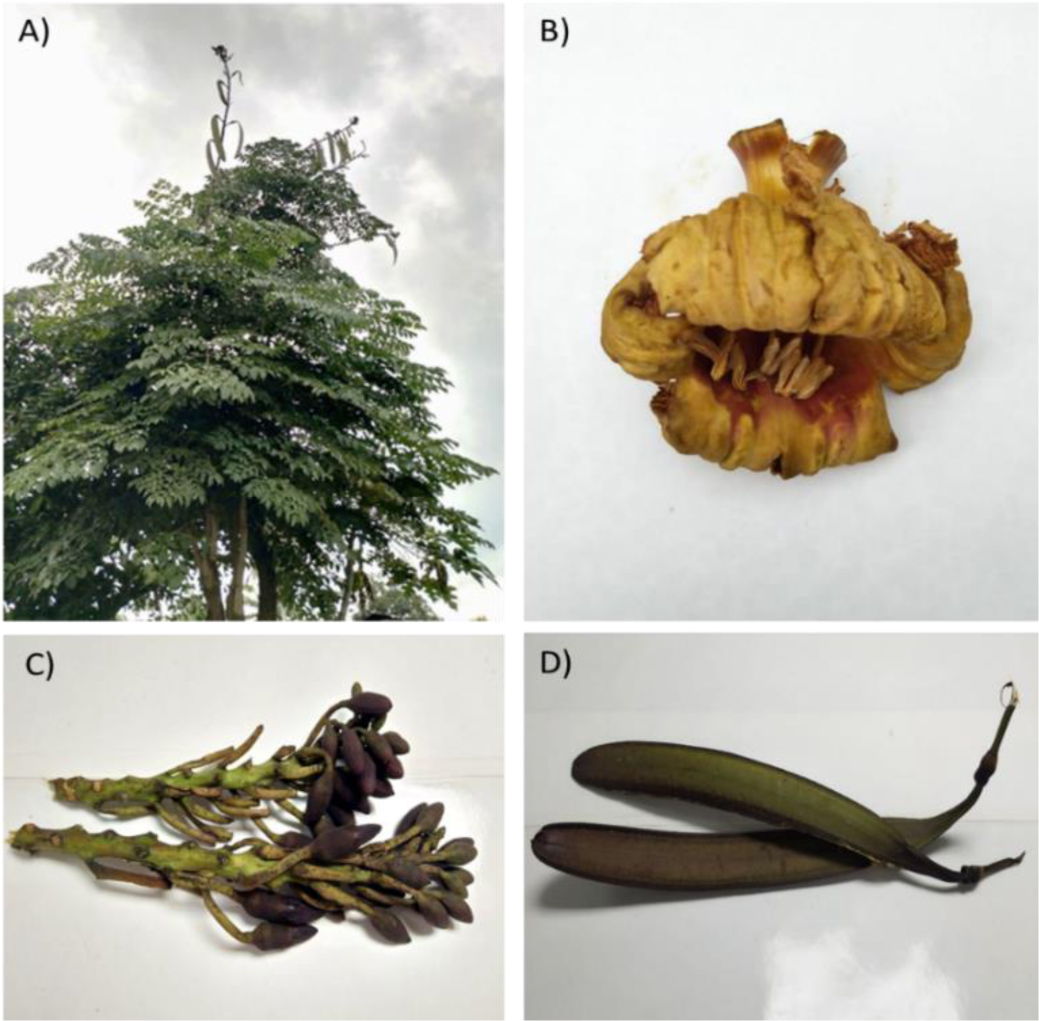
In recent years, numerous studies have discovered the potential of natural compounds extracted from medicinal plants as a promising alternative treatment for ischemic stroke (IS). Oroxylum indicum is among the plants that has been widely proven to contain a beneficial flavonoid compound known as baicalein. However, the therapeutic potential of this compound for IS disease has yet to be demonstrated. In this study, a baicalein-enriched fraction (BEF) was harvested from the leaves of O. indicum and tested on neural stem cells (NSCs) to determine its effects on the cell viability and gene expression prior to an in vivo animal study. NSCs were chosen because they are the primitive brain cells that play important roles in neurogenesis after IS injury. It was found that NSCs treated with BEF at concentration of 3.125 µg/mL for 48 hours showed a significant higher cell proliferation rate. Moreover, these cells expressed high NSC markers (Nestin and SOX2) and genes responsible for antioxidant (SOD2) and angiogenesis (ANGPT1) mechanisms, indicating that the BEF treatment could enhance the progenitor brain cells viability and the expression of cytokines that are beneficial to reduce oxidative stress and reinstate blood flow in an ischemic brain. Subsequently, the in vivo therapeutic effects of BEF for IS disease was tested by administering 50 mg/kg b.wt of BEF to male Sprague Dawley (SD) rats via oral gavage before and after induction of IS. Assessments of neurological impairments were performed using a modified neurological severity score (mNSS) and the quantification of the total infarct volume was evaluated as the endpoint of this study. Results showed that an oral administration of BEF improved the behavioral scoring for motor functions (forelimb flexion and forelimb twisting), contralateral sensory functions (paw-whiskers test), motor coordination, balance functions (beam balance test) and reflex functions (pinna, corneal, startle and tail reflex) within 24–72 h; alternatively, the non-treated rats showed continuously lower scores for all the tests throughout the study, indicating that the BEF-treated rats exhibited a faster recovery rate as compared to the non-treated rats. Such improvements were observed up to two weeks. In addition, histological assessment also revealed a reduction of infarct volume in the BEF-treated rats as compared to the control rats. In summary, the present study demonstrated the potential of BEF extracted from the O. indicum as a supplement to improve preclinical IS models.
Citation: Farah Amna Othman, Nik Nur Hakimah Nik Salleh, Suat Cheng Tan. Oral administration of baicalein-enriched fraction during pre- and post-ischemic stroke alleviated cognitive impairments in rat models[J]. AIMS Molecular Science, 2023, 10(4): 343-362. doi: 10.3934/molsci.2023020
In recent years, numerous studies have discovered the potential of natural compounds extracted from medicinal plants as a promising alternative treatment for ischemic stroke (IS). Oroxylum indicum is among the plants that has been widely proven to contain a beneficial flavonoid compound known as baicalein. However, the therapeutic potential of this compound for IS disease has yet to be demonstrated. In this study, a baicalein-enriched fraction (BEF) was harvested from the leaves of O. indicum and tested on neural stem cells (NSCs) to determine its effects on the cell viability and gene expression prior to an in vivo animal study. NSCs were chosen because they are the primitive brain cells that play important roles in neurogenesis after IS injury. It was found that NSCs treated with BEF at concentration of 3.125 µg/mL for 48 hours showed a significant higher cell proliferation rate. Moreover, these cells expressed high NSC markers (Nestin and SOX2) and genes responsible for antioxidant (SOD2) and angiogenesis (ANGPT1) mechanisms, indicating that the BEF treatment could enhance the progenitor brain cells viability and the expression of cytokines that are beneficial to reduce oxidative stress and reinstate blood flow in an ischemic brain. Subsequently, the in vivo therapeutic effects of BEF for IS disease was tested by administering 50 mg/kg b.wt of BEF to male Sprague Dawley (SD) rats via oral gavage before and after induction of IS. Assessments of neurological impairments were performed using a modified neurological severity score (mNSS) and the quantification of the total infarct volume was evaluated as the endpoint of this study. Results showed that an oral administration of BEF improved the behavioral scoring for motor functions (forelimb flexion and forelimb twisting), contralateral sensory functions (paw-whiskers test), motor coordination, balance functions (beam balance test) and reflex functions (pinna, corneal, startle and tail reflex) within 24–72 h; alternatively, the non-treated rats showed continuously lower scores for all the tests throughout the study, indicating that the BEF-treated rats exhibited a faster recovery rate as compared to the non-treated rats. Such improvements were observed up to two weeks. In addition, histological assessment also revealed a reduction of infarct volume in the BEF-treated rats as compared to the control rats. In summary, the present study demonstrated the potential of BEF extracted from the O. indicum as a supplement to improve preclinical IS models.

| [1] | Mergenthaler P, Dirnagl U, Kunz A (2021) Ischemic Stroke: Basic pathophysiology and clinical implication. Neuroscience in the 21st Century . New York, NY.: Springer 1-12. |
| [2] | Zhang S, Wang D, Li L (2023) Recombinant tissue-type plasminogen activator (rt-PA) effectively restores neurological function and improves prognosis in acute ischemic stroke. Am J Transl Res 15: 3460-3467. |
| [3] |
Mei T, Kim A, Vong LB, et al. (2019) Encapsulation of tissue plasminogen activator in pH-sensitive self-assembled antioxidant nanoparticles for ischemic stroke treatment–Synergistic effect of thrombolysis and antioxidant. Biomaterials 215: 119209. https://doi.org/10.1016/j.biomaterials.2019.05.020 
|
| [4] |
Tamta BP, Kumar R, Uniyal S, et al. (2022) Exploration and selection of elite germplasm of Oroxylum indicum (L.) Vent. (Shyonak) in the forest divisions of Punjab, India. Curr Sci 122: 1401-1406. https://doi.org/10.18520/cs/v122/i12/1401-1406
|
| [5] |
Abdulha F, Reduan MFH, Hisam AH, et al. (2022) LC-TOF-MS/MS and GC-MS based phytochemical pro fi ling and evaluation of wound healing activity of Oroxylum indicum (L.) Kurz (Beka). Front Pharmacol 13: 1050453. https://doi.org/10.3389/fphar.2022.1050453
|
| [6] |
Nik Salleh NNH, Othman FA, Kamarudin NA, et al. (2020) The biological activities and therapeutic potentials of baicalein extracted from Oroxylum indicum: A systematic review. Molecules 25: 5677. https://doi.org/10.3390/molecules25235677
|
| [7] |
Jagetia GC (2021) A review on the medicinal and pharmacological properties of traditional ethnomedicinal plant sonapatha, Oroxylum indicum. Sinusitis 5: 71-89. https://doi.org/10.3390/sinusitis5010009
|
| [8] |
Sowndhararajan K, Deepa P, Kim M, et al. (2017) Baicalein as a potent neuroprotective agent: A review. Biomed Pharmacother 95: 1021-1032. https://doi.org/10.1016/j.biopha.2017.08.135
|
| [9] |
Li Q, Li QQ, Jia JN, et al. (2019) Baicalein exerts neuroprotective effects in FeCl3-induced posttraumatic epileptic seizures via suppressing ferroptosis. Front Pharmacol 10: 638. https://doi.org/10.3389/fphar.2019.00638
|
| [10] |
Dey A, Nath De J (2015) Possible anti-Parkinson's disease therapeutics from nature: A review. Studies in natural products chemistry : 447-520. https://doi.org/10.1016/B978-0-444-63460-3.00009-2
|
| [11] |
Ji Y, Han J, Lee N, et al. (2020) Neuroprotective effects of baicalein, wogonin, and oroxylin A on amyloid beta-induced toxicity via NF-κB/MAPK pathway modulation. Molecules 25: 5087. https://doi.org/10.3390/molecules25215087
|
| [12] |
Wang CX, Xie GB, Zhou CH, et al. (2015) Baicalein alleviates early brain injury after experimental subarachnoid hemorrhage in rats: possible involvement of TLR4/NF-κB-mediated inflammatory pathway. Brain Res 1594: 245-255. https://doi.org/10.1016/j.brainres.2014.10.014
|
| [13] |
Kang IN, Salleh NNHN, Chung WJ, et al. (2019) Baicalein-enriched fraction extracted from Oroxylum indicum (L.) Benth. ex Kurz leaves exerts antioxidant and inhibitory effects against glioblastoma multiforme. Processes 7: 963. https://doi.org/10.3390/pr7120963
|
| [14] |
Ottoboni L, von Wunster B, Martino G (2020) Therapeutic plasticity of neural stem cells. Front Neurol 11: 148. https://doi.org/10.3389/fneur.2020.00148
|
| [15] |
Tornero D (2022) Neuronal circuitry reconstruction after stem cell therapy in damaged brain. Neural Regen Res 17: 1959-1960. https://doi.org/10.4103/1673-5374.335145
|
| [16] |
Guo X, Jin X, Han K, et al. (2022) Iron promotes neurological function recovery in mice with ischemic stroke through endogenous repair mechanisms. Free Radic Biol Med 182: 59-72. https://doi.org/10.1016/j.freeradbiomed.2022.02.017
|
| [17] |
Hamblin MH, Lee JP (2021) Neural stem cells for early ischemic stroke. Int J Mol Sci 22: 7703. https://doi.org/10.3390/ijms22147703
|
| [18] |
Flynn JM, Melovn S (2013) SOD2 in mitochondrial dysfunction and neurodegeneration. Free Radic Biol Med 62: 4-12. https://doi.org/10.1016/j.freeradbiomed.2013.05.027
|
| [19] |
Moxon JV, Trollope AF, Dewdney B, et al. (2019) The effect of angiopoietin-1 upregulation on the outcome of acute ischaemic stroke in rodent models: A meta-analysis. J Cereb Blood Flow Metab 39: 2343-2354. https://doi.org/10.1177/0271678X19876876
|
| [20] |
Chittiboina P, Ganta V, Monceaux CP, et al. (2013) Angiopoietins as promising biomarkers and potential therapeutic targets in brain injury. Pathophysiology 20: 15-21. https://doi.org/10.1016/j.pathophys.2012.02.004
|
| [21] |
Kang IN, Lee CY, Tan SC (2019) Selection of best reference genes for qRT-PCR analysis of human neural stem cells preconditioned with hypoxia or baicalein-enriched fraction extracted from Oroxylum indicum medicinal plant. Heliyon 5: e02156. https://doi.org/10.1016/j.heliyon.2019.e02156
|
| [22] |
Pavón-Fuentes N, Marín-Prida J, Llópiz-Arzuaga A, et al. (2020) Phycocyanobilin reduces brain injury after endothelin-1-induced focal cerebral ischaemia. Clin Exp Pharmacol Physiol 47: 383-392. https://doi.org/10.1111/1440-1681.13214
|
| [23] |
Mundugaru R, Sivanesan S, Popa-Wagner A, et al. (2018) Pluchea lanceolata protects hippocampal neurons from endothelin-1 induced ischemic injury to ameliorate cognitive deficits. J Chem Neuroanat 94: 75-85. https://doi.org/10.1016/j.jchemneu.2018.09.002
|
| [24] | Ansari S, Azari H, Caldwell KJ, et al. (2013) Endothelin-1 induced middle cerebral artery occlusion model for ischemic stroke with laser Doppler flowmetry guidance in rat. J Vis Exp 16: 50014. https://doi.org/10.3791/50014 |
| [25] |
Yamamoto PK, de Souza TA, Antiorio ATFB, et al. (2019) Genetic and behavioral characterization of a Kmt2d mouse mutant, a new model for Kabuki Syndrome. Genes Brain Behav 18: e12568. https://doi.org/10.1111/gbb.12568
|
| [26] |
Shaikh KT, Yang A, Youshin E, et al. (2015) Transgenic LRRK2 (R1441G) rats—A model for Parkinson disease. Peer J 3: e945. https://doi.org/10.7717/peerj.945
|
| [27] | Doeppner TR, Kaltwasser B, Bähr M, et al. (2014) Effects of neural progenitor cells on post-stroke neurological impairment—A detailed and comprehensive analysis of behavioral tests. Front Cell Neurosci 8: 338. https://doi.org/10.3389/fncel.2014.00338 |
| [28] |
Yarnell AM, Barry ES, Mountney A, et al. (2016) The revised neurobehavioral severity scale (NSS-R) for rodents. Curr Protoc Neurosci 75: 9.52.1-9.52.16. https://doi.org/10.1002/cpns.10
|
| [29] | Roth JM (2011) Recombinant tissue plasminogen activator for the treatment of acute ischemic stroke. Proc (Bayl Univ Med Cent) 24: 257-259. https://doi.org/10.1080/08998280.2011.11928729 |
| [30] |
Yperzeele L, Van Hooff RJ, De Smedt A, et al. (2014) Prehospital stroke care: Limitations of current interventions and focus on new developments. Cerebrovasc Dis 38: 1-9. https://doi.org/10.1159/000363617
|
| [31] |
Wang Y, Wei N, Li X (2020) Preclinical evidence and possible mechanisms of baicalein for rats and mice with Parkinson's disease: A systematic review and meta-analysis. Front Aging Neurosci 12: 277. https://doi.org/10.3389/fnagi.2020.00277
|
| [32] |
Jadhav R, Kulkarni YA (2023) Effects of baicalein with memantine on aluminium chloride-induced neurotoxicity in Wistar rats. Front pharmacol 14: 1034620. https://doi.org/10.3389/fphar.2023.1034620
|
| [33] |
Liang W, Huang X, Chen W (2017) The effects of baicalin and baicalein on cerebral ischemia: A review. Aging Dis 8: 850-867. https://doi.org/10.14336/ad.2017.0829
|
| [34] |
Li Y, Chen Q, Ran D, et al. (2019) Changes in the levels of 12/15-lipoxygenase, apoptosis-related proteins and inflammatory factors in the cortex of diabetic rats and the neuroprotection of baicalein. Free Radic Biol Med 134: 239-247. https://doi.org/10.1016/j.freeradbiomed.2019.01.019
|
| [35] |
Bernal A, Arranz L (2018) Nestin-expressing progenitor cells: function, identity and therapeutic implications. Cell Mol Life Sci 75: 2177-2195. https://doi.org/10.1007/s00018-018-2794-z
|
| [36] |
Vona R, Pallotta L, Cappelletti M, et al. (2021) The impact of oxidative stress in human pathology: focus on gastrointestinal disorders. Antioxidants 10: 201. https://doi.org/10.3390/antiox10020201
|
| [37] |
Kim Y, Cho AY, Kim HC, et al. (2022) Effects of natural polyphenols on oxidative stress-mediated blood-brain barrier dysfunction. Antioxidants 11: 197. https://doi.org/10.3390/antiox11020197
|
| [38] |
Lugano R, Ramachandran M, Dimberg A (2020) Tumor angiogenesis: causes, consequences, challenges and opportunities. Cell Mol Life Sci 77: 1745-1770. https://doi.org/10.1007/s00018-019-03351-7
|
| [39] |
Eklund L, Kangas J, Saharinen P (2017) Angiopoietin-Tie signalling in the cardiovascular and lymphatic systems. Clin Sci 131: 87-103. https://doi.org/10.1042/CS20160129
|
| [40] |
Pauzi NAM, Cheema MS, Ismail A, et al. (2021) Safety assessment of natural products in Malaysia: current practices, challenges, and new strategies. Rev Environ Health 37: 169-179. https://doi.org/10.1515/reveh-2021-0072
|
| [41] | National Pharmaceutical Regulatory AgencyGuidelines for control of cosmetic products in Malaysia (2022). |
| [42] |
Sarkar P, Nath K, Banu S (2019) Modulatory effect of baicalein on gene expression and activity of antioxidant enzymes in streptozotocin-nicotinamide induced diabetic rats. Braz J Pharm Sci 55: e18201. http://doi.org/10.1590/s2175-97902019000118201
|
| [43] |
Ren M, Zhao Y, He Z, et al. (2021) Baicalein inhibits inflammatory response and promotes osteogenic activity in periodontal ligament cells challenged with lipopolysaccharides. BMC Complement Med Ther 21: 43. http://doi.org/10.1186/s12906-021-03213-5
|
| [44] | Wang K, Zhang J, Li C, et al. (2019) Baicalin promotes angiogenesis of myocardial tissues in myocardial infarction rats. Int J Clin Exp Med 12: 8358-8365. |
| [45] |
Pawluk H, Woźniak A, Grześk G, et al. (2020) The role of selected pro-inflammatory cytokines in pathogenesis of ischemic stroke. Clin Interv Aging 15: 469-484. http://doi.org/10.2147/CIA.S233909
|
| [46] |
Liu HT, Lin YN, Tsai MC, et al. (2022) Baicalein exerts therapeutic effects against endotoxin-induced depression-like behavior in mice by decreasing inflammatory cytokines and increasing brain-derived neurotrophic factor levels. Antioxidants 11: 947. http://doi.org/10.3390/antiox11050947
|
| [47] |
Zheng ZV, Cheung CY, Hao L, et al. (2019) Baicalein enhances the effect of low dose Levodopa on the gait deficits and protects dopaminergic neurons in experimental Parkinsonism. J Clin Neurosci 64: 242-251. https://doi.org/10.1016/j.jocn.2019.02.005
|
| [48] |
Jadhav R, Kulkarni YA (2023) The combination of baicalein and memantine reduces oxidative stress and protects against β-amyloid-induced Alzheimer's disease in rat model. Antioxidants 12: 707. https://doi.org/10.3390/antiox12030707
|
| [49] | Pan L, Cho K-S, Irvin Y, et al. (2021) Baicalein, baicalin, and wogonin: Protective effects against ischemia-induced neurodegeneration in the brain and retina. Oxid Med Cell Longev 2021: 8377362. https://doi.org/10.1155/2021/8377362 |
| [50] |
Lin R, Lin J, Li S, et al. (2018) Effects of the traditional Chinese medicine baicalein on the viability of random pattern skin flaps in rats. Drug Des Devel Ther 12: 2267-2276. https://doi.org/10.2147/DDDT.S173371
|
| [51] |
Han J, Ji Y, Youn K, et al. (2019) Baicalein as a potential inhibitor against BACE1 and AChE: Mechanistic comprehension through in vitro and computational approaches. Nutrients 11: 2694. https://doi.org/10.3390/nu11112694
|
| [52] |
Kadry H, Noorani B, Cucullo L (2020) A blood–brain barrier overview on structure, function, impairment, and biomarkers of integrity. Fluids Barriers CNS 17: 69. https://doi.org/10.1186/s12987-020-00230-3
|
| [53] |
Teleanu RI, Preda MD, Niculescu AG, et al. (2022) Current strategies to enhance delivery of drugs across the blood–brain barrier. Pharmaceutics 14: 987. https://doi.org/10.3390/pharmaceutics14050987
|


Figures(7) / Tables(2)

Farah Amna Othman, Nik Nur Hakimah Nik Salleh, Suat Cheng Tan. Oral administration of baicalein-enriched fraction during pre- and post-ischemic stroke alleviated cognitive impairments in rat models[J]. AIMS Molecular Science, 2023, 10(4): 343-362. doi: 10.3934/molsci.2023020


 DownLoad: