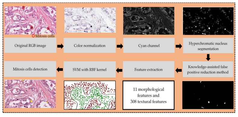
Based on the Nottingham Histopathology Grading (NHG) system, mitosis cells detection is one of the important criteria to determine the grade of breast carcinoma. Mitosis cells detection is a challenging task due to the heterogeneous microenvironment of breast histopathology images. Recognition of complex and inconsistent objects in the medical images could be achieved by incorporating domain knowledge in the field of interest. In this study, the strategies of the histopathologist and domain knowledge approach were used to guide the development of the image processing framework for automated mitosis cells detection in breast histopathology images. The detection framework starts with color normalization and hyperchromatic nucleus segmentation. Then, a knowledge-assisted false positive reduction method is proposed to eliminate the false positive (i.e., non-mitosis cells). This stage aims to minimize the percentage of false positive and thus increase the F1-score. Next, features extraction was performed. The mitosis candidates were classified using a Support Vector Machine (SVM) classifier. For evaluation purposes, the knowledge-assisted detection framework was tested using two datasets: a custom dataset and a publicly available dataset (i.e., MITOS dataset). The proposed knowledge-assisted false positive reduction method was found promising by eliminating at least 87.1% of false positive in both the dataset producing promising results in the F1-score. Experimental results demonstrate that the knowledge-assisted detection framework can achieve promising results in F1-score (custom dataset: 89.1%; MITOS dataset: 88.9%) and outperforms the recent works.
Citation: Xiao Jian Tan, Nazahah Mustafa, Mohd Yusoff Mashor, Khairul Shakir Ab Rahman. Automated knowledge-assisted mitosis cells detection framework in breast histopathology images[J]. Mathematical Biosciences and Engineering, 2022, 19(2): 1721-1745. doi: 10.3934/mbe.2022081
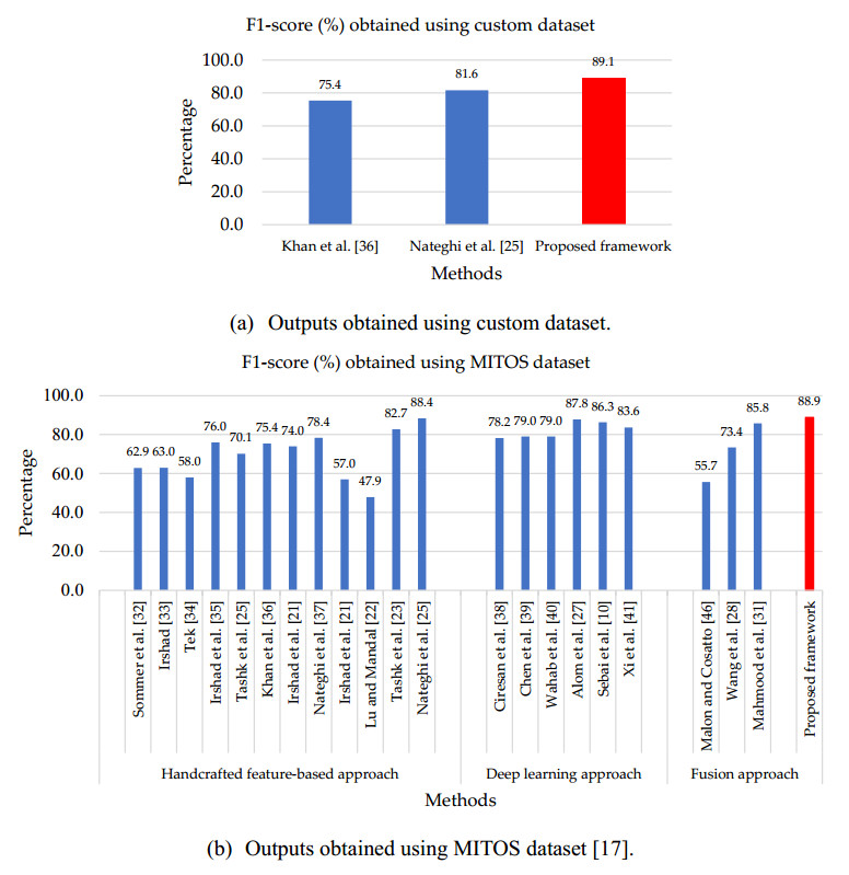
Based on the Nottingham Histopathology Grading (NHG) system, mitosis cells detection is one of the important criteria to determine the grade of breast carcinoma. Mitosis cells detection is a challenging task due to the heterogeneous microenvironment of breast histopathology images. Recognition of complex and inconsistent objects in the medical images could be achieved by incorporating domain knowledge in the field of interest. In this study, the strategies of the histopathologist and domain knowledge approach were used to guide the development of the image processing framework for automated mitosis cells detection in breast histopathology images. The detection framework starts with color normalization and hyperchromatic nucleus segmentation. Then, a knowledge-assisted false positive reduction method is proposed to eliminate the false positive (i.e., non-mitosis cells). This stage aims to minimize the percentage of false positive and thus increase the F1-score. Next, features extraction was performed. The mitosis candidates were classified using a Support Vector Machine (SVM) classifier. For evaluation purposes, the knowledge-assisted detection framework was tested using two datasets: a custom dataset and a publicly available dataset (i.e., MITOS dataset). The proposed knowledge-assisted false positive reduction method was found promising by eliminating at least 87.1% of false positive in both the dataset producing promising results in the F1-score. Experimental results demonstrate that the knowledge-assisted detection framework can achieve promising results in F1-score (custom dataset: 89.1%; MITOS dataset: 88.9%) and outperforms the recent works.

| [1] |
T. Mathew, J. R. Kini, J. Rajan, Computational methods for automated mitosis detection in histopathology images: A review, Biocybern. Biomed. Eng., 41 (2021), 64–82. doi: 10.1016/j.bbe.2020.11.005. doi: 10.1016/j.bbe.2020.11.005

|
| [2] |
P. H. Tan, I. Ellis, K. Allison, E. Brogi, S. B. Fox, S. Lakhani, et al., The 2019 World Health Organization classification of tumours of the breast, Histopathology, 77 (2020), 181–185. doi: 10.1111/his.14091. doi: 10.1111/his.14091
|
| [3] |
E. A. Rakha, J. S. Reis-Filho, F. Baehner, D. J. Dabbs, T. Decker, V. Eusebi, et al., Breast cancer prognostic classification in the molecular era: the role of histological grade, Breast Cancer Res., 12 (2010), 207. doi: 10.1186/bcr2607. doi: 10.1186/bcr2607
|
| [4] |
S. M. Samuel, E. Varghese, S. Varghese, D. Büsselberg, Challenges and perspectives in the treatment of diabetes associated breast cancer, Cancer Treat. Rev., 70 (2018), 98–111. doi: 10.1016/j.ctrv.2018.08.004. doi: 10.1016/j.ctrv.2018.08.004
|
| [5] |
A. D. Shah, A. K. Mehta, N. Talati, R. Brem, L. R. Margolies, Breast tissue markers: Why? What's out there? How do I choose? Clin. Imaging, 52 (2018), 123–136. doi: 10.1016/j.clinimag.2018.07.003. doi: 10.1016/j.clinimag.2018.07.003
|
| [6] |
A. Ramírez-torres, R. Rodríguez-Ramos, F. J. Sabina, C. García-Reimbert, R. Penta, J. Merodio, et al., The role of malignant tissue on the thermal distribution of cancerous breast, J. Theor. Biol., 426 (2017), 152–161. doi: 10.1016/j.jtbi.2017.05.031. doi: 10.1016/j.jtbi.2017.05.031
|
| [7] | J. Dalle, W. K. Leow, D. Racoceanu, A. E. Tutac, T. C. Putti, Automatic breast cancer grading of histopathological images, in 2008 30th Annual International Conference of the IEEE Engineering in Medicine and Biology Society, 2 (2008), 3052–3055. doi: 10.1109/IEMBS.2008.4649847. |
| [8] |
C. Li, X. Wang, W. Liu, L. J. Latecki, DeepMitosis: Mitosis detection via deep detection, verification and segmentation networks, Med. Image Anal., 45 (2018), 121–133. doi: 10.1016/j.media.2017.12.002. doi: 10.1016/j.media.2017.12.002
|
| [9] |
A. Duggento, A. Conti, A. Mauriello, M. Guerrisi, N. Toschi, Deep computational pathology in breast cancer, Semin. Cancer Biol., 72 (2021), 226–237. doi: 10.1016/j.semcancer.2020.08.006. doi: 10.1016/j.semcancer.2020.08.006
|
| [10] |
M. Sebai, X. Wang, T. Wang, MaskMitosis: a deep learning framework for fully supervised, weakly supervised, and unsupervised mitosis detection in histopathology images, Med. Biol. Eng. Comput., 58 (2020), 1603–1623. doi: 10.1007/s11517-020-02175-z. doi: 10.1007/s11517-020-02175-z
|
| [11] |
M. Peikari, M. J. Gangeh, J. Zubovits, G. Clarke, A. L. Martel, Triaging diagnostically relevant regions from pathology whole slides of breast cancer: A texture based approach, IEEE Trans. Med. Imaging, 35 (2016), 307–315. doi: 10.1109/TMI.2015.2470529. doi: 10.1109/TMI.2015.2470529
|
| [12] |
M. Saha, C. Chakraborty, D. Racoceanu, Efficient deep learning model for mitosis detection using breast histopathology images, Comput. Med. Imaging Graph., 64 (2018), 29–40. doi: 10.1016/j.compmedimag.2017.12.001. doi: 10.1016/j.compmedimag.2017.12.001
|
| [13] |
S. Roy, A. k. Jain, S. Lal, J. Kini, A study about color normalization methods for histopathology images, Micron, 114 (2018), 42–61. doi: 10.1016/j.micron.2018.07.005. doi: 10.1016/j.micron.2018.07.005
|
| [14] |
X. Li, K. N. Plataniotis, A complete color normalization approach to histopathology images using color cues computed from saturation-weighted statistics, IEEE Trans. Biomed. Eng., 62 (2015), 1862–1873. doi: 10.1109/TBME.2015.2405791. doi: 10.1109/TBME.2015.2405791
|
| [15] |
Ş. Öztürk, B. Akdemir, Phase classification of mitotic events using selective dictionary learning for stem cell populations, Comput. Electr. Eng., 67 (2018), 25–37. doi: 10.1016/j.compeleceng.2018.03.025. doi: 10.1016/j.compeleceng.2018.03.025
|
| [16] |
B. Plasma, C. Mott, Enigmatic morpho insight: Mitosis at a glance, J. Oral Maxillofac. Pathol., 18 (2014), 2–5. doi: 10.4103/0973-029X.141175. doi: 10.4103/0973-029X.141175
|
| [17] | MITOS–dataset, 2012. Available from: http://ludo17.free.fr/mitos_2012/dataset.html. |
| [18] | AMIDA–dataset, 2013. Available from: http://amida13.isi.uu.nl/?q=node/62. |
| [19] | MITOS-ATYPIA–dataset, 2014. Available from: https://mitos-atypia-14.grand-challenge.org/Dataset/. |
| [20] | TUPAC–dataset, 2016. Available from: http://tupac.tue-image.nl/node/3. |
| [21] |
H. Irshad, A. Gouaillard, L. Roux, D. Racoceanu, Multispectral band selection and spatial characterization: Application to mitosis detection in breast cancer histopathology, Comput. Med. Imaging Graph., 38 (2014), 390–402. doi: 10.1016/j.compmedimag.2014.04.003. doi: 10.1016/j.compmedimag.2014.04.003
|
| [22] |
C. Lu, M. Mandal, Toward automatic mitotic cell detection and segmentation in multispectral histopathological images, IEEE J. Biomed. Heal. Informatics, 18 (2014), 594–605. doi: 10.1109/JBHI.2013.2277837. doi: 10.1109/JBHI.2013.2277837
|
| [23] |
A. Tashk, M. S. Helfroush, H. Danyali, M. Akbarzadeh-jahromi, Automatic detection of breast cancer mitotic cells based on the combination of textural, statistical and innovative mathematical features, Appl. Math. Model., 39 (2015), 6165–6182. doi: 10.1016/j.apm.2015.01.051. doi: 10.1016/j.apm.2015.01.051
|
| [24] |
A. Paul, D. P. Mukherjee, Mitosis detection for invasive breast cancer grading in histopathological images, IEEE Trans. Image Process., 24 (2015), 4041–4054. doi: 10.1109/TIP.2015.2460455. doi: 10.1109/TIP.2015.2460455
|
| [25] |
R. Nateghi, H. Danyali, M. S. Helfroush, Maximized inter-class weighted mean for fast and accurate mitosis cells detection in breast cancer histopathology images, J. Med. Syst., 41 (2017), 146. doi: 10.1007/s10916-017-0773-9. doi: 10.1007/s10916-017-0773-9
|
| [26] | D. Cai, X. Sun, N. Zhou, X. Han, J. Yao, Efficient mitosis detection in breast cancer histology images by RCNN, in 2019 IEEE 16th International Symposium on Biomedical Imaging (ISBI 2019), (2019), 919–922. doi: 10.1109/ISBI.2019.8759461. |
| [27] |
M. Z. Alom, T. Aspiras, T. M. Taha, T. Bowen, V. K. Asari, MitosisNet: End-to-end mitotic cell detection by multi-task learning, IEEE Access, 8 (2020) 68695–68710. doi: 10.1109/ACCESS.2020.2983995. doi: 10.1109/ACCESS.2020.2983995
|
| [28] |
H. Wang, A. C. Roa, A. N. Basavanhally, H. L. Gilmore, N. Shih, M. Feldman, et al., Mitosis detection in breast cancer pathology images by combining handcrafted and convolutional neural network features, J. Med. Imaging, 1 (2014), 034003. doi: 10.1117/1.JMI.1.3.034003. doi: 10.1117/1.JMI.1.3.034003
|
| [29] |
K. S. Beevi, M. S. Nair, G. R. Bindu, A multi-classifier system for automatic mitosis detection in breast histopathology images using deep belief networks, IEEE J. Transl. Eng. Heal. Med., 5 (2017), 1–11. doi: 10.1109/JTEHM.2017.2694004. doi: 10.1109/JTEHM.2017.2694004
|
| [30] |
D. K. Das, P. K. Dutta, Efficient automated detection of mitotic cells from breast histological images using deep convolution neutral network with wavelet decomposed patches, Comput. Biol. Med., 104 (2019), 29–42. doi: 10.1016/j.compbiomed.2018.11.001. doi: 10.1016/j.compbiomed.2018.11.001
|
| [31] |
T. Mahmood, M. Arsalan, M. Owais, M. B. Lee, K. R. Park, Artificial intelligence-based mitosis detection in breast cancer histopathology images using faster R-CNN and deep CNNs, J. Clin. Med., 9 (2020), 749. doi: 10.3390/jcm9030749. doi: 10.3390/jcm9030749
|
| [32] | C. Sommer, L. Fiaschi, F. A. Hamprecht, D. W. Gerlich, Learning-based mitotic cell detection in histopathological images, in Proceedings of the 21st International Conference on Pattern Recognition (ICPR2012), (2012), 2306–2309. |
| [33] |
H. Irshad, Automated mitosis detection in histopathology using morphological and multi-channel statistics features, J. Pathol. Inform., 4 (2013), 10. doi: 10.4103/2153-3539.112695. doi: 10.4103/2153-3539.112695
|
| [34] |
F. B. Tek, Mitosis detection using generic features and an ensemble of cascade adaboosts, J. Pathol. Inform., 4 (2013), 12. doi: 10.4103/2153-3539.112697. doi: 10.4103/2153-3539.112697
|
| [35] |
H. Irshad, S. Jalali, L. Roux, D. Racoceanu, L. J. Hwee, G. L. Naour, et al., Automated mitosis detection using texture, SIFT features and HMAX biologically inspired approach, J. Pathol. Inform., 4 (2013), 12. doi: 10.4103/2153-3539.109870. doi: 10.4103/2153-3539.109870
|
| [36] |
A. M. Khan, H. ElDaly, N. M. Rajpoot, A gamma-gaussian mixture model for detection of mitotic cells in breast cancer histopathology images, J. Pathol. Inform., (2013), 11. doi: 10.4103/2153-3539.112696. doi: 10.4103/2153-3539.112696
|
| [37] | R. Nateghi, H. Danyali, M. S. Helfroush, F. P. Pour, Automatic detection of mitosis cell in breast cancer histopathology images using genetic algorithm, in 2014 21th Iranian Conference on Biomedical Engineering (ICBME), (2014), 1–6. doi: 10.1109/ICBME.2014.7043883. |
| [38] |
D. C. Cireşan, A. Giusti, L. M. Gambardella, J. Schmidhuber, Mitosis detection in breast cancer histology images with deep neural networks, Lect. Notes Comput. Sci., 8150 (2013), 411–418. doi: 10.1007/978-3-642-40763-5_51. doi: 10.1007/978-3-642-40763-5_51
|
| [39] | H. Chen, X. Wang, P. A. Heng, Automated mitosis detection with deep regression networks, in 2016 IEEE 13th International Symposium on Biomedical Imaging (ISBI), (2016), 1204–1207. doi: 10.1109/ISBI.2016.7493482. |
| [40] |
N. Wahab, A. Khan, Y. S. Lee, Two-phase deep convolutional neural network for reducing class skewness in histopathological images based breast cancer detection, Comput. Biol. Med., 85 (2017), 86–97. doi: 10.1016/j.compbiomed.2017.04.012. doi: 10.1016/j.compbiomed.2017.04.012
|
| [41] |
X. Lu, Z. You, M. Sun, J. Wu, Z. Zhang, Breast cancer mitotic cell detection using cascade convolutional neural network with U-Net, Math. Biosci. Eng., 18 (2020), 673–695. doi: 10.3934/MBE.2021036. doi: 10.3934/MBE.2021036
|
| [42] |
S. Albarqouni, C. Baur, F. Achilles, V. Belagiannis, S. Demirci, N. Navab, AggNet: Deep learning from crowds for mitosis detection in breast cancer histology images, IEEE Trans. Med. Imaging, 35 (2016), 1313–1321. doi: 10.1109/TMI.2016.2528120. doi: 10.1109/TMI.2016.2528120
|
| [43] | T. Wollmann, K. Rohr, Deep residual hough voting for mitotic cell detection in histopathology images, in 2017 IEEE 14th International Symposium on Biomedical Imaging (ISBI 2017), (2017), 341–344. doi: 10.1109/ISBI.2017.7950533. |
| [44] |
D. Romo-Bucheli, A. Janowczyk, H. Gilmore, E. Romero, A. Madabhushi, A deep learning based strategy for identifying and associating mitotic activity with gene expression derived risk categories in estrogen receptor positive breast cancers, Cytometry Part A, 91 (2017), 566–573.doi: 10.1002/cyto.a.23065. doi: 10.1002/cyto.a.23065
|
| [45] | H. Chen, J. Qin, Q. Dou, X. Wang, P. A. Heng, Mitosis detection in breast cancer histology images via deep cascaded networks, in Proceedings of the Thirtieth AAAI Conference on Artificial Intelligence, (2016), 1160–1166. |
| [46] |
C. D. Malon, E. Cosatto, Classification of mitotic figures with convolutional neural networks and seeded blob features, J. Pathol. Inform., 4 (2013), 9. doi: 10.4103/2153-3539.112694. doi: 10.4103/2153-3539.112694
|
| [47] |
X. J. Tan, N. Mustafa, M. Y. Mashor, K. S. Ab Rahman, A novel quantitative measurement method for irregular tubules in breast carcinoma, Eng. Sci. Technol. Int. J., (2021). doi: 10.1016/j.jestch.2021.08.008. doi: 10.1016/j.jestch.2021.08.008
|
| [48] |
A. Basavanhally, E. Yu, J. Xu, S. Ganesan, M. Feldman, J. Tomaszewski, et al., Incorporating domain knowledge for tubule detection in breast histopathology using O'Callaghan neighborhoods, SPIE Med. Imaging, 7963 (2011), 796310–796315. doi: 10.1117/12.878092. doi: 10.1117/12.878092
|
| [49] |
D. Racoceanu, F. Capron, Towards semantic-driven high-content image analysis: An operational instantiation for mitosis detection in digital histopathology, Comput. Med. Imaging Graph., 42 (2015), 2–15. doi: 10.1016/j.compmedimag.2014.09.004. doi: 10.1016/j.compmedimag.2014.09.004
|
| [50] |
M. C. Clark, L. O. Hall, D. B. Goldgof, R. Velthuizen, F. R. Murtagh, M. S. Silbiger, Automatic tumor segmentation using knowledge-based techniques, IEEE Trans. Med. Imaging, 17 (1998), 187–201. doi: 10.1109/42.700731. doi: 10.1109/42.700731
|
| [51] |
A. Madabhushi, D. N. Metaxas, Combining low-, high-level and empirical domain knowledge for automated segmentation of ultrasonic breast lesions, IEEE Trans. Med. Imaging, 22 (2003), 155–169. doi: 10.1109/TMI.2002.808364. doi: 10.1109/TMI.2002.808364
|
| [52] |
H. Pan, J. Li, W. Zhang, Incorporating domain knowledge into medical image clustering, Appl. Math. Comput., 185 (2007), 844–856. doi: 10.1016/j.amc.2006.06.083. doi: 10.1016/j.amc.2006.06.083
|
| [53] | S. Naik, S. Doyle, S. Agner, A. Madabhushi, M. Feldman, J. Tomaszewski, Automated gland and nuclei segmentation for grading of prostate and breast cancer histopathology, in 2008 5th IEEE International Symposium on Biomedical Imaging: From Nano to Macro, (2008), 284–287. doi: 10.1109/ISBI.2008.4540988. |
| [54] | M. Macenko, M. Niethammer, J. S. Marron, D. Borland, J. T. Woosley, X. Guan, et al., A method for normalizing histology slides for quantitative analysis, in 2009 IEEE International Symposium on Biomedical Imaging: From Nano to Macro, (2009), 1107–1110. doi: 10.1109/ISBI.2009.5193250. |
| [55] |
J. Wang, M. J. Slattery, M. H. Hoskins, S. Liang, C. Dong, Q. Du, Monte carlo simulation of heterotypic cell aggregation in nonlinear shear flow, Math. Biosci. Eng., 3 (2006), 683–696. doi: 10.3934/mbe.2006.3.683. doi: 10.3934/mbe.2006.3.683
|
| [56] |
J. C. Han, F. Shang, P. Li, B. Li, Y. Zhou, Y. Huang, Coupling bayesian-monte carlo simulations with substance flow analysis for efficient pollutant management: A case study of phosphorus flows in China, Resour. Conserv. Recycl., 169 (2021), 105550. doi: 10.1016/j.resconrec.2021.105550. doi: 10.1016/j.resconrec.2021.105550
|
| [57] |
Q. Li, Y. Wang, H. Liu, X. He, D. Xu, J. Wang, et al., Leukocyte cells identification and quantitative morphometry based on molecular hyperspectral imaging technology, Comput. Med. Imaging Graph., 38 (2014), 171–178. doi: 10.1016/j.compmedimag.2013.12.008. doi: 10.1016/j.compmedimag.2013.12.008
|
| [58] | M. M. Saleck, A. Elmoutaouakkil, M. Moucouf, Tumor detection in mammography images using fuzzy C-means and GLCM texture features, in 2017 14th International Conference on Computer Graphics, Imaging and Visualization, (2017), 122–125. doi: 10.1109/CGiV.2017.22. |
| [59] |
B. Abraham, M. S. Nair, Computer-aided classification of prostate cancer grade groups from MRI images using texture features and stacked sparse autoencoder, Comput. Med. Imaging Graph., 69 (2018), 60–68. doi: 10.1016/j.compmedimag.2018.08.006. doi: 10.1016/j.compmedimag.2018.08.006
|
| [60] |
I. Qabajeh, F. Thabtah, F. Chiclana, A recent review of conventional vs. automated cybersecurity anti-phishing techniques, Comput. Sci. Rev., 29 (2018), 44–55. doi: 10.1016/j.cosrev.2018.05.003. doi: 10.1016/j.cosrev.2018.05.003
|
| [61] | H. Amitha, I. Selvamani, D. A. S. Dhas, Developement of computer aided system for detection and classification of mitosis using SVM, in 2017 International Conference on Inventive Computing and Informatics (ICICI), (2017), 954–958. doi: 10.1109/ICICI.2017.8365278. |
| [62] | J. MacQueen, Some Methods for classification and snalysis of multivariate observations, in Proceedings of 5th Berkeley Symposium on Mathematical Statistics and Probability, (1967), 281–297. |
| [63] | E. Forgy, Cluster analysis of multivariate data: Efficiency vs. interpretability of classification, Biometrics, 21 (1965), 768. |
| [64] | D. Arthur, S. Vassilvitskii, K-means++: The advantages of careful seeding, in Proceedings of the eighteenth Annual ACM-SIAM Symposium on Discrete Algorithms, (2006), 1027–1035. |
| [65] | A. Bhattacharya, J. Eube, H. Röglin, M. Schmidt, Noisy, greedy and not so greedy k-means++, preprint, arXiv: 1912.00653v1. |
| [66] |
P. Perona, J. Malik, Scale-space and edge detection using anisotropic diffusion, IEEE Trans. Pattern Anal. Mach. Intell., 12 (1990), 629–639. doi: 10.1109/34.56205. doi: 10.1109/34.56205
|
| [67] |
L. Xu, C. Lu, Y. Xu, J. Jia, Image smoothing via L0 gradient minimization, ACM Trans. Graph., 30 (2011), 1–12. doi: 10.1145/2024156.2024208. doi: 10.1145/2024156.2024208
|

Figures(10) / Tables(12)

Xiao Jian Tan, Nazahah Mustafa, Mohd Yusoff Mashor, Khairul Shakir Ab Rahman. Automated knowledge-assisted mitosis cells detection framework in breast histopathology images[J]. Mathematical Biosciences and Engineering, 2022, 19(2): 1721-1745. doi: 10.3934/mbe.2022081


 DownLoad: