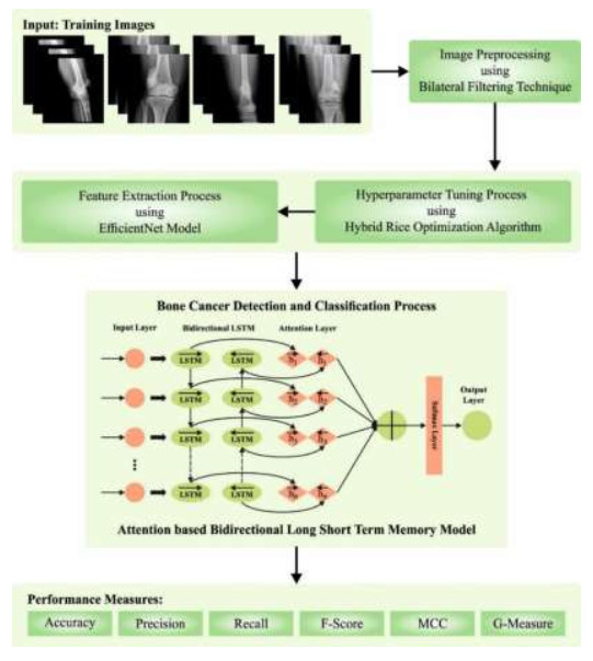
Bone cancer detection is an essential region of medical analysis but developments in medical imaging and artificial intelligence (AI) are vital. Using approaches, namely deep learning (DL) and machine learning (ML), radiologists and medical staff can examine X-ray, CT, and MRI scans to identify bone cancer and abnormalities. These technologies support earlier diagnosis, correct diagnosis, and treatment planning, enhancing patient solutions. The combination of AI-driven image analysis and the knowledge of medical practitioners improves the speed and precision of bone cancer detection, contributing to more effectual clinical activities. DL algorithms, particularly CNNs, are exposed to great performance in image classification tasks and are extremely utilized for medical image analysis. We offer a Hybrid Rice Optimization Algorithm with DL-Assisted Bone Cancer Detection (HROADL-BCD) technique on medical X-ray images. The major intention of the HROADL-BCD method is to examine the X-ray images for the recognition of bone cancer. In the presented HROADL-BCD method, a bilateral filtering (BF) process was performed to remove the noise. To derive feature vectors, the HROADL-BCD technique applied the EfficientNet model. The HROADL-BCD technique involved the HROA for hyperparameter tuning of the EfficientNet model. Last, the bone cancer detection and classification process were executed by the attention-based bidirectional long short-term memory (ABiLSTM) approach. A wide range of simulations could be applied for the simulation result analysis of the HROADL-BCD algorithm. The extensive outcome of the HROADL-BCD approach inferred the superior outcome of 97.62% outcome concerning various aspects.
Citation: Thavavel Vaiyapuri, Prasanalakshmi Balaji, S. Shridevi, Santhi Muttipoll Dharmarajlu, Nourah Ali AlAseem. An attention-based bidirectional long short-term memory based optimal deep learning technique for bone cancer detection and classifications[J]. AIMS Mathematics, 2024, 9(6): 16704-16720. doi: 10.3934/math.2024810
Bone cancer detection is an essential region of medical analysis but developments in medical imaging and artificial intelligence (AI) are vital. Using approaches, namely deep learning (DL) and machine learning (ML), radiologists and medical staff can examine X-ray, CT, and MRI scans to identify bone cancer and abnormalities. These technologies support earlier diagnosis, correct diagnosis, and treatment planning, enhancing patient solutions. The combination of AI-driven image analysis and the knowledge of medical practitioners improves the speed and precision of bone cancer detection, contributing to more effectual clinical activities. DL algorithms, particularly CNNs, are exposed to great performance in image classification tasks and are extremely utilized for medical image analysis. We offer a Hybrid Rice Optimization Algorithm with DL-Assisted Bone Cancer Detection (HROADL-BCD) technique on medical X-ray images. The major intention of the HROADL-BCD method is to examine the X-ray images for the recognition of bone cancer. In the presented HROADL-BCD method, a bilateral filtering (BF) process was performed to remove the noise. To derive feature vectors, the HROADL-BCD technique applied the EfficientNet model. The HROADL-BCD technique involved the HROA for hyperparameter tuning of the EfficientNet model. Last, the bone cancer detection and classification process were executed by the attention-based bidirectional long short-term memory (ABiLSTM) approach. A wide range of simulations could be applied for the simulation result analysis of the HROADL-BCD algorithm. The extensive outcome of the HROADL-BCD approach inferred the superior outcome of 97.62% outcome concerning various aspects.

| [1] | S. Breden, F. Hinterwimmer, S. Consalvo, J. Neumann, C. Knebel, R. Eisenhart-Rothe, et al., Deep learning-based detection of bone tumors around the Knee in X-rays of children, J. Clin. Med., 12 (2023), 1–9. |
| [2] | K. Furuo, K. Morita, T. Hagi, T. Nakamura, T. Wakabayashi, Automatic benign and malignant estimation of bone tumors using deep learning, 2021 5th IEEE International Conference on Cybernetics (CYBCONF), Sendai, Japan, 2021,030–033, https://doi.org/10.1109/CYBCONF51991.2021.9464132 |
| [3] | M. H. Mazumder, M. P. Singh, Bone cancer detection using deep learning. In International Conference on Innovations in Computer Science and Engineering, 285–296, 2022. Singapore: Springer Nature Singapore. |
| [4] | B. S. Vandana, S. R. Alva, Deep Learning Based Automated tool for cancer diagnosis from bone histopathology images, 2021 International Conference on Intelligent Technologies (CONIT), Hubli, India, 2021, 1–8. https://doi.org/10.1109/CONIT51480.2021.9498367 |
| [5] | A. Shukla, A. Patel, Bone cancer detection from X-ray and MRI images through image segmentation techniques, Int. J. Recent Technol. Eng., 8 (2020), 273–278. |
| [6] | S. Gawade, A. Bhansali, K. Patil, D. Shaikh, Application of the convolutional neural networks and supervised deep-learning methods for osteosarcoma bone cancer detection, Health. Anal., 3 (2023), 100153. |
| [7] | T. Zimbalist, R. Rosen, K. Peri-Hanania, Y. Caspi, B. Rinott, Detecting bone lesions in X-Ray under diverse acquisition conditions, arXiv preprint arXiv: 2212.07792., 2022. https://doi.org/10.1117/1.JMI.11.2.024502 |
| [8] | H. X. Huynh, H. B. T. Nguyen, C. A. Phan, H. T. Nguyen, Abnormality Bone Detection in X-Ray Images Using Convolutional Neural Network, In Context-Aware Systems and Applications, and Nature of Computation and Communication: 9th EAI International Conference, ICCASA 2020, and 6th EAI International Conference, ICTCC 2020, Thai Nguyen, Vietnam, Proceedings 9, 31–43, 2021. Springer International Publishing. |
| [9] | G. Suganeshwari, R. Balakumar, K. Karuppanan, S. B. Prathiba, S. Anbalagan, DTBV: A deep transfer-based bone cancer diagnosis system using VGG16 feature extraction, Diagnostics, 13 (2023), 757. |
| [10] |
F. R. Eweje, B. Bao, J. Wu, D. Dalal, W. H. Liao, Y. He, et al., Deep learning for classification of bone lesions on routine MRI, EBioMedicine, 68, 2021. https://doi.org/10.1016/j.ebiom.2021.103402 doi: 10.1016/j.ebiom.2021.103402

|
| [11] |
A. Rehman, A. Raza, F. S. Alamri, B. Alghofaily, T. Saba, Transfer learning-based smart features engineering for osteoarthritis diagnosis from knee X-Ray images, IEEE Access, 11 (2023), 71326–71338, 2023. https://doi.org/10.1109/ACCESS.2023.3294542 doi: 10.1109/ACCESS.2023.3294542
|
| [12] | N. T. Do, S. T. Jung, H. J. Yang, S. H. Kim, Multi-level seg-unet model with global and patch-based X-ray images for knee bone tumor detection, Diagnostics, 11 (2021), 691. |
| [13] |
A. K. Bharodiya, A. M. Gonsai, An intelligent assistive algorithm for bone tumor detection from human X-ray images based on binary Blob analysis, Int. J. Inf. Technol., 1–7, 2020. https://doi.org/10.1007/s41870-020-00539-0 doi: 10.1007/s41870-020-00539-0
|
| [14] | Z. Xu, K. Niu, S. Tang, T. Song, Y. Rong, W. Guo, et al., Bone tumor necrosis rate detection in few-shot X-rays based on deep learning, Comput. Med. Imag. Grap., 102 (2022), 102141. |
| [15] | I. M. Wani, S. Arora, Osteoporosis diagnosis in knee X-rays by transfer learning based on convolution neural network, Multimed. Tools Appl., 82 (2023), 14193–14217. |
| [16] | M. Singh, M. Angurala, M. Bala, Bone tumour detection using feature extraction with classification by deep learning techniques, Res. J. Comput. Syst. Eng., 1 (2020), 23–27. |
| [17] | Z. Xie, K. Zhao, X. Yan, S. Wu, J. Mei, H. Lu, Merged U-Net for Bone Tumors X-Ray Images Segmentation, 2022 IEEE International Conference on Image Processing (ICIP), Bordeaux, France, 2022. 1276–1280. https://doi.org/10.1109/ICIP46576.2022.9897539 |
| [18] | Y. Gül, S. Yaman, D. Avcı, A. H. Çilengir, M. Balaban, H. Güler, A novel deep transfer learning-based approach for automated Pes Planus diagnosis using X-ray image, Diagnostics, 13 (2023), 1662. |
| [19] |
H. Li, X. L. Duan, SAR Ship Image Speckle Noise Suppression Algorithm based on adaptive bilateral filter, Wirel. Commun. Mob. Comput., 2022. https://doi.org/10.1155/2022/9392648 doi: 10.1155/2022/9392648
|
| [20] |
K. Yousaf, T. Nawaz, A. Habib, Using two-stream EfficientNet-BiLSTM network for multiclass classification of disturbing YouTube videos, Multimed. Tools Appl., 1–28, 2023. https://doi.org/10.1007/s11042-023-15774-3 doi: 10.1007/s11042-023-15774-3
|
| [21] |
A. Z. Ye, B. R. Li, C. W. Zhou, D. M. Wang, E. M. Mei, F. Shu, et al., High-Dimensional feature selection based on improved Binary Ant Colony Optimization combined with Hybrid Rice Optimization Algorithm, Int. J. Intell. Syst., 2023. https://doi.org/10.1155/2023/1444938 doi: 10.1155/2023/1444938
|
| [22] | W. Zeng, K. Wang, J. Zhou, R. Cheng, Traffic Flow Prediction based on hybrid deep learning models considering missing data and multiple factors, Sustainability, 15 (2023), 11092. |
| [23] |
E. Alabdulkreem, M. K. Saeed, S. S. Alotaibi, R. Allafi, A. Mohamed, M. A. Hamza, Bone cancer detection and Classification Using Owl Search Algorithm with deep learning on X-Ray images, IEEE Access, 11 (2023), 109095–109103. DOI:10.1109/ACCESS.2023.3319293 doi: 10.1109/ACCESS.2023.3319293
|


Figures(10) / Tables(4)

Thavavel Vaiyapuri, Prasanalakshmi Balaji, S. Shridevi, Santhi Muttipoll Dharmarajlu, Nourah Ali AlAseem. An attention-based bidirectional long short-term memory based optimal deep learning technique for bone cancer detection and classifications[J]. AIMS Mathematics, 2024, 9(6): 16704-16720. doi: 10.3934/math.2024810


 DownLoad: