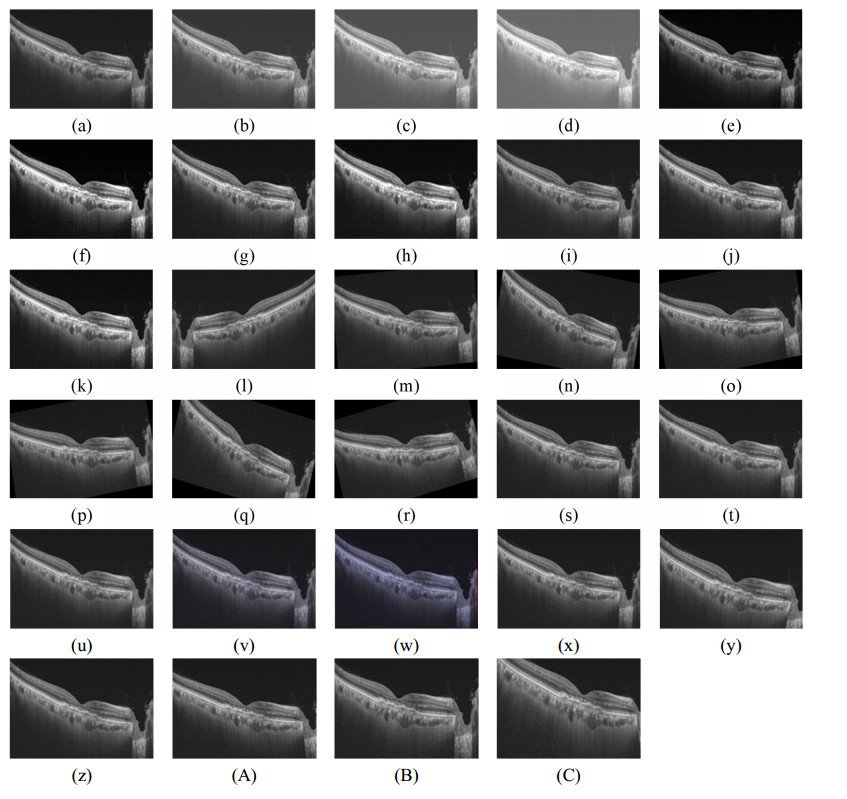
Optical coherence tomography (OCT) is a noninvasive, high-resolution imaging technique widely used in clinical practice to depict the structure of the retina. Over the past few decades, ophthalmologists have used OCT to diagnose, monitor, and treat retinal diseases. However, manual analysis of the complicated retinal layers using two colors, black and white, is time consuming. Although ophthalmologists have more experience, their results may be prone to erroneous diagnoses. Therefore, in this study, we propose an automatic method for diagnosing five retinal diseases based on the use of hybrid and ensemble deep learning (DL) methods. DL extracts a thousand constitutional features from images as features for training classifiers. The machine learning method classifies the extracted features and fuses the outputs of the two classifiers to improve classification performance. The distribution probabilities of two classifiers of the same class are aggregated; then, class prediction is made using the class with the highest probability. The limited dataset is resolved by the fine-tuning of classification knowledge and generating augmented images using transfer learning and data augmentation. Multiple DL models and machine learning classifiers are used to access a suitable model and classifier for the OCT images. The proposed method is trained and evaluated using OCT images collected from a hospital and exhibits a classification accuracy of 97.68% (InceptionResNetV2, ensemble: Extreme gradient boosting (XG-Boost) and k-nearest neighbor (k-NN). The experimental results show that our proposed method can improve the OCT classification performance; moreover, in the case of a limited dataset, the proposed method is critical to develop accurate classifications.
Citation: Kuntha Pin, Jung Woo Han, Yunyoung Nam. Retinal diseases classification based on hybrid ensemble deep learning and optical coherence tomography images[J]. Electronic Research Archive, 2023, 31(8): 4843-4861. doi: 10.3934/era.2023248
Optical coherence tomography (OCT) is a noninvasive, high-resolution imaging technique widely used in clinical practice to depict the structure of the retina. Over the past few decades, ophthalmologists have used OCT to diagnose, monitor, and treat retinal diseases. However, manual analysis of the complicated retinal layers using two colors, black and white, is time consuming. Although ophthalmologists have more experience, their results may be prone to erroneous diagnoses. Therefore, in this study, we propose an automatic method for diagnosing five retinal diseases based on the use of hybrid and ensemble deep learning (DL) methods. DL extracts a thousand constitutional features from images as features for training classifiers. The machine learning method classifies the extracted features and fuses the outputs of the two classifiers to improve classification performance. The distribution probabilities of two classifiers of the same class are aggregated; then, class prediction is made using the class with the highest probability. The limited dataset is resolved by the fine-tuning of classification knowledge and generating augmented images using transfer learning and data augmentation. Multiple DL models and machine learning classifiers are used to access a suitable model and classifier for the OCT images. The proposed method is trained and evaluated using OCT images collected from a hospital and exhibits a classification accuracy of 97.68% (InceptionResNetV2, ensemble: Extreme gradient boosting (XG-Boost) and k-nearest neighbor (k-NN). The experimental results show that our proposed method can improve the OCT classification performance; moreover, in the case of a limited dataset, the proposed method is critical to develop accurate classifications.

| [1] |
I. Pearce, A. Clemens, M. H. Brent, L. Lu, R. Gallego-Pinazo, A. M. Minnella, et al., Real-world outcomes with ranibizumab in branch retinal vein occlusion: The prospective, global, luminous study, PloS One., 15 (2020), 0234739. https://doi.org/10.1371/journal.pone.0234739 doi: 10.1371/journal.pone.0234739

|
| [2] |
F. Tang, F. Xu, H. Zhong, X. Zhao, M. Lv, K. Yang, et al., Comparison of subfoveal choroidal thickness in eyes with crvo and brvo, BMC Ophthalmol., 19 (2019), 1–7. https://doi.org/10.1186/s12886-019-1143-9 doi: 10.1186/s12886-019-1143-9
|
| [3] |
S. Ţălu, S. D. Nicoara., Malfunction of outer retinal barrier and choroid in the occurrence and progression of diabetic macular edema, World J. Diabetes, 12 (2021), 437. https://doi.org/10.4239/wjd.v12.i4.437 doi: 10.4239/wjd.v12.i4.437
|
| [4] |
I. J. Orlando, B. S. Gerendas, S. Riedl, C. Grechenig, A. Breger, M. Ehler, et al., Automated quantification of photoreceptor alteration in macular disease using optical coherence tomography and deep learning, Sci. Rep., 10 (2020), 1–12. https://doi.org/10.1038/s41598-020-62329-9 doi: 10.1038/s41598-020-62329-9
|
| [5] |
A. F. Borkenstein, E. Borkenstein, Cataract surgery with implantation of a high-add intraocular lens lentis® max ls-313 mf80 in end-stage, age-related macular degeneration: A case report of magnifying surgery, Clin. Case Rep., 7 (2019), 74–78. https://doi.org/10.1002/ccr3.1912 doi: 10.1002/ccr3.1912
|
| [6] |
S. Rogers, R. L. McIntosh, N. Cheung, L. Lim, J. J. Wang, P. Mitchell, et al., The prevalence of retinal vein occlusion: pooled data from population studies from the United States, Europe, Asia, and Australia, Ophthalmology, 117 (2010), 313–319. https://doi.org/10.1016/j.ophtha.2009.07.017 doi: 10.1016/j.ophtha.2009.07.017
|
| [7] |
F. Xu, S. Liu, Y. Xiang, Z. Lin, C. Li, L. Zhou, et al., Deep learning for detecting subretinal fluid and discerning macular status by fundus images in central serous chorioretinopathy, Front. Bioeng. Biotechnol., 9 (2021), 651340. https://doi.org/10.3389/fbioe.2021.651340 doi: 10.3389/fbioe.2021.651340
|
| [8] | R. Dandona, L. Dandona, R. K. John, C. A. McCarty, G. N. Rao, Awareness of eye diseases in an urban population in southern india, Bull. World Health Organ., 79 (2001), 96–102. |
| [9] |
N. Eladawi, M. Elmogy, M. Ghazal, O. Helmy, A. Aboelfetouh, A. Riad, et al., Classification of retinal diseases based on OCT images, Front. Biosci.-Landmark, 23 (2018), 247–264. https://doi.org/10.2741/4589 doi: 10.2741/4589
|
| [10] |
N. Rajagopalan, V. Narasimhan, S. K. Vinjimoor, J. Aiyer, Deep CNN framework for retinal disease diagnosis using optical coherence tomography images, J. Ambient Intell. Human. Comput., 12 (2021), 7569–7580. https://doi.org/10.1007/s12652-020-02460-7 doi: 10.1007/s12652-020-02460-7
|
| [11] |
B. Keerthiveena, S. Esakkirajan, K. Selvakumar, T. Yogesh, Computer-aided diagnosis of retinal diseases using multidomain feature fusion, Int. J. Imaging Syst. Technol., 30 (2020), 367–379. https://doi.org/10.1002/ima.22379 doi: 10.1002/ima.22379
|
| [12] |
T. Kurmann, S. Yu, P. Márquez-Neila, A. Ebneter, M. Zinkernagel, M. R. Munk, et al., Expert-level automated biomarker identification in optical coherence tomography scans, Sci. Rep., 9 (2019), 1–9. https://doi.org/10.1038/s41598-019-49740-7 doi: 10.1038/s41598-019-49740-7
|
| [13] |
J. Han, S. Choi, J. I. Park, J. S. Hwang, J. M. Han, H. J. Lee, et al., Classifying neovascular age-related macular degeneration with a deep convolutional neural network based on optical coherence tomography images, Sci. Rep., 12 (2022), 1–10. https://doi.org/10.1038/s41598-022-05903-7 doi: 10.1038/s41598-022-05903-7
|
| [14] |
S. Sotoudeh-Paima, A. Jodeiri, F. Hajizadeh, H. Soltanian-Zadeh, Multi-scale convolutional neural network for automated amd classification using retinal OCT images, Comput. Biol. Med., 144 (2022), 105368. https://doi.org/10.1016/j.compbiomed.2022.105368 doi: 10.1016/j.compbiomed.2022.105368
|
| [15] |
M. A. Elaziz, A. Mabrouk, A. Dahou, S. A. Chelloug, Medical image classification utilizing ensemble learning and levy flight-based honey badger algorithm on 6g-enabled internet of things, Comput. Intell. Neurosci., 2022 (2022). https://doi.org/10.1155/2022/5830766 doi: 10.1155/2022/5830766
|
| [16] |
X. Liu, Y. Bai, J. Cao, J. Yao, Y. Zhang, M. Wang, Joint disease classification and lesion segmentation via one-stage attention-based convolutional neural network in OCT images, Biomed. Signal Process. Control, 71 (2022), 103087. https://doi.org/10.1016/j.bspc.2021.103087 doi: 10.1016/j.bspc.2021.103087
|
| [17] |
A. Minagi, H. Hirano, K. Takemoto, Natural images allow universal adversarial attacks on medical image classification using deep neural networks with transfer learning, J. Imaging, 8 (2022), 38. https://doi.org/10.3390/jimaging8020038 doi: 10.3390/jimaging8020038
|
| [18] |
A. Tayal, J. Gupta, A. Solanki, K. Bisht, A. Nayyar, M. Masud, DL-CNN-based approach with image processing techniques for diagnosis of retinal diseases, Multimedia Syst., (2021). https://doi.org/10.1007/s00530-021-00791-9 doi: 10.1007/s00530-021-00791-9
|
| [19] |
P. Cao, X. Li, K. Mao, F. Lu, G. Ning, L. Fang, et al., A novel data augmentation method to enhance deep neural networks for detection of atrial fibrillation, Biomed. Signal Process. Control, 56 (2020), 101675. https://doi.org/10.1016/j.bspc.2019.101675 doi: 10.1016/j.bspc.2019.101675
|
| [20] | K. Kong, G. Li, M. Ding, Z. Wu, C. Zhu, B. Ghanem, et al., Robust optimization as data augmentation for large-scale graphs, in 2022 IEEE/CVF Conference on Computer Vision and Pattern Recognition (CVPR), (2022), 60–69. https://doi.org/10.1109/CVPR52688.2022.00016 |
| [21] |
C. Shorten, T. M. Khoshgoftaar, B. Furht, Text data augmentation for deep learning, J. Big Data, 8 (2021). https://doi.org/10.1186/s40537-021-00492-0 doi: 10.1186/s40537-021-00492-0
|
| [22] |
T. Le, M. T. Vo, T. Kieu, E. Hwang, S. Rho, W. S. Baik, Multiple electric energy consumption forecasting using a cluster-based strategy for transfer learning in smart building, Sensors, 20 (2020), 2668. https://doi.org/10.3390/s20092668 doi: 10.3390/s20092668
|
| [23] |
M. O. El Zein, M. M. Soliman, A. K. Elkholy, N. I. Ghali, Transfer learning based model for pneumonia detection in chest x-ray images, Int. J. Intell. Eng. Syst., 14 (2021), 56–66. https://doi.org/10.22266/ijies2021.1031.06 doi: 10.22266/ijies2021.1031.06
|
| [24] |
T. Kaur, T. K. Gandhi, Deep convolutional neural networks with transfer learning for automated brain image classification, Mach. Vision Appl., 31 (2020), 1–16. https://doi.org/10.1007/s00138-020-01069-2 doi: 10.1007/s00138-020-01069-2
|
| [25] |
S. Elmuogy, A. N. Hikal, E. Hassan, An efficient technique for CT scan images classification of COVID-19, J. Intell. Fuzzy Syst., 40 (2021), 5225–5238. https://doi.org/10.3233/JIFS-201985 doi: 10.3233/JIFS-201985
|
| [26] |
S. A. A. Ismael, A. Mohammed, H. Hefny, An enhanced deep learning approach for brain cancer mri images classification using residual networks, Artif. Intell. Med., 102 (2020), 101779. https://doi.org/10.1016/j.artmed.2019.101779 doi: 10.1016/j.artmed.2019.101779
|
| [27] |
D. Theckedath, R. R. Sedamkar, Detecting affect states using vgg16, resnet50 and se-resnet50 networks, SN Comput. Sci., 1 (2020). https://doi.org/10.1007/s42979-020-0114-9 doi: 10.1007/s42979-020-0114-9
|
| [28] |
M. Bansal, M. Kumar, M. Sachdeva, A. Mittal, Transfer learning for image classification using vgg19: Caltech-101image data set, J. Ambient Intell. Humanized Comput., (2021), 1–12. https://doi.org/10.1007/s12652-021-03488-z doi: 10.1007/s12652-021-03488-z
|
| [29] | A. Mechelli, S. Vieira, Machine learning Methods and Applications to Brain Disorders, Academic Press, London, 2019. |
| [30] |
C. Wang, Y. Long, W. Li, W. Dai, S. Xie, Y. Liu, et al., Exploratory study on classification of lung cancer subtypes through a combined k-nearest neighbor classifier in breathomics, Sci. Rep., 10 (2020), 5880. https://doi.org/10.1038/s41598-020-62803-4 doi: 10.1038/s41598-020-62803-4
|
| [31] |
A. Suresh, R. Udendhran, M. Balamurgan, Hybridized neural network and decision tree based classifier for prognostic decision making in breast cancers, Soft Comput., 24 (2020), 7947–7953. https://doi.org/10.1007/s00500-019-04066-4 doi: 10.1007/s00500-019-04066-4
|
| [32] |
A. Subudhi, M. Dash, S. Sabut, Automated segmentation and classification of brain stroke using expectation-maximization and random forest classifier, Biocybern. Biomed. Eng., 40 (2020), 277–289. https://doi.org/10.1016/j.bbe.2019.04.004 doi: 10.1016/j.bbe.2019.04.004
|
| [33] |
V. R. Balaji, S. T. Suganthi, R. Rajadevi, V. K. Kumar, B. S. Balaji, S. Pandiyan, Skin disease detection and segmentation using dynamic graph cut algorithm and classification through naive bayes classifier, Measurement, 163 (2020), 107922. https://doi.org/10.1016/j.measurement.2020.107922 doi: 10.1016/j.measurement.2020.107922
|
| [34] |
Y. X. Liew, N. Hameed, J. Clos, An investigation of xgboost-based algorithm for breast cancer classification, Mach. Learn. Appl., 6 (2021), 100154. https://doi.org/10.1016/j.mlwa.2021.100154 doi: 10.1016/j.mlwa.2021.100154
|
| [35] |
S. Kumari, D. Kumar, M. Mittal, An ensemble approach for classification and prediction of diabetes mellitus using soft voting classifier, Int. J. Cognit. Comput. Eng., 2 (2021), 40–46. https://doi.org/10.1016/j.ijcce.2021.01.001 doi: 10.1016/j.ijcce.2021.01.001
|


Figures(6) / Tables(8)
Kuntha Pin, Jung Woo Han, Yunyoung Nam. Retinal diseases classification based on hybrid ensemble deep learning and optical coherence tomography images[J]. Electronic Research Archive, 2023, 31(8): 4843-4861. doi: 10.3934/era.2023248


 DownLoad: