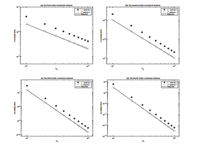
The stability and convergence of the Fourier pseudo-spectral method are analyzed for the three dimensional incompressible Navier-Stokes equation, coupled with a variety of time-stepping methods, of up to fourth order temporal accuracy. An aliasing error control technique is applied in the error estimate for the nonlinear convection term, while an a-priori assumption for the numerical solution at the previous time steps will also play an important role in the analysis. In addition, a few multi-step temporal discretization is applied to achieve higher order temporal accuracy, while the numerical stability is preserved. These semi-implicit numerical schemes use a combination of explicit Adams-Bashforth extrapolation for the nonlinear convection term, as well as the pressure gradient term, and implicit Adams-Moulton interpolation for the viscous diffusion term, up to the fourth order accuracy in time. Optimal rate convergence analysis and error estimates are established in details. It is proved that, the Fourier pseudo-spectral method coupled with the carefully designed time-discretization is stable provided only that the time-step and spatial grid-size are bounded by two constants over a finite time. Some numerical results are also presented to verify the established convergence rates of the proposed schemes.
Citation: Cheng Wang. Convergence analysis of Fourier pseudo-spectral schemes for three-dimensional incompressible Navier-Stokes equations[J]. Electronic Research Archive, 2021, 29(5): 2915-2944. doi: 10.3934/era.2021019
The stability and convergence of the Fourier pseudo-spectral method are analyzed for the three dimensional incompressible Navier-Stokes equation, coupled with a variety of time-stepping methods, of up to fourth order temporal accuracy. An aliasing error control technique is applied in the error estimate for the nonlinear convection term, while an a-priori assumption for the numerical solution at the previous time steps will also play an important role in the analysis. In addition, a few multi-step temporal discretization is applied to achieve higher order temporal accuracy, while the numerical stability is preserved. These semi-implicit numerical schemes use a combination of explicit Adams-Bashforth extrapolation for the nonlinear convection term, as well as the pressure gradient term, and implicit Adams-Moulton interpolation for the viscous diffusion term, up to the fourth order accuracy in time. Optimal rate convergence analysis and error estimates are established in details. It is proved that, the Fourier pseudo-spectral method coupled with the carefully designed time-discretization is stable provided only that the time-step and spatial grid-size are bounded by two constants over a finite time. Some numerical results are also presented to verify the established convergence rates of the proposed schemes.

| [1] |
The spectral accuracy of a fully-discrete scheme for a nonlinear third order equation. Computing (1990) 44: 187-196. 
|
| [2] |
A second order projection method for the incompressible Navier-Stokes equations. J. Comput. Phys. (1989) 85: 257-283.
|
| [3] |
On the solution of the Navier-Stokes equations using projection schemes with third- order accuracy in time. Comput. Fluids (1997) 26: 107-116.
|
| [4] |
An implicit/explcit spectral method for Burgers' equation. Calcolo (1986) 23: 265-284.
|
| [5] |
Approximation results for orthogonal polynomials in Sobolev spaces. Math. Comp. (1982) 38: 67-86.
|
| [6] |
A linear energy stable scheme for a thin film model without slope selection. J. Sci. Comput. (2012) 52: 546-562.
|
| [7] |
A stabilized second order ETD multistep method for thin film growth model without slope selection. ESAIM Math. Model. Numer. Anal. (2020) 54: 727-750.
|
| [8] |
W. Chen, W. Li, C. Wang, S. Wang and X. Wang, Energy stable higher order linear ETD multi-step methods for gradient flows: Application to thin film epitaxy, Res. Math. Sci., 7 (2020), Paper No. 13, 27 pp. doi: 10.1007/s40687-020-00212-9
|
| [9] |
W. Chen, C. Wang, S. Wang, X. Wang and S. M. Wise, Energy stable numerical schemes for ternary Cahn-Hilliard system, J. Sci. Comput., 84 (2020), Paper No. 27, 36 pp. doi: 10.1007/s10915-020-01276-z
|
| [10] |
A linear iteration algorithm for a second-order energy stable scheme for a thin film model without slope selection.. J. Sci. Comput. (2014) 59: 574-601.
|
| [11] |
A Fourier pseudospectral method for the "Good" Boussinesq equation with second-order temporal accuracy. Numer. Methods Partial Differential Equations (2015) 31: 202-224.
|
| [12] |
A third order exponential time differencing numerical scheme for no-slope-selection epitaxial thin film model with energy stability. J. Sci. Comput. (2019) 81: 154-185.
|
| [13] |
Long time stability of high order multi-step numerical schemes for two-dimensional incompressible Navier-Stokes equations. SIAM J. Numer. Anal. (2016) 54: 3123-3144.
|
| [14] | Q. Cheng and C. Wang, Error estimate of a second order accurate scalar auxiliary variable (SAV) scheme for the thin film epitaxial equation, Adv. Appl. Math. Mech., Accepted and in press. |
| [15] |
An energy stable BDF2 Fourier pseudo-spectral numerical scheme for the square phase field crystal equation. Commun. Comput. Phys. (2019) 26: 1335-1364.
|
| [16] |
K. Cheng, C. Wang and S. M. Wise, A weakly nonlinear energy stable scheme for the strongly anisotropic Cahn-Hilliard system and its convergence analysis, J. Comput. Phys., 405 (2020), 109109, 28 pp. doi: 10.1016/j.jcp.2019.109109
|
| [17] |
A second-order, weakly energy-stable pseudo-spectral scheme for the Cahn-Hilliard equation and its solution by the homogeneous linear iteration method. J. Sci. Comput. (2016) 69: 1083-1114.
|
| [18] |
Numerical solution of Navier-Stokes equations. Math. Comp. (1968) 22: 745-762.
|
| [19] |
Une méthode multipas implicite-explicite pour l'approximation des équations d'évolution paraboliques. Numer. Math. (1980) 35: 257-276.
|
| [20] |
Pseudo-spectral method for the "Good" boussinesq equation. Math. Comp. (1991) 57: 109-122.
|
| [21] |
Convergence of Fourier methods for Navier-Stokes equations. SIAM J. Numer. Anal. (1993) 30: 650-674.
|
| [22] |
Convergence of spectral methods for the {Burgers'} equation. SIAM J. Numer. Anal. (1992) 29: 1520-1541.
|
| [23] |
Projection method I: Convergence and numerical boundary layers. SIAM J. Numer. Anal. (1995) 32: 1017-1057.
|
| [24] |
Gauge finite element method for incompressible flows. Int. J. Num. Meth. Fluids (2000) 34: 701-710.
|
| [25] |
Gauge method for viscous incompressible flows. Commu. Math. Sci. (2003) 1: 317-332.
|
| [26] | D. Gottlieb and S. A. Orszag, Numerical Analysis of Spectral Methods, Theory and Applications, SIAM, Philadelphia, PA, 1977. |
| [27] |
Long time stability of a classical efficient scheme for two dimensional Navier-Stokes equations. SIAM J. Numer. Anal. (2012) 50: 126-150.
|
| [28] |
Stability and convergence analysis of fully discrete Fourier collocation spectral method for 3-D viscous Burgers' equation. J. Sci. Comput. (2012) 53: 102-128.
|
| [29] |
A spectral method for the vorticity equation on the surface. Math. Comp. (1995) 64: 1067-1079.
|
| [30] |
Mixed Jacobi-Spherical harmonic spectral method for Navier-Stokes equations. Appl. Numer. Math. (2007) 57: 939-961.
|
| [31] |
Fourier spectral projection method and nonlinear convergence analysis for Navier-Stokes equation. J. Math. Anal. Appl. (2003) 282: 766-791.
|
| [32] |
A third order BDF energy stable linear scheme for the no-slope-selection thin film model. Commun. Comput. Phys. (2021) 29: 905-929.
|
| [33] |
A finite difference scheme for incompressible flow based on local pressure boundary conditions. J. Comput. Phys. (2002) 180: 120-154.
|
| [34] |
Accurate, stable and efficient Navier-Stokes solvers based on explicit treatment of the pressure term. J. Comput. Phys. (2004) 199: 221-259.
|
| [35] |
High-order splitting methods for the incompressible Navier-Stokes equations. J. Comput. Phys. (1991) 97: 414-443.
|
| [36] |
Application of a fractional-step method to incompressible Navier-Stokes equations. J. Comput. Phys. (1985) 59: 308-323.
|
| [37] |
Spectral and pseudospectral approximation of the Navier-Stokes equations. SIAM J. Numer. Anal. (1982) 19: 761-780.
|
| [38] |
Artificial regularization parameter analysis for the no-slope-selection epitaxial thin film model. CSIAM Trans. Appl. Math. (2020) 1: 441-462.
|
| [39] |
Boundary conditions for incompressible flows. J. Sci. Comput. (1986) 1: 75-111.
|
| [40] |
R. Peyret, Spectral Methods for Incompressible Viscous Flow, Springer-Verlag, New York, 2002. doi: 10.1007/978-1-4757-6557-1
|
| [41] |
The exponential accuracy of Fourier and Chebyshev differencing methods. SIAM J. Numer. Anal. (1986) 23: 1-10.
|
| [42] |
Convergence of spectral methods to nonlinear conservation laws. SIAM J. Numer. Anal. (1989) 26: 30-44.
|
| [43] |
Shock capturing by the spectral viscosity method. Comput. Methods Appl. Mech. Engrg. (1990) 80: 197-208.
|
| [44] |
Sur l'approximation de la Solution Des équation de Navier-Stokes par la Méthode Des Fractionnarires II. Arch. Rational Mech. Anal. (1969) 33: 377-385.
|
| [45] |
R. Temam, Navier-Stokes Equations: Theory and Numerical Analysis, American Mathematical Society, Providence, Rhode Island, 2001. doi: 10.1090/chel/343
|
| [46] | M. Wang, Q. Huang and C. Wang, A second order accurate scalar auxiliary variable (SAV) numerical method for the square phase field crystal equation, J. Sci. Comput., Accepted and in press. |
| [47] |
Convergence of gauge method for incompressible flow. Math. Comp. (2000) 69: 1385-1407.
|
| [48] |
A second order operator splitting numerical scheme for the "Good" Boussinesq equation. Appl. Numer. Math. (2017) 119: 179-193.
|
Figures(1)
Cheng Wang. Convergence analysis of Fourier pseudo-spectral schemes for three-dimensional incompressible Navier-Stokes equations[J]. Electronic Research Archive, 2021, 29(5): 2915-2944. doi: 10.3934/era.2021019


 DownLoad: