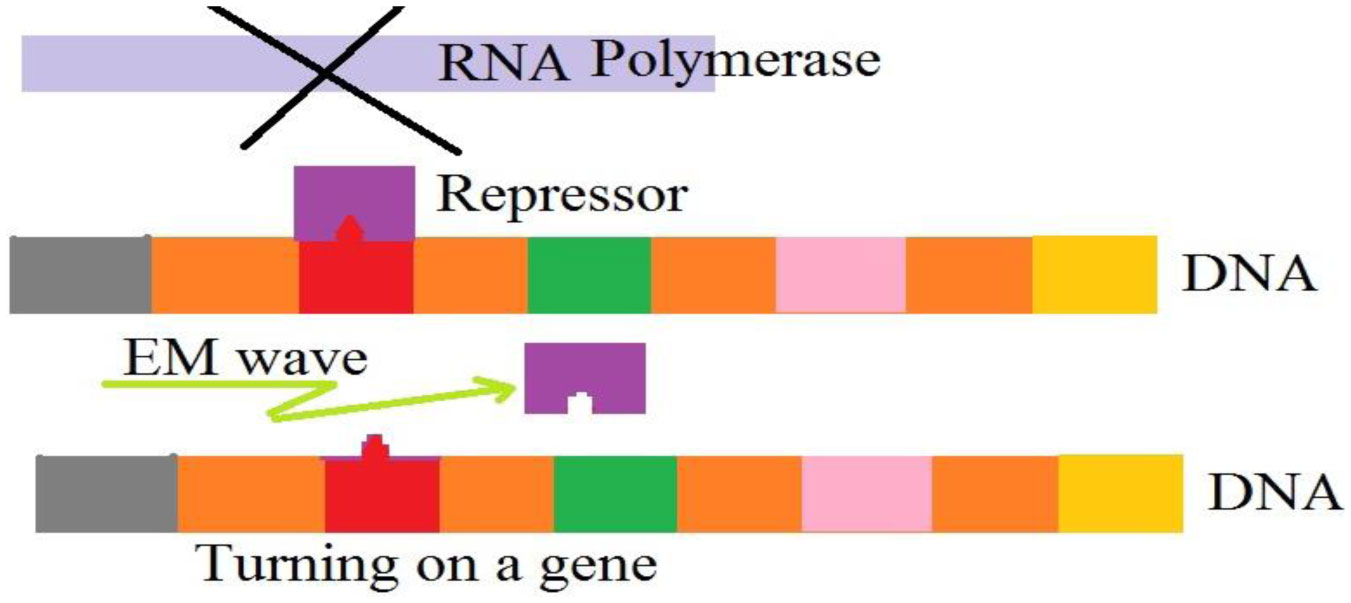
Previous experiments have shown that extremely low frequency electromagnetic fields could cause serious effects on the evolution of cells. We propose a mathematical model which confirms those results. In our model, electromagnetic waves could cause the motions of ions and charges and the emergence of some currents around and in the interior of cells. These currents produce some waves which interact with the DNAs and remove or attach some repressors. Consequently, some genes could be turned on or off, and cells could obtain some properties or lose them. The frequency of the external waves should be close to the frequency of the exchanged waves between the repressors and DNAs or even bigger than them. We test this idea and did some experiments on quail embryonic cells. We connected a sample of these cells to a battery and considered their evolution. We observed that after connecting the battery and the production of electrical current, some rings around the quail embryonic cells emerged. Maybe, these rings are the response of the cells to changes in electromagnetic waves and electrical currents.
Citation: Massimo Fioranelli, Maria Grazia Roccia, Aroonkumar Beesham, Dana Flavin, M. Ghaeni, Faissal AZIZ. A model for considering effects of extremely low frequency electromagnetic fields on quail embryonic cells[J]. AIMS Biophysics, 2022, 9(3): 198-207. doi: 10.3934/biophy.2022017
Previous experiments have shown that extremely low frequency electromagnetic fields could cause serious effects on the evolution of cells. We propose a mathematical model which confirms those results. In our model, electromagnetic waves could cause the motions of ions and charges and the emergence of some currents around and in the interior of cells. These currents produce some waves which interact with the DNAs and remove or attach some repressors. Consequently, some genes could be turned on or off, and cells could obtain some properties or lose them. The frequency of the external waves should be close to the frequency of the exchanged waves between the repressors and DNAs or even bigger than them. We test this idea and did some experiments on quail embryonic cells. We connected a sample of these cells to a battery and considered their evolution. We observed that after connecting the battery and the production of electrical current, some rings around the quail embryonic cells emerged. Maybe, these rings are the response of the cells to changes in electromagnetic waves and electrical currents.

| [1] |
Zhang Y, Yan J, Xu H, et al. (2018) Extremely low frequency electromagnetic fields promote mesenchymal stem cell migration by increasing intracellular Ca2+ and activating the FAK/Rho GTPases signaling pathways in vitro. Stem Cell Res Ther 9: 1-10. https://doi.org/10.1186/s13287-018-0883-4 
|
| [2] |
Ross CL, Siriwardane M, Almeida-Porada G, et al. (2015) The effect of low-frequency electromagnetic field on human bone marrow stem/progenitor cell differentiation. Stem Cell Res 15: 96-108. https://doi.org/10.1016/j.scr.2015.04.009
|
| [3] |
Chernov AS, Reshetnikov DA, Ristsov GK, et al. (2019) Influence of electromagnetic waves, with maxima in the green or red range, on the morphofunctional properties of multipotent stem cells. J Biol Phys 45: 317-334. https://doi.org/10.1007/s10867-019-09531-7
|
| [4] |
Bai W, Li M, Xu W, et al. (2021) Comparison of effects of high-and low-frequency electromagnetic fields on proliferation and differentiation of neural stem cells. Neurosci Lett 741: 135463. https://doi.org/10.1016/j.neulet.2020.135463
|
| [5] |
Parate D, Franco-Obregón A, Fröhlich J, et al. (2017) Enhancement of mesenchymal stem cell chondrogenesis with short-term low intensity pulsed electromagnetic fields. Sci Rep 7: 9421. https://doi.org/10.1038/s41598-017-09892-w
|
| [6] |
Tu C, Xiao Y, Ma Y, et al. (2018) The legacy effects of electromagnetic fields on bone marrow mesenchymal stem cell self-renewal and multiple differentiation potential. Stem Cell Res Ther 9: 215. https://doi.org/10.1186/s13287-018-0955-5
|
| [7] |
Chen J, Tu C, Tang X, et al. (2019) The combinatory effect of sinusoidal electromagnetic field and VEGF promotes osteogenesis and angiogenesis of mesenchymal stem cell-laden PCL/HA implants in a rat subcritical cranial defect. Stem Cell Res Ther 10: 379. https://doi.org/10.1186/s13287-019-1464-x
|
| [8] |
Maziarz A, Kocan B, Bester M, et al. (2016) How electromagnetic fields can influence adult stem cells: positive and negative impacts. Stem Cell Res Ther 7: 54. https://doi.org/10.1186/s13287-016-0312-5
|
| [9] |
He N, Wang Y, Zhang C, et al. (2018) Wnt signaling pathway regulates differentiation of chicken embryonic stem cells into spermatogonial stem cells via Wnt5a. J Cell Biochem 119: 1689-1701. https://doi.org/10.1002/jcb.26329
|
| [10] |
Giotis ES, Montillet G, Pain B, et al. (2019) Chicken embryonic-stem cells are permissive to poxvirus recombinant vaccine vectors. Genes 10: 237. https://doi.org/10.3390/genes10030237
|
| [11] |
Xiong C, Wang M, Ling W, et al. (2020) Advances in isolation and culture of chicken embryonic stem cells in vitro. Cell Reprogram 22: 43-54. https://doi.org/10.1089/cell.2019.0080
|
| [12] |
Zuo Q, Jin K, Zhang Y, et al. (2017) Dynamic expression and regulatory mechanism of TGF-β signaling in chicken embryonic stem cells differentiating into spermatogonial stem cells. Biosci Rep 37: BSR20170179. https://doi.org/10.1042/BSR20170179
|


Figures(7)

Massimo Fioranelli, Maria Grazia Roccia, Aroonkumar Beesham, Dana Flavin, M. Ghaeni, Faissal AZIZ. A model for considering effects of extremely low frequency electromagnetic fields on quail embryonic cells[J]. AIMS Biophysics, 2022, 9(3): 198-207. doi: 10.3934/biophy.2022017


 DownLoad: