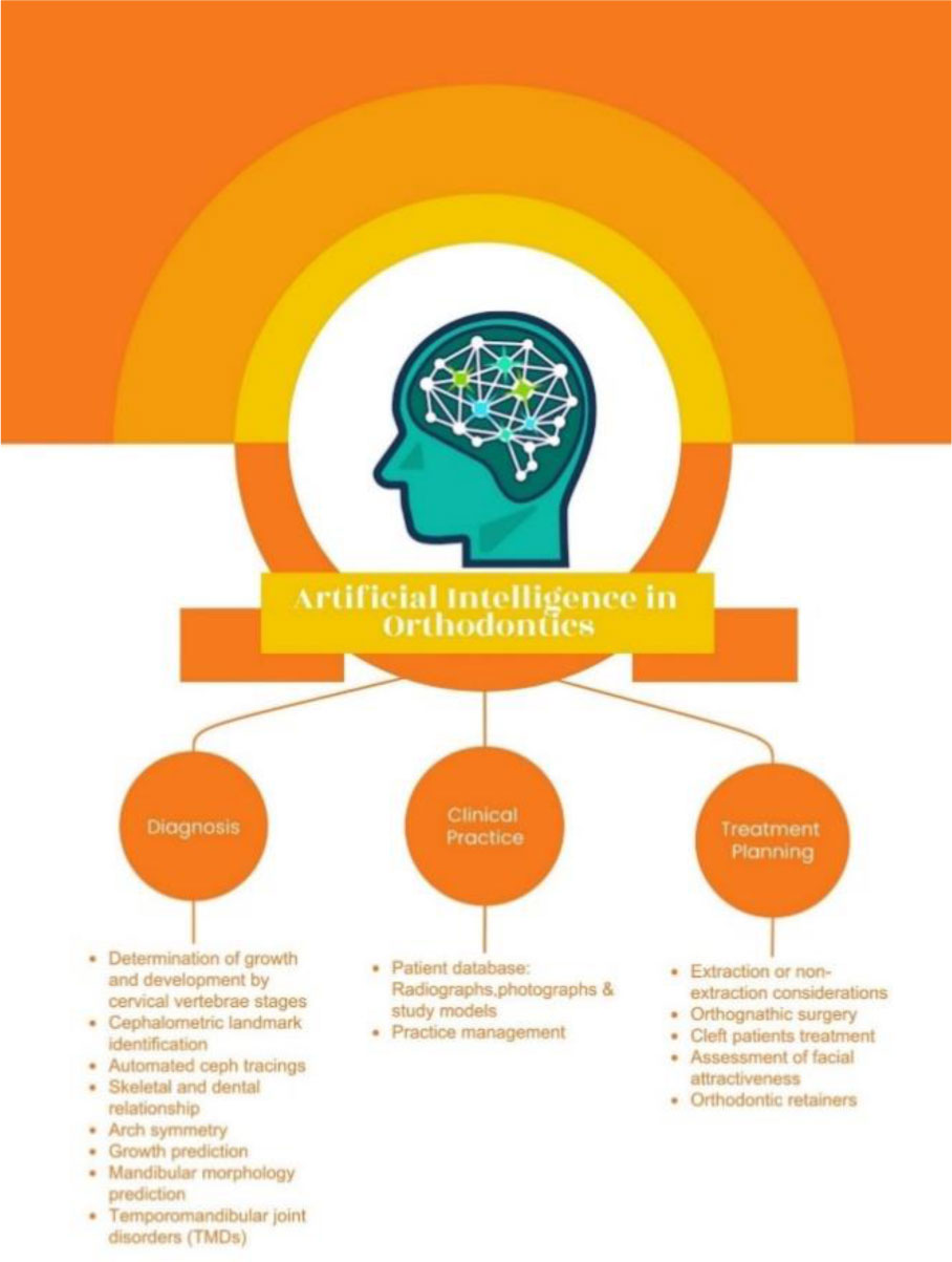
New digital technologies, like in other fields, have revolutionized the health care field and orthodontic practice in the 21st century. They can assist the health care professionals in working more efficiently by saving time and improving patient care. Recent advances in artificial intelligence (AI) and 3D printing technology are useful for improving diagnosis and treatment planning, creating algorithms and manufacturing customized orthodontic appliances. AI accomplishes the task of human beings with the help of machines and technology. In orthodontics, AI-based models have been used for diagnosis, treatment planning, clinical decision-making and prognosis prediction. It minimizes the required workforce and speeds up the diagnosis and treatment procedure. In addition, the 3D printing technology is used to fabricate study models, clear aligner models, surgical guides for inserting mini-implants, clear aligners, lingual appliances, wires components for removable appliances and occlusal splints. This paper is a review of the future and scope of AI and 3D printing technology in orthodontics.
Citation: Mahamad Irfanulla Khan, Laxmikanth SM, Tarika Gopal, Praveen Kumar Neela. Artificial intelligence and 3D printing technology in orthodontics: future and scope[J]. AIMS Biophysics, 2022, 9(3): 182-197. doi: 10.3934/biophy.2022016
New digital technologies, like in other fields, have revolutionized the health care field and orthodontic practice in the 21st century. They can assist the health care professionals in working more efficiently by saving time and improving patient care. Recent advances in artificial intelligence (AI) and 3D printing technology are useful for improving diagnosis and treatment planning, creating algorithms and manufacturing customized orthodontic appliances. AI accomplishes the task of human beings with the help of machines and technology. In orthodontics, AI-based models have been used for diagnosis, treatment planning, clinical decision-making and prognosis prediction. It minimizes the required workforce and speeds up the diagnosis and treatment procedure. In addition, the 3D printing technology is used to fabricate study models, clear aligner models, surgical guides for inserting mini-implants, clear aligners, lingual appliances, wires components for removable appliances and occlusal splints. This paper is a review of the future and scope of AI and 3D printing technology in orthodontics.

| [1] | Negnevitsky M (2011) Artificial intelligence: a guide to intelligent systems. Canada: Pearson Education. |
| [2] | Khanna S (2010) Artificial intelligence: contemporary applications and future compass. Int Dent J 60: 269-272. https://doi.org/10.1922/IDJ_2422Khanna04 |
| [3] | Deshmukh SV (2018) Artificial intelligence in dentistry. J Int Clin Dent Res Organ 10: 47-48. https://doi.org/10.4103/jicdro.jicdro_17_18 |
| [4] | Scerri M, Grech V (2020) Artificial intelligence in medicine. Early Hum Dev 20: 105017. https://doi.org/10.1016/j.earlhumdev.2020.105017 |
| [5] | Ahmed N, Abbasi MS, Zuberi F, et al. (2021) Artificial intelligence techniques: analysis, application, and outcome in dentistry—a systematic review. Biomed Res Int 2021: 9751564. https://doi.org/10.1155/2021/9751564 |
| [6] | Hung HC, Wang YC, Wang YC (2020) Applications of artificial intelligence in orthodontics. Taiwan J Orthod 32: 85-92. https://doi.org/10.38209/2708-2636.1005 |
| [7] | Bichu YM, Hansa I, Bichu AY, et al. (2021) Applications of artificial intelligence and machine learning in orthodontics: a scoping review. Prog Orthod 22: 18. https://doi.org/10.1186/s40510-021-00361-9 |
| [8] | Schwendicke F, Samek W, Krois J (2020) Artificial intelligence in dentistry: chances and challenges. J Dent Res 99: 769-774. https://doi.org/10.1177/0022034520915714 |
| [9] | Wong KV, Hernandez A (2012) A review of additive manufacturing. ISRN Mech Eng 2012: 208760. https://doi.org/10.5402/2012/208760 |
| [10] | Andonović V, Vrtanoski G (2010) Growing rapid prototyping as a technology in dental medicine. Mech Eng Sci J 29: 31-39. |
| [11] | Beguma Z, Chhedat P (2014) Rapid prototyping—when virtual meets reality rapid prototyping—virtuelltrifftrealität. Int J Comput Dent 17: 297-306. |
| [12] | Hofmann M (2014) 3D printing gets a boost and opportunities with polymer materials. ACS Macro Lett 3: 382-386. https://doi.org/10.1021/mz4006556 |
| [13] | Mahamood S, Khader MA, Ali H (2016) Applications of 3-D printing in orthodontics: a review. Int J Sci Stud 3: 267-270. https://doi.org/10.17354/ijss/2016/99 |
| [14] | Jiang F, Jiang Y, Zhi H, et al. (2017) Artificial intelligence in healthcare: past, present, and future. Stroke Vasc Neurol 2: 230-243. https://doi.org/10.1136/svn-2017-000101 |
| [15] | Leonardi R, Giordano D, Maiorana F (2009) An evaluation of cellular neural networks for the automatic identification of cephalometric landmarks on digital images. J Biomed Biotechnol 2009: 717102. https://doi.org/10.1155/2009/717102 |
| [16] | Schwendicke F, Golla T, Dreher M, et al. (2019) Convolutional neural networks for dental image diagnostics: a scoping review. J Dent 91: 103226. https://doi.org/10.1016/j.jdent.2019.103226 |
| [17] | Gupta A, Kharbanda OP, Sardana V, et al. (2016) Accuracy of 3D cephalometric measurements based on an automatic knowledge-based landmark detection algorithm. Int J Comput Ass Rad 11: 1297-1309. https://doi.org/10.1007/s11548-015-1334-7 |
| [18] | Brickley MR, Shepherd JP, Armstrong RA (1998) Neural networks: a new technique for development of decision support systems in dentistry. J Dent 26: 305-309. https://doi.org/10.1016/S0300-5712(97)00027-4 |
| [19] | Katne T, Kanaparthi A, Gotoor S, et al. (2019) Artificial intelligence: demystifying dentistry—the future and beyond. Int J Contemp Med SurgRadiol 4: D6-D9. http://dx.doi.org/10.21276/ijcmsr.2019.4.4.2 |
| [20] | Faber J, Faber C, Faber P (2019) Artificial intelligence in orthodontics. APOS Trends Orthod 9: 201-205. https://doi.org/10.25259/APOS_123_2019 |
| [21] | Auconi P, Caldarelli G, Scala A, et al. (2011) A network approach to orthodontic diagnosis. Orthod Craniofac Res 14: 189-197. https://doi.org/10.1111/j.1601-6343.2011.01523.x |
| [22] | Xie X, Wang L, Wang A (2010) Artificial neural network modeling for deciding if extractions are necessary prior to orthodontic treatment. Angle Orthod 80: 262-266. https://doi.org/10.2319/111608-588.1 |
| [23] | Jung SK, Kim TW (2016) New approach for the diagnosis of extractions with neural network machine learning. Am J Orthod Dentofac 149: 127-133. https://doi.org/10.1016/j.ajodo.2015.07.030 |
| [24] | Jheon AH, Oberoi S, Solem RC, et al. (2017) Moving towards precision orthodontics: an evolving paradigm shift in the planning and delivery of customized orthodontic therapy. Orthod Craniofac Res 20: 106-113. https://doi.org/10.1111/ocr.12171 |
| [25] | Niño-Sandoval TC, Pérez SVG, González FA, et al. (2017) Use of automated learning techniques for predicting mandibular morphology in skeletal class I, II and III. Forensic Sci Int 281: 187.e1-187.e7. https://doi.org/10.1016/j.forsciint.2017.10.004 |
| [26] | Choi HI, Jung SK, Baek SH, et al. (2019) Artificial intelligent model with neural network machine learning for the diagnosis of orthognathic surgery. J Craniofac Surg 30: 1986-1989. https://doi.org/10.1097/SCS.0000000000005650 |
| [27] | Weichel F, Eisenmann U, Richter S, et al. (2019) A computer-assisted optimization approach for orthognathic surgery planning. Curr Dir Biomed Eng 5: 41-44. https://doi.org/10.1515/cdbme-2019-0011 |
| [28] | Patcas R, Timofte R, Volokitin A, et al. (2019) Facial attractiveness of cleft patients: a direct comparison between artificial-intelligence-based scoring and conventional rater groups. Eur J Orthodont 41: 428-433. https://doi.org/10.1093/ejo/cjz007 |
| [29] | Makaremi M, Lacaule C, Mohammad-Djafari A (2019) Deep learning and artificial intelligence for the determination of the cervical vertebra maturation degree from lateral radiography. Entropy 21: 1222. https://doi.org/10.3390/e21121222 |
| [30] | Kök H, Acilar AM, İzgi MS (2019) Usage and comparison of artificial intelligence algorithms for determination of growth and development by cervical vertebrae stages in orthodontics. Prog Orthod 20: 41. https://doi.org/10.1186/s40510-019-0295-8 |
| [31] | Shoukri B, Prieto JC, Ruellas A, et al. (2019) Minimally invasive approach for diagnosing TMJ osteoarthritis. J Dent Res 98: 1103-1111. https://doi.org/10.1177/0022034519865187 |
| [32] | Castle E, Chung P, Behfar MH, et al. (2019) Compliance monitoring via a Bluetooth-enabled retainer: a prospective clinical pilot study. Orthod Craniofac Res 22: 149-153. https://doi.org/10.1111/ocr.12263 |
| [33] | Lee KS, Kwak HJ, Oh JM, et al. (2020) Automated detection of TMJ osteoarthritis based on artificial intelligence. J Dent Res 99: 1363-1367. https://doi.org/10.1177/0022034520936950 |
| [34] | Akdeniz S, Tosun ME (2021) A review of the use of artificial intelligence in orthodontics. J Exp Clin Med 38: 157-162. https://doi.org/10.52142/omujecm.38.si.dent.13 |
| [35] | Kök H, Izgi MS, Acilar AM (2021) Determination of growth and development periods in orthodontics with artificial neural network. Orthod Craniofac Res 24: 76-83. https://doi.org/10.1111/ocr.12443 |
| [36] | Littlewood SJ, Dalci O, Dolce C, et al. (2021) Orthodontic retention: What's on the horizon? Brit Dent J 230: 760-764. https://doi.org/10.1038/s41415-021-2937-8 |
| [37] | Jung SK, Kim TW (2016) New approach for the diagnosis of extractions with neural network machine learning. Am J Orthod Dentofac 149: 127-133. https://doi.org/10.1016/j.ajodo.2015.07.030 |
| [38] | Hutton TJ, Cunningham S, Hammond P (2000) An evaluation of active shape models for the automatic identification of cephalometric landmarks. Eur J Orthodont 22: 499-508. https://doi.org/10.1093/ejo/22.5.499 |
| [39] | Tanikawa C, Yagi M, Takada K (2009) Automated cephalometry: System performance reliability using landmark-dependent criteria. Angle Orthod 79: 1037-1046. https://doi.org/10.2319/092908-508R.1 |
| [40] | Montúfar J, Romero M, Scougall-Vilchis RJ (2018) Automatic 3-dimensional cephalometric landmarking based on active shape models in related projections. Am J Orthod Dentofac 153: 449-458. https://doi.org/10.1016/j.ajodo.2017.06.028 |
| [41] | Lee H, Tajmir S, Lee J, et al. (2017) Fully automated deep learning system for bone age assessment. J Digit Imaging 30: 427-441. https://doi.org/10.1007/s10278-017-9955-8 |
| [42] | Kunz F, Stellzig-Eisenhauer A, Zeman F, et al. (2020) Artificial intelligence in orthodontics. J Orofac Orthop 81: 52-68. https://doi.org/10.1007/s00056-019-00203-8 |
| [43] | Nishimoto S, Sotsuka Y, Kawai K, et al. (2019) Personal computer-based cephalometric landmark detection with deep learning, using cephalograms on the internet. J Craniofac Surg 30: 91-95. https://doi.org/10.1097/SCS.0000000000004901 |
| [44] | Hwang HW, Park JH, Moon JH, et al. (2020) Automated identification of cephalometric landmarks: Part 2—Might it be better than human? Angle Orthod 90: 69-76. https://doi.org/10.2319/022019-129.1 |
| [45] | Park JH, Hwang HW, Moon JH, et al. (2019) Automated identification of cephalometric landmarks: Part 1—Comparisons between the latest deep-learning methods YOLOV3 and SSD. Angle Orthod 89: 903-909. https://doi.org/10.2319/022019-127.1 |
| [46] | Ribarevski R, Vig P, Vig KD, et al. (1996) Consistency of orthodontic extraction decisions. Eur J Orthodont 18: 77-80. https://doi.org/10.1093/ejo/18.1.77 |
| [47] | Bouletreau P, Makaremi M, Ibrahim B, et al. (2019) Artificial intelligence: applications in orthognathic surgery. J Stomatol Oral Maxi 120: 347-354. https://doi.org/10.1016/j.jormas.2019.06.001 |
| [48] | Pascal E, Majoufre C, Bondaz M, et al. (2018) Current status of surgical planning and transfer methods in orthognathic surgery. J Stomatol Oral Maxi 119: 245-248. https://doi.org/10.1016/j.jormas.2018.02.001 |
| [49] | Cevidanes LHS, Bailey LJ, Tucker Jr GR, et al. (2005) Superimposition of 3D cone-beam CT models of orthognathic surgery patients. Dentomaxillofac Rad 34: 369-375. https://doi.org/10.1259/dmfr/17102411 |
| [50] | Yu X, Liu B, Pei Y, et al. (2014) Evaluation of facial attractiveness for patients with malocclusion: a machine-learning technique employing Procrustes. Angle Orthod 84: 410-416. https://doi.org/10.2319/071513-516.1 |
| [51] | Patcas R, Bernini DAJ, Volokitin A, et al. (2019) Applying artificial intelligence to assess the impact of orthognathic treatment on facial attractiveness and estimated age. Int J Oral Max Surg 48: 77-83. https://doi.org/10.1016/j.ijom.2018.07.010 |
| [52] | Harrar H, Myers S, Ghanem AM (2018) Art or science? An evidence-based approach to human facial beauty a quantitative analysis towards an informed clinical aesthetic practice. Aesthet Plast Surg 42: 137-146. https://doi.org/10.1007/s00266-017-1032-7 |
| [53] | Zhang SJ, Meng P, Zhang J, et al. (2018) Machine learning models for genetic risk assessment of infants with non-syndromic orofacial cleft. Genom Proteom Bioinf 16: 354-364. https://doi.org/10.1016/j.gpb.2018.07.005 |
| [54] | Prescher A, Meyers A, von Keyserlingk DG (2005) Neural net applied to anthropological material: a methodical study on the human nasal skeleton. Ann Anat 187: 261-269. https://doi.org/10.1016/j.aanat.2005.01.003 |
| [55] | Al-Moghrabi D, Pandis N, McLaughlin K, et al. (2020) Evaluation of the effectiveness of a tailored mobile application in increasing the duration of wear of thermoplastic retainers: a randomized controlled trial. Eur J Orthodont 42: 571-579. https://doi.org/10.1093/ejo/cjz088 |
| [56] | Cole D, Bencharit S, Carrico CK, et al. (2019) Evaluation of fit for 3D-printed retainers compared with thermoform retainers. Am J Orthod Dentofac 155: 592-599. https://doi.org/10.1016/j.ajodo.2018.09.011 |
| [57] | Dietrich CA, Ender A, Baumgartner S, et al. (2017) A validation study of reconstructed rapid prototyping models produced by two technologies. Angle Orthod 87: 782-787. https://doi.org/10.2319/01091-727.1 |
| [58] | Mohd Tahir N, Wan Hassan WN, Saub R (2019) Comparing retainers constructed on conventional stone models and on 3D printed models: a randomized crossover clinical study. Eur J Orthodont 41: 370-380. https://doi.org/10.1093/ejo/cjy063 |
| [59] | Ligon SC, Liska R, Stampfl J, et al. (2017) Polymers for 3D printing and customized additive manufacturing. Chem Rev 117: 10212-10290. https://doi.org/10.1021/acs.chemrev.7b00074 |
| [60] | Dodziuk H (2016) Applications of 3D printing in healthcare. Kardiochir Torakochi 13: 283-293. https://doi.org/10.5114/kitp.2016.62625 |
| [61] | Bhargav A, Sanjairaj V, Rosa V, et al. (2018) Applications of additive manufacturing in dentistry: a review. J Biomed Mater Res B 106: 2058-2064. https://doi.org/10.1002/jbm.b.33961 |
| [62] | Liaw CY, Guvendiren M (2017) Current and emerging applications of 3D printing in medicine. Biofabrication 9: 024102. https://doi.org/10.1088/1758-5090/aa7279 |
| [63] | Wiechmann D, Rummel V, Thalheim A, et al. (2003) Customized brackets and archwires for lingual orthodontic treatment. Am J Orthod Dentofac 124: 593-599. https://doi.org/10.1016/j.ajodo.2003.08.008 |
| [64] | Faber J, Berto PM, Quaresma M (2006) Rapid prototyping as a tool for diagnosis and treatment planning for maxillary canine impaction. Am J Orthod Dentofac 129: 583-589. https://doi.org/10.1016/j.ajodo.2005.12.015 |
| [65] | Wiechmann D, Schwestka-Polly R, Hohoff A (2008) Herbst appliance in lingual orthodontics. Am J Orthod Dentofac 134: 439-446. https://doi.org/10.1016/j.ajodo.2007.09.015 |
| [66] | Tsui WK, Chua HDP, Cheung LK (2012) Bone anchor systems for orthodontic application: a systematic review. Int J Oral Max Surg 41: 1427-1438. https://doi.org/10.1016/j.ijom.2012.05.011 |
| [67] | Salmi M, Paloheimo KS, Tuomi J, et al. (2013) A digital process for additive manufacturing of occlusal splints: a clinical pilot study. J R Soc Interface 10: 20130203. https://doi.org/10.1098/rsif.2013.0203 |
| [68] | Nasef AA, El-Beialy AR, Mostafa YA (2014) Virtual techniques for designing and fabricating a retainer. Am J Orthod Dentofac 146: 394-398. https://doi.org/10.1016/j.ajodo.2014.01.025 |
| [69] | Wang YT, Yu JH, Lo LJ, et al. (2017) Developing customized dental miniscrew surgical template from thermoplastic polymer material using image superimposition, CAD system, and 3D printing. Biomed Res Int 2017: 1906197. https://doi.org/10.1155/2017/1906197 |
| [70] | Sherman SL, Kadioglu O, Currier GF, et al. (2020) Accuracy of digital light processing printing of 3-dimensional dental models. Am J Orthod Dentofac 157: 422-428. https://doi.org/10.1016/j.ajodo.2019.10.012 |
| [71] | Al-Moghrabi D, Barber S, Fleming PS (2021) Removable retention: enhancing adherence and the remit of shared decision-making. Brit Dent J 230: 765-769. https://doi.org/10.1038/s41415-021-2951-x |
| [72] | Cousley RRJ (2020) Introducing 3D printing in your orthodontic practice. J Orthod 47: 265-272. https://doi.org/10.1177/1465312520936704 |
| [73] | Bartkowiak T, Walkowiak-Śliziuk A (2018) 3D printing technology in orthodontics—review of current applications. J Stomatol 71: 356-364. https://doi.org/10.5114/jos.2018.83410 |
| [74] | Lee KY, Cho JW, Chang NY, et al. (2015) Accuracy of three-dimensional printing for manufacturing replica teeth. Korean J Orthod 45: 217-225. https://doi.org/10.4041/kjod.2015.45.5.217 |
| [75] | Kim SY, Shin YS, Jung HD, et al. (2018) Precision and trueness of dental models manufactured with different 3-dimensional printing techniques. Am J Orthod Dentofac 153: 144-153. https://doi.org/10.1016/j.ajodo.2017.05.025 |
| [76] | Martorelli M, Gerbino S, Giudice M, et al. (2013) A comparison between customized clear and removable orthodontic appliances manufactured using RP and CNC techniques. Dent Mater 29: e1-e10. https://doi.org/10.1016/j.dental.2012.10.011 |
| [77] | Al Mortadi N, Eggbeer D, Lewis J, et al. (2012) CAD/CAM/AM applications in the manufacture of dental appliances. Am J Orthod Dentofac 142: 727-733. https://doi.org/10.1016/j.ajodo.2012.04.023 |
| [78] | Salmi M, Tuomi J, Sirkkanen R, et al. (2012) Rapid tooling method for soft customized removable oral appliances. Open Dent J 6: 85-89. https://doi.org/10.2174/1874210601206010085 |
| [79] | Antoszewska J, Papadopoulos MA, Park HS, et al. (2009) Five-year experience with orthodontic miniscrew implants: a retrospective investigation of factors influencing success rates. Am J Orthod Dentofac 136: 158.e1-158.e10. https://doi.org/10.1016/j.ajodo.2009.03.032 |
| [80] | Liu H, Liu DX, Wang G, et al. (2010) Accuracy of surgical positioning of orthodontic miniscrews with a computer-aided design and manufacturing template. Am J Orthod Dentofac 137: 728.e1-728.e10. https://doi.org/10.1016/j.ajodo.2009.12.025 |
| [81] | Hourfar J, Kanavakis G, Goellner P, et al. (2014) Fully customized placement of orthodontic miniplates: a novel clinical technique. Head Face Med 10: 14. https://doi.org/10.1186/1746-160X-10-14 |
| [82] | Neumeister A, Schulz L, Glodecki C (2017) Investigations on the accuracy of 3D-printed drill guides for dental implantology Untersuchungen zur Genauigkeit 3-D-gedruckter Bohrschablonen für die dentale Implantologie. Int J Comput Dent 20: 35-51. |
| [83] | Richert R, Goujat A, Venet L, et al. (2017) Intraoral scanner technologies: a review to make a successful impression. J Healthc Eng 2017: 8427595. https://doi.org/10.1155/2017/8427595 |

Figures(2) / Tables(2)

Mahamad Irfanulla Khan, Laxmikanth SM, Tarika Gopal, Praveen Kumar Neela. Artificial intelligence and 3D printing technology in orthodontics: future and scope[J]. AIMS Biophysics, 2022, 9(3): 182-197. doi: 10.3934/biophy.2022016


 DownLoad: