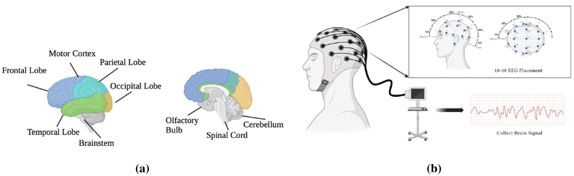
This study presented a new approach to seizure classification utilizing electroencephalogram (EEG) data. We introduced the NeuroWave-Net, an innovative hybrid model that seamlessly integrates convolutional neural networks (CNN) and long short-term memory (LSTM) architectures. Unlike conventional methods, our model capitalized on CNN's proficiency in feature extraction and LSTM's prowess in classifying seizure. The key strength of the NeuroWave-Net lies in its ability to combine these distinct architectures, synergizing their capabilities for enhanced accuracy in identifying seizure conditions within EEG data. Our proposed model exhibited outstanding performance, achieving a classification accuracy of 99.48%. This study contributed to the advancement of seizure classification models, providing a robust and streamlined approach for accurate categorization within EEG datasets. NeuroWave-Net stands as a testament to the potential of hybrid neural network architectures in neurological diagnostics.
Citation: Md. Mehedi Hassan, Rezuana Haque, Sheikh Mohammed Shariful Islam, Hossam Meshref, Roobaea Alroobaea, Mehedi Masud, Anupam Kumar Bairagi. NeuroWave-Net: Enhancing epileptic seizure detection from EEG brain signals via advanced convolutional and long short-term memory networks[J]. AIMS Bioengineering, 2024, 11(1): 85-109. doi: 10.3934/bioeng.2024006
This study presented a new approach to seizure classification utilizing electroencephalogram (EEG) data. We introduced the NeuroWave-Net, an innovative hybrid model that seamlessly integrates convolutional neural networks (CNN) and long short-term memory (LSTM) architectures. Unlike conventional methods, our model capitalized on CNN's proficiency in feature extraction and LSTM's prowess in classifying seizure. The key strength of the NeuroWave-Net lies in its ability to combine these distinct architectures, synergizing their capabilities for enhanced accuracy in identifying seizure conditions within EEG data. Our proposed model exhibited outstanding performance, achieving a classification accuracy of 99.48%. This study contributed to the advancement of seizure classification models, providing a robust and streamlined approach for accurate categorization within EEG datasets. NeuroWave-Net stands as a testament to the potential of hybrid neural network architectures in neurological diagnostics.
Electroencephalogram
Convolutional neural network
Long short-term memory
Random forest
Support vector machine
Particle swarm optimization
1-Dimensional convolutional neural networks
2-Dimensional convolutional neural networks
Deep neural network
Intracranial electroencephalography
Contextual long short-term memory
Bidirectional long short-term memory
Rectified linear unit
1D convolution layer

| [1] |
Fisher RS, van Emde Boas W, Blume W, et al. (2005) Response: Definitions proposed by the international league against epilepsy (ILAE) and the international bureau for epilepsy (IBE). Epilepsia 46: 1701-1702. https://doi.org/10.1111/j.1528-1167.2005.00273_4.x 
|
| [2] |
Juan E, Górska U, Kozma C, et al. (2005) Distinct signatures of loss of consciousness in focal impaired awareness versus tonic-clonic seizures. Brain 146: 109-123. https://doi.org/10.1093/brain/awac291
|
| [3] |
Schwartz PJ, Ackerman MJ, Antzelevitch C, et al. (2020) Inherited cardiac arrhythmias. Nat Rev Dis Primers 6: 58. https://doi.org/10.1038/s41572-020-0188-7
|
| [4] |
Lemoine É, Toffa D, Pelletier-Mc DG, et al. (2023) Machine-learning for the prediction of one-year seizure recurrence based on routine electroencephalography. Sci Rep 13: 12650. https://doi.org/10.1038/s41598-023-39799-8
|
| [5] |
McKee JL, Kaufman MC, Gonzalez AK, et al. (2023) Leveraging electronic medical record-embedded standardised electroencephalogram reporting to develop neonatal seizure prediction models: A retrospective cohort study. Lancet Digit Health 5: e217-e226. https://doi.org/10.1016/S2589-7500(23)00004-3
|
| [6] |
Pinto MF, Batista J, Leal A, et al. (2023) The goal of explaining black boxes in eeg seizure prediction is not to explain models' decisions. Epilepsia Open 8: 285-297. https://doi.org/10.1002/epi4.12748
|
| [7] | Khare SK, Khan AM, Bajaj V, et al. (2023) Introduction to smart healthcare and the role of cognitive sensors. Cognitive Sensors . UK: IOP Publishing Bristol 1-21. |
| [8] |
Hernandez-Pavon JC, Veniero D, Bergmann TO, et al. (2023) TMS combined with EEG: Recommendations and open issues for data collection and analysis. Brain Stimul 6: 567-593. https://doi.org/10.1016/j.brs.2023.02.009
|
| [9] | Chiarion G, Sparacino L, Antonacci Y, et al. (2023) Connectivity analysis in EEG data: A tutorial review of the state of the art and emerging trends. Bioeng 10: 372. https://doi.org/10.3390/bioengineering10030372 |
| [10] |
López-Arango G, Deguire F, Agbogba K, et al. (2023) Impact of macrocephaly, as an isolated trait, on EEG signal as measured by spectral power and multiscale entropy during the first year of life. Dev Neurosci 45: 210-222. https://doi.org/10.1159/000529722
|
| [11] | Zhao W, Zhao WB, Wang WF, et al. (2020) A novel deep neural network for robust detection of seizures using eeg signals. Comput Math Method M 2020: 9689821. https://doi.org/10.1155/2020/9689821 |
| [12] |
Nishad A, Pachori RB (2020) Classification of epileptic electroencephalogram signals using tunable-Q wavelet transform based filter-bank. J Amb Intel Hum Comp 15: 877-891. https://doi.org/10.1007/s12652-020-01722-8
|
| [13] | Nithya K, Sharma S, Sharma RR (2023) Eigenvalues of hankel matrix based epilepsy detection using EEG signals. In 2023 2nd International Conference on Paradigm Shifts in Communications Embedded Systems, Machine Learning and Signal Processing (PCEMS) . New York: IEEE 1-6. |
| [14] |
Hemachandira VS, Viswanathan R (2022) A framework on performance analysis of mathematical model-based classifiers in detection of epileptic seizure from EEG signals with efficient feature selection. J Healthc Eng 2022: 7654666. https://doi.org/10.1155/2022/7654666
|
| [15] | Cao JW, Hu DH, Wang YM, et al. (2021) Epileptic classification with deep-transfer-learning-based feature fusion algorithm. IEEE T Cogn Dev Syst 14: 684-695. https://doi.org/10.1109/TCDS.2021.3064228 |
| [16] |
Wang YP, Dai Y, Liu ZM, et al. (2021) Computer-aided intracranial EEG signal identification method based on a multi-branch deep learning fusion model and clinical validation. Brain Sci 11: 615. https://doi.org/10.3390/brainsci11050615
|
| [17] |
Liu Y, Huang YX, Zhang XX, et al. (2020) Deep C-LSTM neural network for epileptic seizure and tumor detection using high-dimension EEG signals. IEEE Access 8: 37495-37504. https://doi.org/10.1109/ACCESS.2020.2976156
|
| [18] |
Beeraka SM, Kumar A, Sameer M, et al. (2022) Accuracy enhancement of epileptic seizure detection: A deep learning approach with hardware realization of STFT. Circ Syst Signal Pr 41: 461-484. https://doi.org/10.1007/s00034-021-01789-4
|
| [19] |
Ryu S, Joe I (2021) A hybrid DenseNET-LSTM model for epileptic seizure prediction. Appl Sci 11: 7661. https://doi.org/10.3390/app11167661
|
| [20] | Srivastava A, Singh A, Tiwari AK (2022) An efficient hybrid approach for the prediction of epilepsy using CNN with LSTM. Int J Artif Intell Soft Comput 7: 179-193. https://doi.org/10.1504/IJAISC.2022.126336 |
| [21] |
Wang XH, Gong GH, Li N (2019) Detection analysis of epileptic EEG using a novel random forest model combined with grid search optimization. Front Hum Neurosci 13: 52. https://doi.org/10.3389/fnhum.2019.00052
|
| [22] |
Sharma RR, Pachori RB (2018) Time–frequency representation using IEVDHM-HT with application to classification of epileptic EEG signals. Iet Sci Meas Technol 12: 72-82. https://doi.org/10.1049/iet-smt.2017.0058
|
| [23] | Sharma RR, Varshney P, Pachori RB, et al. (2018) Automated system for epileptic EEG detection using iterative filtering. IEEE Sensors Letters 2: 1-4. https://doi.org/10.1109/LSENS.2018.2882622 |
| [24] |
San-Segundo R, Gil-Martin M, D'Haro-Enrquez LF, et al. (2019) Classification of epileptic eeg recordings using signal transforms and convolutional neural networks. Comput Biol Med 109: 148-158. https://doi.org/10.1016/j.compbiomed.2019.04.031
|
| [25] |
Andrzejak RG, Lehnertz K, Mormann F, et al. (2001) Indications of nonlinear deterministic and finite-dimensional structures in time series of brain electrical activity: Dependence on recording region and brain state. Phy Rev E 64: 061907. https://doi.org/10.1103/PhysRevE.64.061907
|
| [26] |
Ahmed AA, Ali W, Abdullah TA, et al. (2023) Classifying cardiac arrhythmia from ECG signal using 1D CNN deep learning model. Mathematics 11: 562. https://doi.org/10.3390/math11030562
|
| [27] |
Xiong QS, Kong QZ, Xiong HB, et al. (2024) Physics-informed deep 1D CNN compiled in extended state space fusion for seismic response modeling. Comput Struct 291: 107215. https://doi.org/10.1016/j.compstruc.2023.107215
|
| [28] |
Moussavou Boussougou MK, Park DJ (2023) Attention-based 1D CNN-BILSTM hybrid model enhanced with fasttext word embedding for korean voice phishing detection. Mathematics 11: 3217. https://doi.org/10.3390/math11143217
|
| [29] | Phukan N, Manikandan MS, Pachori RB (2023) Afibri-net: A lightweight convolution neural network based atrial fibrillation detector. IEEE T Circuits-1 70: 4962-4974. https://doi.org/10.1109/TCSI.2023.3303936 |
| [30] |
Hassan W, Joolee JB, Jeon S (2023) Establishing haptic texture attribute space and predicting haptic attributes from image features using 1D-CNN. Sci Rep 13: 11684. https://doi.org/10.1038/s41598-023-38929-6
|
| [31] |
Iyer A, Das SS, Teotia R (2023) CNN and LSTM based ensemble learning for human emotion recognition using EEG recordings. Multimed Tools Appl 82: 4883-4896. https://doi.org/10.1007/s11042-022-12310-7
|
| [32] | Li DK (2023) Multivariate time series prediction based on quantum enhanced LSTM models. Second International Conference on Electronic Information Technology (EIT 2023) . USA: SPIE 491-497. https://doi.org/10.1117/12.2685468 |
| [33] |
Mohammed AYA, Yaw CT, Koh SP, et al. (2023) Detection of corona faults in switchgear by using 1D-CNN, LSTM, and 1D-CNN-LSTM methods. Sensors 23: 3108. https://doi.org/10.3390/s23063108
|
| [34] |
Albaqami H, Hassan GM, Datta A (2023) MP-seiznet: A multi-path cnn BI-LSTM network for seizure-type classification using EEG. Biomed Signal Proces 84: 104780. https://doi.org/10.1016/j.bspc.2023.104780
|
| [35] |
Shoeibi A, Ghassemi N, Alizadehsani R, et al. (2021) A comprehensive comparison of handcrafted features and convolutional autoencoders for epileptic seizures detection in EEG signals. Expert Syst Appl 163: 113788. https://doi.org/10.1016/j.eswa.2020.113788
|
| [36] | Qureshi MB, Afzaal M, Qureshi MS, et al. (2022) Fuzzy-based automatic epileptic seizure detection framework. Comput Mater Contin 7: 5601-5630. https://doi.org/10.32604/cmc.2022.020348 |


Figures(14) / Tables(5)

Md. Mehedi Hassan, Rezuana Haque, Sheikh Mohammed Shariful Islam, Hossam Meshref, Roobaea Alroobaea, Mehedi Masud, Anupam Kumar Bairagi. NeuroWave-Net: Enhancing epileptic seizure detection from EEG brain signals via advanced convolutional and long short-term memory networks[J]. AIMS Bioengineering, 2024, 11(1): 85-109. doi: 10.3934/bioeng.2024006


 DownLoad: