
Citation: Masahiro Yasunaga, Shino Manabe, Masaru Furuta, Koretsugu Ogata, Yoshikatsu Koga, Hiroki Takashima, Toshirou Nishida, Yasuhiro Matsumura. Mass spectrometry imaging for early discovery and development of cancer drugs[J]. AIMS Medical Science, 2018, 5(2): 162-180. doi: 10.3934/medsci.2018.2.162
[1] | Aslak Tveito, Glenn T. Lines, Pan Li, Andrew McCulloch . Defining candidate drug characteristics for Long-QT (LQT3) syndrome. Mathematical Biosciences and Engineering, 2011, 8(3): 861-873. doi: 10.3934/mbe.2011.8.861 |
[2] | Virginia González-Vélez, Amparo Gil, Iván Quesada . Minimal state models for ionic channels involved in glucagon secretion. Mathematical Biosciences and Engineering, 2010, 7(4): 793-807. doi: 10.3934/mbe.2010.7.793 |
[3] | Hongtao Liu, Shuqin Liu, Xiaoxu Ma, Yunpeng Zhang . A numerical model applied to the simulation of cardiovascular hemodynamics and operating condition of continuous-flow left ventricular assist device. Mathematical Biosciences and Engineering, 2020, 17(6): 7519-7543. doi: 10.3934/mbe.2020384 |
[4] | Siqi Chen, Ran Su . An autonomous agent for negotiation with multiple communication channels using parametrized deep Q-network. Mathematical Biosciences and Engineering, 2022, 19(8): 7933-7951. doi: 10.3934/mbe.2022371 |
[5] | Jiuju Shen . EM-TSA: An ensemble machine learning-based transient stability assessment approach for operation of power systems. Mathematical Biosciences and Engineering, 2023, 20(5): 8226-8240. doi: 10.3934/mbe.2023358 |
[6] | Yue Li, Wusheng Xu, Wei Li, Ang Li, Zengjin Liu . Research on hybrid intrusion detection method based on the ADASYN and ID3 algorithms. Mathematical Biosciences and Engineering, 2022, 19(2): 2030-2042. doi: 10.3934/mbe.2022095 |
[7] | Cong Zhao, Na Deng . An actor-critic framework based on deep reinforcement learning for addressing flexible job shop scheduling problems. Mathematical Biosciences and Engineering, 2024, 21(1): 1445-1471. doi: 10.3934/mbe.2024062 |
[8] | T. J. Newman . Modeling Multicellular Systems Using Subcellular Elements. Mathematical Biosciences and Engineering, 2005, 2(3): 613-624. doi: 10.3934/mbe.2005.2.613 |
[9] | Jimin Yu, Jiajun Yin, Shangbo Zhou, Saiao Huang, Xianzhong Xie . An image super-resolution reconstruction model based on fractional-order anisotropic diffusion equation. Mathematical Biosciences and Engineering, 2021, 18(5): 6581-6607. doi: 10.3934/mbe.2021326 |
[10] | Na Pang . Nonlinear neural networks adaptive control for a class of fractional-order tuberculosis model. Mathematical Biosciences and Engineering, 2023, 20(6): 10464-10478. doi: 10.3934/mbe.2023461 |
Since advanced computing technology makes the computational models of cardiac electrophysiology getting mature, the computational models become an important basis for clinical applications [1,2]. Moreover, cardiac cells up to an order of one billion and the internal processes of each cell are also quite complex in the human heart [3]. Therefore, the computational speed, accuracy, and stability are key hotspots in whole heart simulation. Firstly, advanced computer parallelization greatly reduces computing cost of tissue-scale cardiac simulations [4,5,6,7,8]. Secondly, the adaptive algorithms of time-steps and space-steps are a powerful tool for improving the accuracy of computation [6,9,10]. Thirdly, stiffness is ubiquitous in cardiac cell models, which easily leads to unstable computation [11]. Aiming to solve the stiff model of a single cell, the advanced numerical methods of ODEs system, Exponential Adams-Bashforth integrators [12] and Gating-enhanced implicit-explicit linear multistep methods [13] were proposed. In addition, Sundials CVODE [14] is a useful solver for ODEs system. The backward-differentiation-formula (BDF) method in CVODE with low tolerances can obtain a highly converged solution for stiff problems. Due to linear multistep methods consuming high computation cost for high precision solutions, using one-step methods to study the tradeoff between speedup and stability has been a new hot-spot in recent years.
The stability of stiff Markov model for ion channels is our main concern. In recent years, Markovian model for ion channels has been widely used in cardiac arrhythmia [15,16,17] and pharmacologic screening for anti-arrhythmic drugs [18,19]. The introduction of Markov model makes the cell model become quite stiff, resulting in unstable simulation as large time-step is used to accelerate the simulation. Therefore, the confrontation between the computational speed and stability of a single cell is a noticeable problem. To ensure high simulation stability, Johnny Moreira et al. [20] compared UNI with Forward Euler (FE), Rush-Larsen, First-Order Sundnes et al. (SAST1), Second-Order Sundnes et al. (SAST2), and Second-Order Runge-Kutta (RK2) methods and found that UNI could substantially increase the stability of simple first-order (fixed time-step) methods but with very low speedup. Stary et al. [21] proposed the HOS method in comparison to FE and Matrix Rush-Larsen (MRL) methods to show that HOS method could also increase stability with moderate speedup. While, CCL algorithm [22] has been verified to be an efficient method for the stability of the action potential simulation. And our previous work [23] confirmed that CCL algorithm combined with HOS or UNI method could save markedly computation cost and increased the stability of adaptive methods for time-step from 0.001 to 1 millisecond.
For the simulation of cardiac action potential, Marsh et al. [24] proposed that Mixed Root Mean Square(MRMS) errors should be less than 5% to meet the requirement of clinical applications. The simulation results by Chen et al. [23] even with high speed could not meet the accuracy requirement of clinical applications (less than 5% errors). Due to CCL algorithm only adjusts time-steps according to the first and second derivatives of membrane potential, during the period of slow voltage change (from phase 3 to phase 4), the time-step increases rapidly, causing simulation instability of the state variables for the WT-9 model as the state variables still rapidly change their values, resulting in over 5% MRMS errors at simulations of membrane potential and sodium current. For instance, using fixed time-step HOS method as a reference, CCL+HOS solver can get 295 times speedup, but with 22% and 6.9% MRMS errors for potential and sodium current, respectively. The big errors come from large time-step causing the state occupancies of WT-9 model to be simulated unstably at fast inactivation, closed-inactivation and the close states. To eliminate this problem, we propose a multi-variable CCL method that takes the first and second derivatives of membrane potential and state occupancies as input parameters, and then generates the appropriate time-step based on the quadratic equation solution of time-step.
The Markov model of the Wild-Type (WT) sodium channel [18,25] is integrated into the dynamic Luo-Rudy (LRd) model [26]. To simulate the ventricular action potential, the formula is given by,
$ dV/dt=−(1/Cm)(Iion+Ist) $
|
(2.1) |
$ Iion=INa+INa,b+ICa(L),Na+Ins,Na+3INaK+3INaCa+IK+IKi+IKp+ICa(L),K−2INaK+Ins,K+ICa(L)+ICa,b+Ip(Ca)−2INaCa $
|
(2.2) |
where $ I_{st} $ is an external stimulus current, with time-step 0.01 or 0.001 ms with duration 0.5 ms for WT-9 or WT-8 model, respectively. $ I_{Na}, I_{Na, b}, I_{Ca(L), Na}, I_{ns, Na}, 3I_{NaK}, 3I_{NaCa} $ are sodium channel currents. $ I_{K}, I_{Ki}, I_{Kp}, I_{Ca(L), K}, 2I_{NaK}, I_{ns, K} $ are potassium channel currents. $ I_{Ca(L)}, I_{Ca, b}, I_{p(Ca)}, 2I_{NaCa} $ are calcium channel currents. For the detailed descriptions, please see LRd model [26].
The WT-9 Markovian model of the sodium channel includes nine states [25]. Each state is represented by the proportion of channels with respect to the total number of channels called state occupancy. A Markovian model of the WT sodium channel (Figure 1) is composed of one conducting open state ($ {\rm O} $), three closed states ($ {\rm C}_1, {\rm C}_2, {\rm C}_3 $), two closed-inactivation states ($ {\rm IC}_3, {\rm IC}_2 $), one fast inactivation state($ {\rm IF} $) and two intermediate inactivation states ($ {\rm IM}_1, {\rm IM}_2 $). According to the principle of microscopic reversibility[27], the change in one state occupancy with continuous-time is equal to the net flow for the transition state. This change can be represented by an ordinary differential equation (ODE). The WT-9 Markovian sodium channel has nine equations that form a linear system known as Kolmogorov equations.
$ d→u/dt=Q(V)→u $
|
(2.3) |
where $ Q(V) $ is the $ 9\times9 $ matrix of transition rates $ ({\rm af}11, {\rm af}12, {\rm af}13, {\rm af}2, {\rm af}3, {\rm af}4, {\rm af}5, {\rm bt}11, {\rm bt}12, {\rm bt}13, {\rm bt}2, {\rm bt}3, {\rm bt}4, {\rm bt}5) $. $ Q(V) $ is a real matrix of nine rows by nine columns, and it contains elements $ q_{ij} $. $ \overrightarrow{u} $is a vector with nine states $ [{\rm O}, {\rm C}_1, {\rm C}_2, {\rm C}_3, {\rm IC}_3, {\rm IC}_2, {\rm IF}, {\rm IM}_1, {\rm IM}_2]^T $.
The formulation of the sodium channel is described by,
$ INa=¯gNa×O×(V−ENa) $
|
(2.4) |
where $ \overline{g_{Na}} $ is the conductance constant of the sodium channel and $ E_{Na} $ is the equilibrium potential.
A WT-8 Markovian model with eight states of the sodium channel [18] is composed of one conducting open state $ ({\rm O}) $, three closed states $ ({\rm C}_1, {\rm C}_2, {\rm C}_3) $, two closed-inactivation states $ ({\rm IC}_3, {\rm IC}_2) $, one fast inactivation state$ ({\rm IF}) $ and slow inactivation state $ ({\rm IS}) $ shown in Figure 2.
$ Q(V) $ in Eq (2.3) is the $ 8\times8 $ matrix of transition rates $ ({\rm af}11, {\rm af}12, {\rm af}13, {\rm af}2, {\rm af}3, {\rm afx}, {\rm bt}11, {\rm bt}12, {\rm bt}13, {\rm bt}2, {\rm bt}3, {\rm btx}) $. $ Q(V) $ is a real matrix of eight rows by eight columns, and it contains elements $ q_{ij} $. The vector $ \overrightarrow{u} $ includes eight states $ [{\rm O}, {\rm C}_1, {\rm C}_2, {\rm C}_3, {\rm IC}_3, {\rm IC}_2, {\rm IF}, {\rm IS}]^T $.
CCL method is applied to change time-step with membrane potential in simulations. The maximum time-step $ \Delta t_{max} = 1 $ ms and the minimum time-step $ \Delta t_{min} = 0.001 $ ms. Time-step is changed in the scope of $ \Delta t_{min}\thicksim\Delta t_{max} $. To create a fine adaptive time-step, the new approximated membrane potential with second-order Taylor expansion is given by, $ V(t_{n+1}) \cong V(t_n) + dV/dt \times \Delta t + 1/2 \times d^2 V/dt^2 \times \Delta t^2 $. Which can be a quadratic $ \Delta t $ equation used to update time-steps adaptively. For a detailed description, please see Chen et al. [22]. The equation is given by,
$ 1/2×a×Δt2+b×Δt−c≅0,where a=d2V/dt2,b=dV/dt,c=V(tn+1)−V(tn)=±0.1mV $
|
(2.5) |
Let $ D = b^2+4 \times a/2 \times c $, The solutions of Eq (2.5) are shown,
$ Δt=(−b+√D)/a, if b≥0,D≥0, where Δt∈(0.001∼1)ms,c=0.1mV $
|
(2.6) |
$ Δt=(−b−√D)/a, if b<0,D≥0, where Δt∈(0.001∼1)ms,c=−0.1mV $
|
(2.7) |
$ Δt=−b/a, if D<0, where Δt∈(0.001∼1)ms $
|
(2.8) |
$ Δtnew=Minimum(Δt,Δtmax), where Δtmax=Minimum(2×Δtold,Δtmax) $
|
(2.9) |
In addition to membrane potential, the variables control in MCCL method is expanded to multiple variables given by,
$ 1/2×a×Δt2i+b×Δti−c≅0, where a=d2Xi/dt2,b=dXi/dt $
|
(2.10) |
$ c $ is the offset of variable $ X_i $, $ X_i $ is one of the variables $ V_n, {\rm O}, {\rm C}_1, {\rm C}_2, {\rm C}_3, {\rm IC}_3, {\rm IC}_2, {\rm IF}, {\rm IS}, {\rm IM}_1, {\rm IM}_2 $ at $ i^{th} $ time. $ V_n $ is the normalized membrane potential given by, $ V_n = (V-V_{rest})/N_V, {\rm where}\ N_V = |V_{rest}-V_{reversal} | $. $ V_{rest} $ is the membrane potential of rest state, $ V_{reversal} $ is the reversal potential of sodium channel. $ V_{rest} = -88.65 $ mV and $ V_{reversal} = 70.50 $ mV are set respectively. Let $ c = X_i (t_{n+1})-X_i (t_n) = ±0.1/N_V $ and $ D = b^2+4 \times a/2 \times c $, then The solutions of Eq (2.10) are shown,
$ Δti=(−b+√D)/a, if b≥0,D≥0,where Δti∈(Δtmin∼Δtmax)ms,c=0.1/NV $
|
(2.11) |
$ Δti=(−b−√D)/a, if b<0,D≥0,where Δti∈(Δtmin∼Δtmax)ms,c=−0.1/NV $
|
(2.12) |
$ Δti=−b/a, if D<0,where Δti∈(Δtmin∼Δtmax)ms $
|
(2.13) |
$ Δti=Minimum(Δti,Δtmax),where Δtmax=Minimum(2×Δtold,Δtmax) $
|
(2.14) |
$ For WT−9 model,Δtnew=Minimum(ΔtVn,ΔtO,ΔtC1,ΔtC2,ΔtC3,ΔtIC3,ΔtIC2,ΔtIF,ΔtIM1,ΔtIM2) $
|
(2.15) |
$ Or, for WT−8 model,Δtnew=Minimum(ΔtVn,ΔtO,ΔtC1,ΔtC2,ΔtC3,ΔtIC3,ΔtIC2,ΔtIF,ΔtIS) $
|
(2.16) |
Four methods [23] are mainly adopted to solve transient solutions of Eq (2.3) as shown below,
1) Forward Euler method ("FE") and FE method is given by,
$ →u(tn+Δt)=→u(tn)+ΔtQ(V(tn))→u(tn) $
|
(2.17) |
2) Taylor series expansion ("TAL") and second-order Taylor series method is given by,
$ →u(tn+Δt)=→u(tn)+ΔtQ(V(tn))1!→u(tn)+(ΔtQ(V(tn)))22!→u(tn) $
|
(2.18) |
3) Uniformization ("UNI") and the uniformization method by Sidje et al.[28] is given by,
$ →u(tn+Δt)=exp(ΔtQ(V(tn)))→u(tn)≈→u(tn)+ΔtQ(V(tn))1!→u(tn)+⋅⋅⋅+(ΔtQ(V(tn)))ll!→u(tn) $
|
(2.19) |
$ →u(tn+Δt)=exp(qΔtQ(V(tn))∗−I)→u(tn)=exp(−qΔt)exp(qΔtQ(V(tn))∗)→u(tn) $
|
(2.20) |
$ ˉ→u(tn+Δt)=l∑k=0exp(−qΔt)(qΔt)kk!(Q(V(tn))∗)k→u(tn) $
|
(2.21) |
$ l∑k=0exp(−qΔt)(qΔt)kk!≥1−εtol $
|
(2.22) |
$ ˉ→u(tn+Δt)=(exp(−qΔt)exp(qΔtQ(V(tn))∗))m→u(tn) $
|
(2.23) |
where $ m = \lceil\frac{q\Delta t}{\theta}\rceil, h = \frac{\Delta t}{m} $, $ Q(V(t_n))^\ast = I+\frac{Q(V(t_n))}{q}, q = \max|q_{ii}| $. $ q $ is the maximum diagonal element, in absolute value, of matrix $ Q $. $ \varepsilon_{tol} $ denotes the prescribed error tolerance, $ \theta $ and $ m $ are parameters to partition the integral domain for overflow prevention. Two parameters are set in advance, the default $ \varepsilon_{tol} $ is set $ 10^{-11} $ and $ \theta = 1 $ unless otherwise specified. The truncation point $ l $ is determined by Eq (2.22).
4) Hybrid Operator Splitting ("HOS") and HOS method is given by,
$ Q(V(tn))=Q0(V(tn))+Q1(V(tn))+Q2(V(tn)) $
|
(2.24) |
$ →un+1/3=exp(ΔtQ0(V(tn)))→un $
|
(2.25) |
$ →un+2/3=exp(ΔtQ1(V(tn)))→un+1/3 $
|
(2.26) |
$ →un+1=exp(ΔtQ2(V(tn)))→un+2/3 $
|
(2.27) |
For a detailed description, please see Stary et al. [21].
To compare the performance and accuracy between CCL and MCCL methods combined with Markov solver, we have tested eight hybrid solvers, in which each of four methods (FE, TAL, UNI, HOS) is combined with CCL or MCCL method as listed in Table 1. The reference solver is fourth-order four-stage Runge-Kutta (RK4) method used to solve Markov model with fixed time-step ($ \Delta t = 0.001 $ ms). For the very sharply stiff model, BDF (backward-differentiation formula) with tight tolerance and fixed time-step 0.001 ms in Sundials CVODE as a benchmark is an alternative good choice due to high stability even with high computation cost. RK4 with fixed step 0.001 ms has less than 0.6% MRMSE deviation and 10 times smaller computation cost in comparison to BDF with tight tolerance and fixed time-step 0.001 ms in Sundials CVODE.
Solver | Time-steps | Solver | Time-steps |
CCL+FE | (0.001~1 ms) | MCCL+FE | (0.001~1 ms) |
CCL+TAL | (0.001~1 ms) | MCCL+TAL | (0.001~1 ms) |
CCL+UNI | (0.001~1 ms) | MCCL+UNI | (0.001~1 ms) |
CCL+HOS | (0.001~1 ms) | MCCL+HOS | (0.001~1 ms) |
We implement the numerical experiments with Visual Studio 2013 C/C++ compiler. All the numerical tests are performed on a desktop computer equipped with an Intel(R) Core (TM) i7-7700 CPU running 64-bit Windows10 $ \times $ 64. The fixed time-step scheme with RK4 is used as reference solution to evaluate the performance of adaptive time-step schemes with Markov solvers. The Mixed Root Mean Square Error (MRMSE) [24] is used as a metric of quantitative error. It is given by,
$ MRMSE≐√N∑i=1(Xi−ˆXi1+|ˆXi|)2N $
|
(2.28) |
Where $ X_i $ is the numerical solution, $ \hat{X}_i $ is the reference solution, $ N $ is the number of temporal points. All of the numerical schemes run 3000 basic cycle length (BCL) or beats with 1000 ms per BCL or beat. The simulation data are taken from the $ 1^{st} $, $ 500^{th} $, $ 1000^{th} $, $ 1500^{th} $, $ 2500^{th} $, and $ 3000^{th} $ beats.
Table 2 shows the MRMSE and computational performance of the eight schemes for the WT-9 model.
Solvers | MRMSE-V | MRMSE-I | Speedup | Cost(ms) |
DT+RK4 | 0.0 | 0.0 | 1.0X | $ 1.0 \times 10^7 \pm 9.8 \times 10^4 $ |
CCL+FE | 0.016 | 0.588 | 109.9X | $ 9.5 \times 10^4 \pm 4.0 \times 10^2 $ |
CCL+TAL | 0.745 | 1.158 | 33.0X | $ 3.2 \times 10^5 \pm 1.2 \times 10^3 $ |
CCL+UNI | 0.056 | 0.012 | 16.4X | $ 6.4 \times 10^5 \pm 8.4 \times 10^2 $ |
CCL+HOS | 0.236 | 0.064 | 412.3X | $ 2.6 \times 10^4 \pm 3.2 \times 10^2 $ |
MCCL+FE | 0.022 | 0.065 | 96.9X | $ 1.1 \times 10^5 \pm 2.7 \times 10^2 $ |
MCCL+TAL | 0.015 | 0.124 | 29.8X | $ 3.5 \times 10^5 \pm 1.4 \times 10^3 $ |
MCCL+UNI | 0.021 | 0.009 | 14.8X | $ 7.0 \times 10^5 \pm 3.4 \times 10^3 $ |
MCCL+HOS | 0.006 | 0.002 | 17.2X | $ 6.1 \times 10^5 \pm 3.7 \times 10^3 $ |
Note: MRMSE-V: MRMSE of voltage in comparison to the reference. MRMSE-I: MRMSE of sodium current in comparison to the reference. Speedup: The computation cost ratio of one of eight schemes to the reference solution DT+RK4. X, times speedup. Ten simulations for each solver. Cost in Mean ± Standard deviation. |
To meet the requirement of clinical applications, Marsh et al. [24] suggested that MRMSE of simulations should be less than 5%. Table 2 shows that all of CCL combined any Markov solver (FE, TAL, UNI, HOS) do not satisfy the requirement, nor MCCL+FE or MCCL+TAL solver. Only MCCL+UNI and MCCL+HOS solvers can meet the 5% requirement. Particularly, MCCL+HOS solver has less than 1% MRMSEs in voltage and sodium current with 17.2 times (17.2X) speedup which is greater than 14.8X for MCCL+UNI solver. It is suggested that the optimal solver is MCCL+HOS solver among eight solvers in Table 2 for the WT-9 model.
Figure 3 shows the long-term stability (3000 beats) of CCL method or MCCL method combined Markov solvers for Vmax and APD. Figure 3a, e show that both Vmax and APD of MCCL+FE solver have some unstable fine fluctuation, but they are closer to the reference solution than those of CCL+FE solver. Figure 3b, f show that Vmax and APD of MCCL+TAL solver are close to the reference solution with tiny unstable fluctuations, while Vmax and APD of CCL+TAL solver not only have some tiny unstable oscillation but also deviates far from the reference solution. Importantly, Figure 3c, g show that Vmax of CCL+UNI and that of MCCL+UNI solver have a slight error, but remain stable. Figure 3d, h show that the Vmax and APD of MCCL+HOS solver are closer to the reference solution than that of CCL+HOS solver. It is indicated that MCCL method can guarantee long-term stability and obtain more accurate solutions about Vmax and APD than CCL method.
Figure 4 shows the occupancy of nine states of Markov model for CCL or MCCL combined HOS solver at first beat. Figure 4a, c, f–i show that all these state occupancies ($ {\rm O}, {\rm C}_1, {\rm C}_3, {\rm IC}_3, {\rm IM}_1, {\rm IM}_2 $) are close to the reference solution very well for CCL+HOS and MCCL+HOS solvers. While, in Figure 4b shows that the state occupancy $ {\rm IF} $ of MCCL+HOS solver has tiny unstable fluctuation (inset in Figure 4b), but $ {\rm IF} $ of CCL+HOS solver shows a small deviation near 200 ms. Similarly, in Figure 4d, e state $ {\rm C}_2 $ and $ {\rm IC}_2 $ occupancies of MCCL+HOS solver only have very minor fluctuation (insets in Figure 4d, e), while that of CCL+HOS solver presents a deviation error with rapidly descending towards abscissa near 200 ms. It is suggested that MCCL+HOS solver enhances the instability of CCL+HOS solver.
Table 3 shows the MRMSE and computational performance of the eight simulate schemes for the WT-8 model.
Solvers | MRMSE-V | MRMSE-I | Speedup | Cost(ms) |
DT+RK4 | 0.0 | 0.0 | 1.0X | $ 1.1 \times 10^7 \pm 6.0 \times 10^4 $ |
CCL+FE | - - | - - | - - | failure |
CCL+TAL | - - | - - | - - | failure |
CCL+UNI | 0.019 | 0.008 | 1.2X | $ 8.9 \times 10^6 \pm 1.8 \times 10^4 $ |
CCL+HOS | 0.250 | 0.114 | 266.6X | $ 3.3 \times 10^4 \pm 1.3 \times 10^2 $ |
MCCL+FE | 0.103 | 3.345 | 19.8X | $ 5.3 \times 10^5 \pm 2.5 \times 10^3 $ |
MCCL+TAL | - - | - - | - - | failure |
MCCL+UNI | 0.076 | 0.042 | 1.2X | $ 8.9 \times 10^6 \pm 1.1 \times 10^4 $ |
MCCL+HOS | 0.032 | 0.013 | 21.1X | $ 5.0 \times 10^5 \pm 1.8 \times 10^3 $ |
CCL+FE, CCL+TAL and MCCL+TAL solvers are the failure, while MCCL+FE solver is success but with MRMSE higher than 5%. Only CCL+UNI and MCCL+HOS solvers have MRMSE-V and MRMSE-I less than 5%. CCL+UNI solver almost takes no speedup, while MCCL+HOS solver still obtains 21.1 times speedup. For WT-8 model, MCCL+HOS solver is the best solver among eight solvers in Table 3.
Due to an enormous maximum time-step size, $ \Delta t_{max} $, used by CCL and MCCL algorithms, the simulation is prone to instability and even divergence to failure for CCL and MCCL combined with FE or TAL, especially for WT-8 model. It is improper to use FE or TAL combined with adaptive time-step methods unless a smaller maximum time-step size, $ \Delta t_{max} $, is used but then the adaptivity of the method is limited.
In Tables 2 and 3, the speedup performance of MCCL+UNI and CCL+UNI solvers is similar since the UNI algorithm consumes most of the computation time. The formula (2.23) is iterated 260 times with $ q = 260 $ for the WT-8 model to reach the preset tolerant error even time-step is set as 1ms. As a result, there is almost no speedup of MCCL or CCL combined with UNI for WT-8 model.
While, the speedup of MCCL+HOS and CCL+HOS solvers is significant due to HOS being lightweight. CCL+HOS solver gets high over 250 times speedup for WT-8 and WT-9 models while MCCL+HOS only gets about 20 times. However, CCL+HOS solver has large MRMSE for WT-9 model and even 25% MRMSE for WT-8 failing the clinical application requirement. While MCCL+HOS solver has MRMSEs less than 5% clinical application requirement even with lower speedup.
In our previous work [23], we find that CCL+UNI solver is the most stable but very time-consuming while CCL+HOS solver is less stable but more efficient. It is suggested the optimal solver should take tradeoff between stability and acceleration. In this work, the MCCL+HOS solver is more stable, more accurate and faster than MCCL+UNI solver. For the WT-8 model, CCL+UNI solver is the most accurate but has poor speedup performance (only 1.2X). It is concluded that MCCL+HOS solver is a good solver, especially for high stiff Markov model such as WT-8 sodium channel and MCCL instead of CCL can be a good speedup algorithm tradeoff for stability against acceleration.
Both CCL and MCCL have their limitations in which the speedup of MCCL+HOS solver is far less than CCL+HOS while the accuracy of CCL+HOS is far worse than MCCL+HOS. To achieve better accuracy, MCCL+HOS is highly recommended but its speedup performance is only moderate and, importantly, the speedup performance is about the same for moderate stiff WT-9 and high stiff WT-8 models. It is highly recommended to use MCCL+HOS for a high stiff model, but for moderate stiff model MCCL+HOS should be revised to gain higher speedup for the logic of lower stiff higher speedup performance in the near future.
This work was supported by Sun Yat-sen University, China, under Scientific Initiation Project [No.67000-18821109] for High-level Experts and by the Ministry of Science and Technology, Taiwan, under grant 107-2115-M-006-013 separately.
The authors declare that they have no conflict of interest.
[1] |
Gallo JM (2010) Pharmacokinetic/pharmacodynamic-driven drug development. Mt Sinai J Med N Y 77: 381–388. doi: 10.1002/msj.20193
![]() |
[2] |
Chien JY, Friedrich S, Heathman MA, et al. (2005) Pharmacokinetics/pharmacodynamics and the stages of drug development: Role of modeling and simulation. AAPS J 7: E544–E559. doi: 10.1208/aapsj070355
![]() |
[3] | Garralda E, Dienstmann R, Tabernero J (2017) Pharmacokinetic/pharmacodynamic modeling for drug development in oncology. Am Soc Clin Oncol Educ 37: 210–215. |
[4] | Dingemanse J, Krause A (2017) Impact of pharmacokinetic-pharmacodynamic modelling in early clinical drug development. Eur J Pharm Sci 109S: S53–S58. |
[5] | Glassman PM, Balthasar JP (2014) Mechanistic considerations for the use of monoclonal antibodies for cancer therapy. Cancer Biol Med 11: 20–33. |
[6] | Rajasekaran N, Chester C, Yonezawa A, et al. (2015) Enhancement of antibody-dependent cell mediated cytotoxicity: A new era in cancer treatment. ImmunoTargets Ther 4: 91–100. |
[7] | Krishna M, Nadler SG (2016) Immunogenicity to biotherapeutics-the role of anti-drug immune complexes. Front Immunol 7: 21. |
[8] | Kamath AV (2016) Translational pharmacokinetics and pharmacodynamics of monoclonal antibodies. Drug Discov Today Technol S21–22: 75–83. |
[9] |
Gomez-Mantilla JD, Troconiz IF, Parra-Guillen Z, et al. (2014) Review on modeling anti-antibody responses to monoclonal antibodies. J Pharmacokinet Pharmacodyn 41: 523–536. doi: 10.1007/s10928-014-9367-z
![]() |
[10] |
Adams GP, Weiner LM (2005) Monoclonal antibody therapy of cancer. Nat Biotechnol 23: 1147–1157. doi: 10.1038/nbt1137
![]() |
[11] |
Cornett DS, Reyzer ML, Chaurand P, et al. (2007) Maldi imaging mass spectrometry: Molecular snapshots of biochemical systems. Nat Methods 4: 828–833. doi: 10.1038/nmeth1094
![]() |
[12] |
Rompp A, Spengler B (2013) Mass spectrometry imaging with high resolution in mass and space. Histochem Cell Biol 139: 759–783. doi: 10.1007/s00418-013-1097-6
![]() |
[13] |
Calligaris D, Caragacianu D, Liu X, et al. (2014) Application of desorption electrospray ionization mass spectrometry imaging in breast cancer margin analysis. Proc Natl Acad Sci U. S. A 111: 15184–15189. doi: 10.1073/pnas.1408129111
![]() |
[14] |
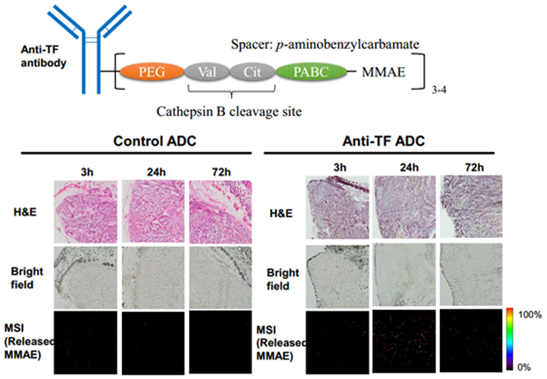
Yasunaga M, Manabe S, Tsuji A, et al. (2017) Development of antibody-drug conjugates using dds and molecular imaging. Bioengineering 4: 78. doi: 10.3390/bioengineering4030078
![]() |
[15] |
Wu C, Dill AL, Eberlin LS, et al. (2013) Mass spectrometry imaging under ambient conditions. Mass Spectrom Rev 32: 218–243. doi: 10.1002/mas.21360
![]() |
[16] |
Murray KK, Seneviratne CA, Ghorai S (2016) High resolution laser mass spectrometry bioimaging. Methods 104: 118–126. doi: 10.1016/j.ymeth.2016.03.002
![]() |
[17] |
Stoeckli M, Chaurand P, Hallahan DE, et al. (2001) Imaging mass spectrometry: A new technology for the analysis of protein expression in mammalian tissues. Nat Med 7: 493–496. doi: 10.1038/86573
![]() |
[18] |
McDonnell LA, Heeren RM (2007) Imaging mass spectrometry. Mass Spectrom Rev 26: 606–643. doi: 10.1002/mas.20124
![]() |
[19] |
Caprioli RM, Farmer TB, Gile J (1997) Molecular imaging of biological samples: Localization of peptides and proteins using maldi-tof ms. Anal Chem 69: 4751–4760. doi: 10.1021/ac970888i
![]() |
[20] |
Wulfkuhle JD, Liotta LA, Petricoin EF (2003) Proteomic applications for the early detection of cancer. Nat Rev Cancer 3: 267–275. doi: 10.1038/nrc1043
![]() |
[21] |
Fujiwara Y, Furuta M, Manabe S, et al. (2016) Imaging mass spectrometry for the precise design of antibody-drug conjugates. Sci Rep 6: 24954. doi: 10.1038/srep24954
![]() |
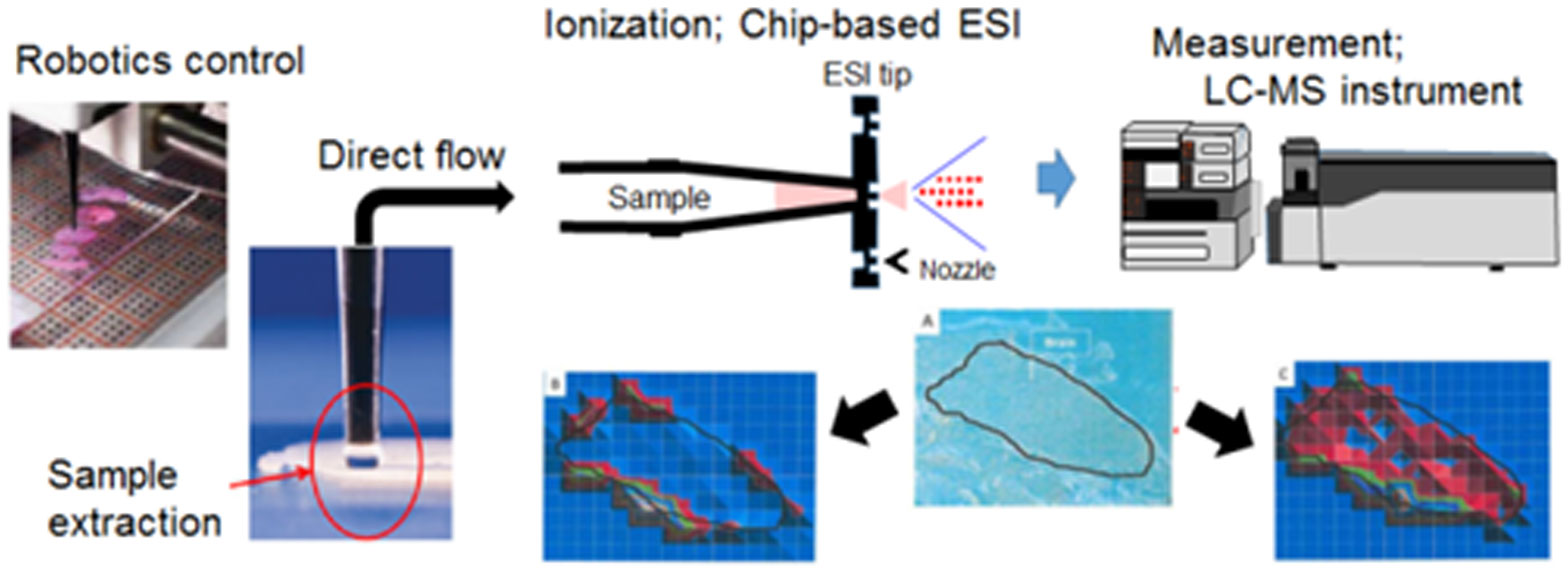
[22] | Yasunaga M, Furuta M, Ogata K, et al. (2013) The significance of microscopic mass spectrometry with high resolution in the visualisation of drug distribution. Sci Rep 3: 3050. |
[23] |
Peletier LA, Gabrielsson J (2012) Dynamics of target-mediated drug disposition: Characteristic profiles and parameter identification. J Pharmacokinet Pharmacodyn 39: 429–451. doi: 10.1007/s10928-012-9260-6
![]() |
[24] | Pineda C, Jacobs IA, Alvarez DF, et al. (2016) Assessing the immunogenicity of biopharmaceuticals. Biodrugs 30: 195–206. |
[25] |
Hampe CS (2012) Protective role of anti-idiotypic antibodies in autoimmunity-lessons for type 1 diabetes. Autoimmunity 45: 320–331. doi: 10.3109/08916934.2012.659299
![]() |
[26] |
Thomas A, Teicher BA, Hassan R (2016) Antibody-drug conjugates for cancer therapy. Lancet Oncol 17: e254–e262. doi: 10.1016/S1470-2045(16)30030-4
![]() |
[27] |
Diamantis N, Banerji U (2016) Antibody-drug conjugates-an emerging class of cancer treatment. Br J Cancer 114: 362–367. doi: 10.1038/bjc.2015.435
![]() |
[28] |
Damelin M, Zhong W, Myers J, et al. (2015) Evolving strategies for target selection for antibody-drug conjugates. Pharm Res 32: 3494–3507. doi: 10.1007/s11095-015-1624-3
![]() |
[29] |
Senter PD, Sievers EL (2012) The discovery and development of brentuximab vedotin for use in relapsed hodgkin lymphoma and systemic anaplastic large cell lymphoma. Nat Biotechnol 30: 631–637. doi: 10.1038/nbt.2289
![]() |
[30] |
Ogitani Y, Aida T, Hagihara K, et al. (2016) Ds-8201a, a novel her2-targeting adc with a novel DNA topoisomerase i inhibitor, demonstrates a promising antitumor efficacy with differentiation from t-dm1. Clin Cancer Res 22: 5097–5108. doi: 10.1158/1078-0432.CCR-15-2822
![]() |
[31] |
Sau S, Alsaab HO, Kashaw SK, et al. (2017) Advances in antibody-drug conjugates: A new era of targeted cancer therapy. Drug Discovery Today 22: 1547–1556. doi: 10.1016/j.drudis.2017.05.011
![]() |
[32] |
Mitsunaga M, Ogawa M, Kosaka N, et al. (2011) Cancer cell-selective in vivo near infrared photoimmunotherapy targeting specific membrane molecules. Nat Med 17: 1685–1691. doi: 10.1038/nm.2554
![]() |
[33] |
Larson SM, Carrasquillo JA, Cheung NK, et al. (2015) Radioimmunotherapy of human tumours. Nat Rev Cancer 15: 347–360. doi: 10.1038/nrc3925
![]() |
[34] | Sau S, Alsaab HO, Kashaw SK, et al. (2017) Advances in antibody-drug conjugates: A new era of targeted cancer therapy. Drug Discovery Today. |
[35] |
Gerber HP, Sapra P, Loganzo F, et al. (2016) Combining antibody-drug conjugates and immune-mediated cancer therapy: What to expect? Biochem Pharmacol 102: 1–6. doi: 10.1016/j.bcp.2015.12.008
![]() |
[36] |
Alsaab HO, Sau S, Alzhrani R, et al. (2017) Pd-1 and pd-l1 checkpoint signaling inhibition for cancer immunotherapy: Mechanism, combinations, and clinical outcome. Front Pharmacol 8: 561. doi: 10.3389/fphar.2017.00561
![]() |
[37] |
Matsumura Y (2014) The drug discovery by nanomedicine and its clinical experience. Jpn J Clin Oncol 44: 515–525. doi: 10.1093/jjco/hyu046
![]() |
[38] |
Nishiyama N, Matsumura Y, Kataoka K (2016) Development of polymeric micelles for targeting intractable cancers. Cancer Sci 107: 867–874. doi: 10.1111/cas.12960
![]() |
[39] | Matsumura Y, Maeda H (1986) A new concept for macromolecular therapeutics in cancer chemotherapy: Mechanism of tumoritropic accumulation of proteins and the antitumor agent smancs. Cancer Res 46: 6387–6392. |
[40] |
Cabral H, Matsumoto Y, Mizuno K, et al. (2011) Accumulation of sub-100 nm polymeric micelles in poorly permeable tumours depends on size. Nat Nanotechnol 6: 815–823. doi: 10.1038/nnano.2011.166
![]() |
[41] |
Oku N (2017) Innovations in liposomal dds technology and its application for the treatment of various diseases. Biol Pharm Bull 40: 119–127. doi: 10.1248/bpb.b16-00857
![]() |
[42] |
Kinoshita R, Ishima Y, Chuang VTG, et al. (2017) Improved anticancer effects of albumin-bound paclitaxel nanoparticle via augmentation of epr effect and albumin-protein interactions using s-nitrosated human serum albumin dimer. Biomaterials 140: 162–169. doi: 10.1016/j.biomaterials.2017.06.021
![]() |
[43] |
Duncan R (2006) Polymer conjugates as anticancer nanomedicines. Nat Rev Cancer 6: 688–701. doi: 10.1038/nrc1958
![]() |
[44] |
Yokoyama M (2014) Polymeric micelles as drug carriers: Their lights and shadows. J Drug Targeting 22: 576–583. doi: 10.3109/1061186X.2014.934688
![]() |
[45] |
Jain RK, Stylianopoulos T (2010) Delivering nanomedicine to solid tumors. Nat Rev Clin Oncol 7: 653–664. doi: 10.1038/nrclinonc.2010.139
![]() |
[46] |
Allen TM, Cullis PR (2013) Liposomal drug delivery systems: From concept to clinical applications. Adv Drug Delivery Rev 65: 36–48. doi: 10.1016/j.addr.2012.09.037
![]() |
[47] |
Bae Y, Nishiyama N, Fukushima S, et al. (2005) Preparation and biological characterization of polymeric micelle drug carriers with intracellular ph-triggered drug release property: Tumor permeability, controlled subcellular drug distribution, and enhanced in vivo antitumor efficacy. Bioconjugate Chem 16: 122–130. doi: 10.1021/bc0498166
![]() |
[48] |
Kraft JC, Freeling JP, Wang Z, et al. (2014) Emerging research and clinical development trends of liposome and lipid nanoparticle drug delivery systems. J Pharm Sci 103: 29–52. doi: 10.1002/jps.23773
![]() |
[49] | Rao W, Pan N, Yang Z (2016) Applications of the single-probe: Mass spectrometry imaging and single cell analysis under ambient conditions. J Visualized Exp JoVE 2016: 53911. |
[50] |
Calligaris D, Feldman DR, Norton I, et al. (2015) Molecular typing of meningiomas by desorption electrospray ionization mass spectrometry imaging for surgical decision-making. Int J Mass Spectrom 377: 690–698. doi: 10.1016/j.ijms.2014.06.024
![]() |
[51] |
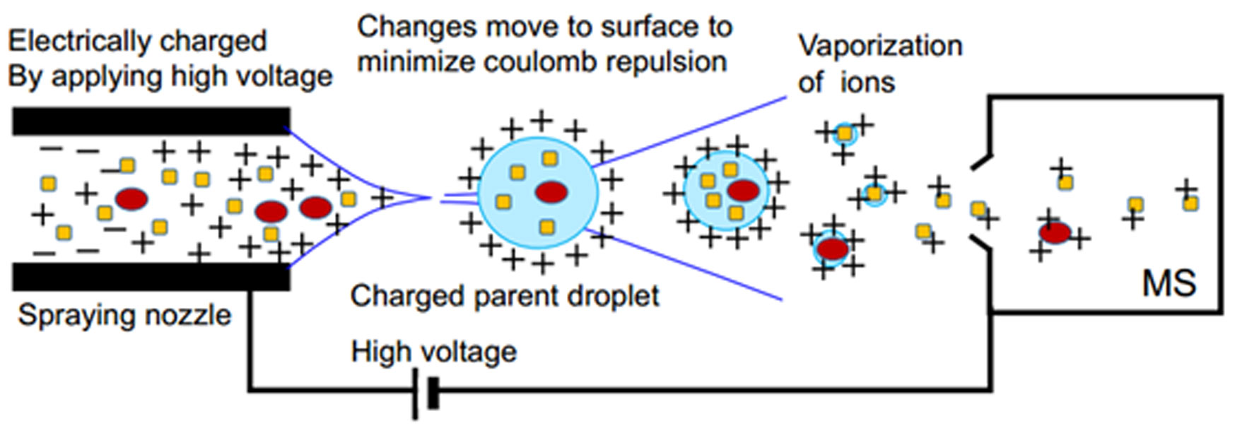
Fenn JB, Mann M, Meng CK, et al. (1989) Electrospray ionization for mass spectrometry of large biomolecules. Science 246: 64–71. doi: 10.1126/science.2675315
![]() |
[52] |
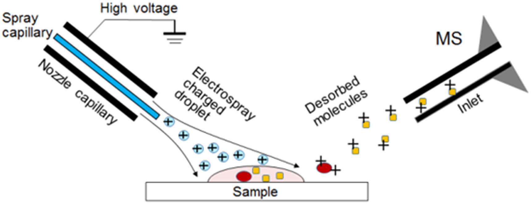
Takats Z, Wiseman JM, Gologan B, et al. (2004) Mass spectrometry sampling under ambient conditions with desorption electrospray ionization. Science 306: 471–473. doi: 10.1126/science.1104404
![]() |
[53] | Parrot D, Papazian S, Foil D, et al. (2018) Imaging the unimaginable: Desorption electrospray ionization-imaging mass spectrometry (desi-ims) in natural product research. Planta Med. |
[54] | Cooks RG, Ouyang Z, Takats Z, et al. (2006) Detection technologies. Ambient mass spectrometry. Science 311: 1566–1570. |
[55] |
Zimmerman TA, Monroe EB, Tucker KR, et al. (2008) Chapter 13: Imaging of cells and tissues with mass spectrometry: Adding chemical information to imaging. Methods Cell Biol 89: 361–390. doi: 10.1016/S0091-679X(08)00613-4
![]() |
[56] | Signor L, Boeri EE (2013) Matrix-assisted laser desorption/ionization time of flight (maldi-tof) mass spectrometric analysis of intact proteins larger than 100 kda. J Visualized Exp JoVE 108: e50635. |
[57] |
Sudhir PR, Wu HF, Zhou ZC (2005) Identification of peptides using gold nanoparticle-assisted single-drop microextraction coupled with ap-maldi mass spectrometry. Anal Chem 77: 7380–7385. doi: 10.1021/ac051162m
![]() |
[58] |
Abdelhamid HN, Wu HF (2012) A method to detect metal-drug complexes and their interactions with pathogenic bacteria via graphene nanosheet assist laser desorption/ionization mass spectrometry and biosensors. Anal Chim Acta 751: 94–104. doi: 10.1016/j.aca.2012.09.012
![]() |
[59] |
Abdelhamid HN, Wu HF (2013) Furoic and mefenamic acids as new matrices for matrix assisted laser desorption/ionization-(maldi)-mass spectrometry. Talanta 115: 442–450. doi: 10.1016/j.talanta.2013.05.050
![]() |
[60] |
Nasser AH, Wu BS, Wu HF (2014) Graphene coated silica applied for high ionization matrix assisted laser desorption/ionization mass spectrometry: A novel approach for environmental and biomolecule analysis. Talanta 126: 27–37. doi: 10.1016/j.talanta.2014.03.016
![]() |
[61] |
Abdelhamid HN, Wu HF (2015) Synthesis of a highly dispersive sinapinic acid@graphene oxide (sa@go) and its applications as a novel surface assisted laser desorption/ionization mass spectrometry for proteomics and pathogenic bacteria biosensing. Analyst 140: 1555–1565. doi: 10.1039/C4AN02158D
![]() |
[62] |
Abdelhamid HN, Wu HF (2016) Gold nanoparticles assisted laser desorption/ionization mass spectrometry and applications: From simple molecules to intact cells. Anal Bioanal Chem 408: 4485–4502. doi: 10.1007/s00216-016-9374-6
![]() |
[63] |
Harada T, Yuba-Kubo A, Sugiura Y, et al. (2009) Visualization of volatile substances in different organelles with an atmospheric-pressure mass microscope. Anal Chem 81: 9153–9157. doi: 10.1021/ac901872n
![]() |
[64] |
Saito Y, Waki M, Hameed S, et al. (2012) Development of imaging mass spectrometry. Biol Pharm Bull 35: 1417–1424. doi: 10.1248/bpb.b212007
![]() |
[65] |
Sugiura Y, Honda K, Suematsu M (2015) Development of an imaging mass spectrometry technique for visualizing localized cellular signaling mediators in tissues. Mass Spectrom 4: A0040. doi: 10.5702/massspectrometry.A0040
![]() |
[66] |
Setou M, Kurabe N (2011) Mass microscopy: High-resolution imaging mass spectrometry. J Electron Microsc 60: 47–56. doi: 10.1093/jmicro/dfq079
![]() |
[67] |
Maeda H (2001) Smancs and polymer-conjugated macromolecular drugs: Advantages in cancer chemotherapy. Adv Drug Delivery Rev 46: 169–185. doi: 10.1016/S0169-409X(00)00134-4
![]() |
[68] |
Barenholz Y (2012) Doxil(r)-the first fda-approved nano-drug: Lessons learned. J Controlled Release 160: 117–134. doi: 10.1016/j.jconrel.2012.03.020
![]() |
[69] |
Giordano G, Pancione M, Olivieri N, et al. (2017) Nano albumin bound-paclitaxel in pancreatic cancer: Current evidences and future directions. World J Gastroenterol 23: 5875–5886. doi: 10.3748/wjg.v23.i32.5875
![]() |
[70] |
Kogure K, Akita H, Yamada Y, et al. (2008) Multifunctional envelope-type nano device (mend) as a non-viral gene delivery system. Adv Drug Delivery Rev 60: 559–571. doi: 10.1016/j.addr.2007.10.007
![]() |
[71] |
Sugaya A, Hyodo I, Koga Y, et al. (2016) Utility of epirubicin-incorporating micelles tagged with anti-tissue factor antibody clone with no anticoagulant effect. Cancer Sci 107: 335–340. doi: 10.1111/cas.12863
![]() |
[72] |
Hiyama E, Ali A, Amer S, et al. (2015) Direct lipido-metabolomics of single floating cells for analysis of circulating tumor cells by live single-cell mass spectrometry. Anal Sci 31: 1215–1217. doi: 10.2116/analsci.31.1215
![]() |
[73] |
Hamaguchi T, Matsumura Y, Suzuki M, et al. (2005) Nk105, a paclitaxel-incorporating micellar nanoparticle formulation, can extend in vivo antitumour activity and reduce the neurotoxicity of paclitaxel. Br J Cancer 92: 1240–1246. doi: 10.1038/sj.bjc.6602479
![]() |
[74] |
Verma S, Miles D, Gianni L, et al. (2012) Trastuzumab emtansine for her2-positive advanced breast cancer. N Engl J Med 367: 1783–1791. doi: 10.1056/NEJMoa1209124
![]() |
[75] |
Younes A, Gopal AK, Smith SE, et al. (2012) Results of a pivotal phase ii study of brentuximab vedotin for patients with relapsed or refractory hodgkin's lymphoma. J Clin Oncol 30: 2183–2189. doi: 10.1200/JCO.2011.38.0410
![]() |
[76] |
Pro B, Advani R, Brice P, et al. (2012) Brentuximab vedotin (sgn-35) in patients with relapsed or refractory systemic anaplastic large-cell lymphoma: Results of a phase ii study. J Clin Oncol 30: 2190–2196. doi: 10.1200/JCO.2011.38.0402
![]() |
[77] |
Doronina SO, Toki BE, Torgov MY, et al. (2003) Development of potent monoclonal antibody auristatin conjugates for cancer therapy. Nat Biotechnol 21: 778–784. doi: 10.1038/nbt832
![]() |
[78] |
Lyon RP, Bovee TD, Doronina SO, et al. (2015) Reducing hydrophobicity of homogeneous antibody-drug conjugates improves pharmacokinetics and therapeutic index. Nat Biotechnol 33: 733–735. doi: 10.1038/nbt.3212
![]() |
[79] |
Hisada Y, Yasunaga M, Hanaoka S, et al. (2013) Discovery of an uncovered region in fibrin clots and its clinical significance. Sci Rep 3: 2604. doi: 10.1038/srep02604
![]() |
[80] |
Takashima H, Tsuji AB, Saga T, et al. (2017) Molecular imaging using an anti-human tissue factor monoclonal antibody in an orthotopic glioma xenograft model. Sci Rep 7: 12341. doi: 10.1038/s41598-017-12563-5
![]() |
[81] |
Koga Y, Manabe S, Aihara Y, et al. (2015) Antitumor effect of antitissue factor antibody-mmae conjugate in human pancreatic tumor xenografts. Int J Cancer 137: 1457–1466. doi: 10.1002/ijc.29492
![]() |
[82] |
Rao W, Celiz AD, Scurr DJ, et al. (2013) Ambient desi and lesa-ms analysis of proteins adsorbed to a biomaterial surface using in-situ surface tryptic digestion. J Am Soc Mass Spectrom 24: 1927–1936. doi: 10.1007/s13361-013-0737-3
![]() |
[83] | Takahashi T, Serada S, Ako M, et al. (2013) New findings of kinase switching in gastrointestinal stromal tumor under imatinib using phosphoproteomic analysis. Int J Cancer 133: 2737–2743. |
[84] |
Emara S, Amer S, Ali A, et al. (2017) Single-cell metabolomics. Adv Exp Med Biol 965: 323–343. doi: 10.1007/978-3-319-47656-8_13
![]() |
[85] |
Matsumura Y (2012) Cancer stromal targeting (cast) therapy. Adv Drug Delivery Rev 64: 710–719. doi: 10.1016/j.addr.2011.12.010
![]() |
[86] |
Yasunaga M, Manabe S, Tarin D, et al. (2011) Cancer-stroma targeting therapy by cytotoxic immunoconjugate bound to the collagen 4 network in the tumor tissue. Bioconjugate Chem 22: 1776–1783. doi: 10.1021/bc200158j
![]() |
[87] |
Yasunaga M, Manabe S, Tarin D, et al. (2013) Tailored immunoconjugate therapy depending on a quantity of tumor stroma. Cancer Sci 104: 231–237. doi: 10.1111/cas.12062
![]() |
[88] |
Yasunaga M, Manabe S, Matsumura Y (2017) Immunoregulation by il-7r-targeting antibody-drug conjugates: Overcoming steroid-resistance in cancer and autoimmune disease. Sci Rep 7: 10735. doi: 10.1038/s41598-017-11255-4
![]() |
1. | Ning He, Changyou Tang, Xianchao Zhou, Shuolin Feng, M Pallikonda Rajasekaran, Diagnostic Value of Magnetic Resonance Image Feature Analysis under Reconstruction Algorithm for Knee Epiphyseal Injury, 2022, 2022, 1875-919X, 1, 10.1155/2022/1783975 |
Solver | Time-steps | Solver | Time-steps |
CCL+FE | (0.001~1 ms) | MCCL+FE | (0.001~1 ms) |
CCL+TAL | (0.001~1 ms) | MCCL+TAL | (0.001~1 ms) |
CCL+UNI | (0.001~1 ms) | MCCL+UNI | (0.001~1 ms) |
CCL+HOS | (0.001~1 ms) | MCCL+HOS | (0.001~1 ms) |
Solvers | MRMSE-V | MRMSE-I | Speedup | Cost(ms) |
DT+RK4 | 0.0 | 0.0 | 1.0X | $ 1.0 \times 10^7 \pm 9.8 \times 10^4 $ |
CCL+FE | 0.016 | 0.588 | 109.9X | $ 9.5 \times 10^4 \pm 4.0 \times 10^2 $ |
CCL+TAL | 0.745 | 1.158 | 33.0X | $ 3.2 \times 10^5 \pm 1.2 \times 10^3 $ |
CCL+UNI | 0.056 | 0.012 | 16.4X | $ 6.4 \times 10^5 \pm 8.4 \times 10^2 $ |
CCL+HOS | 0.236 | 0.064 | 412.3X | $ 2.6 \times 10^4 \pm 3.2 \times 10^2 $ |
MCCL+FE | 0.022 | 0.065 | 96.9X | $ 1.1 \times 10^5 \pm 2.7 \times 10^2 $ |
MCCL+TAL | 0.015 | 0.124 | 29.8X | $ 3.5 \times 10^5 \pm 1.4 \times 10^3 $ |
MCCL+UNI | 0.021 | 0.009 | 14.8X | $ 7.0 \times 10^5 \pm 3.4 \times 10^3 $ |
MCCL+HOS | 0.006 | 0.002 | 17.2X | $ 6.1 \times 10^5 \pm 3.7 \times 10^3 $ |
Note: MRMSE-V: MRMSE of voltage in comparison to the reference. MRMSE-I: MRMSE of sodium current in comparison to the reference. Speedup: The computation cost ratio of one of eight schemes to the reference solution DT+RK4. X, times speedup. Ten simulations for each solver. Cost in Mean ± Standard deviation. |
Solvers | MRMSE-V | MRMSE-I | Speedup | Cost(ms) |
DT+RK4 | 0.0 | 0.0 | 1.0X | $ 1.1 \times 10^7 \pm 6.0 \times 10^4 $ |
CCL+FE | - - | - - | - - | failure |
CCL+TAL | - - | - - | - - | failure |
CCL+UNI | 0.019 | 0.008 | 1.2X | $ 8.9 \times 10^6 \pm 1.8 \times 10^4 $ |
CCL+HOS | 0.250 | 0.114 | 266.6X | $ 3.3 \times 10^4 \pm 1.3 \times 10^2 $ |
MCCL+FE | 0.103 | 3.345 | 19.8X | $ 5.3 \times 10^5 \pm 2.5 \times 10^3 $ |
MCCL+TAL | - - | - - | - - | failure |
MCCL+UNI | 0.076 | 0.042 | 1.2X | $ 8.9 \times 10^6 \pm 1.1 \times 10^4 $ |
MCCL+HOS | 0.032 | 0.013 | 21.1X | $ 5.0 \times 10^5 \pm 1.8 \times 10^3 $ |