Loading [Contrib]/a11y/accessibility-menu.js
Review Special Issues

Probing the evolutionary history of epigenetic mechanisms: what can we learn from marine diatoms

  • Received: 19 May 2015 Accepted: 13 July 2015 Published: 25 January 2015
  • Recent progress made on epigenetic studies revealed the conservation of epigenetic features in deep diverse branching species including Stramenopiles, plants and animals. This suggests their fundamental role in shaping species genomes across different evolutionary time scales. Diatoms are a highly successful and diverse group of phytoplankton with a fossil record of about 190 million years ago. They are distantly related from other super-groups of Eukaryotes and have retained some of the epigenetic features found in mammals and plants suggesting their ancient origin. Phaeodactylum tricornutum and Thalassiosira pseudonana, pennate and centric diatoms, respectively, emerged as model species to address questions on the evolution of epigenetic phenomena such as what has been lost, retained or has evolved in contemporary species. In the present work, we will discuss how the study of non-model or emerging model organisms, such as diatoms, helps understand the evolutionary history of epigenetic mechanisms with a particular focus on DNA methylation and histone modifications.

    Citation: Achal Rastogi, Xin Lin, Bérangère Lombard, Damarys Loew, Leïla Tirichine. Probing the evolutionary history of epigenetic mechanisms: what can we learn from marine diatoms[J]. AIMS Genetics, 2015, 2(3): 173-191. doi: 10.3934/genet.2015.3.173

    Related Papers:

    [1] Sohrab Najafipour, Abdolreza karami, Manoochehr Rasouli, Mehdi Kalani, Seyed-Jalal-Aldin Ashraf-Mansoori, Ali Moravej . Association of interleukin-2 gene variants (positions +114 and −384) and susceptibility to brucellosis in Iranian population. AIMS Molecular Science, 2017, 4(1): 103-109. doi: 10.3934/molsci.2017.1.103
    [2] Viviana Imbua Levi Enoka, Gideon Mutie Kikuvi, Perpetual Wangui Ndung'u . Effect of garlic and onion extract chitosan nanoparticles on selected intestinal bacterial flora in indigenous rainbow rooster chicken in Kenya. AIMS Molecular Science, 2021, 8(2): 98-116. doi: 10.3934/molsci.2021008
    [3] Carlos Gutierrez-Merino, Dorinda Marques-da-Silva, Sofia Fortalezas, Alejandro K. Samhan-Arias . The critical role of lipid rafts nanodomains in the cross-talk between calcium and reactive oxygen and nitrogen species in cerebellar granule neurons apoptosis by extracellular potassium deprivation. AIMS Molecular Science, 2016, 3(1): 12-29. doi: 10.3934/molsci.2016.1.12
    [4] Francisco Les, Zsuzsa Iffiú-Soltész, Josep Mercarder, Christian Carpéné . Tyramine activates lipid accumulation in rat adipocytes: influences of in vitro and in vivo administration. AIMS Molecular Science, 2017, 4(3): 339-351. doi: 10.3934/molsci.2017.3.339
    [5] Charles A. Downs, Abdel A. Alli, Nicholle M. Johnson, My N. Helms . Cigarette smoke extract is a Nox agonist and regulates ENaC in alveolar type 2 cells. AIMS Molecular Science, 2016, 3(3): 439-453. doi: 10.3934/molsci.2016.3.439
    [6] Elisa Isopi, Giuseppe Legname . Pin1 and neurodegeneration: a new player for prion disorders?. AIMS Molecular Science, 2015, 2(3): 311-323. doi: 10.3934/molsci.2015.3.311
    [7] Jian Zou, Fulton T. Crews . Glutamate/NMDA excitotoxicity and HMGB1/TLR4 neuroimmune toxicity converge as components of neurodegeneration. AIMS Molecular Science, 2015, 2(2): 77-100. doi: 10.3934/molsci.2015.2.77
    [8] Genu George, Sumita K. Chellappan, Mandagini Geetha, Padinjaradath S. Appukuttan . Possible molecular basis for macromolecular antigen attachment to host cells: their immune complex with plasma antibodies have unoccupied binding sites enabling binding to smaller ligands. AIMS Molecular Science, 2017, 4(1): 91-102. doi: 10.3934/molsci.2017.1.91
    [9] David Trac, My N. Helms . Nadph oxidase and epithelial sodium channels regulate neonatal mouse lung development. AIMS Molecular Science, 2017, 4(1): 28-40. doi: 10.3934/molsci.2017.1.28
    [10] Noor Riyadh Thiab, Nicola King, Mary McMillan, Amer Almashhadany, Graham L Jones . Age-related protein and mRNA expression of glutathione peroxidases (GPx) and Hsp-70 in different regions of rat kidney with and without stressor. AIMS Molecular Science, 2016, 3(2): 125-137. doi: 10.3934/molsci.2016.2.125
  • Recent progress made on epigenetic studies revealed the conservation of epigenetic features in deep diverse branching species including Stramenopiles, plants and animals. This suggests their fundamental role in shaping species genomes across different evolutionary time scales. Diatoms are a highly successful and diverse group of phytoplankton with a fossil record of about 190 million years ago. They are distantly related from other super-groups of Eukaryotes and have retained some of the epigenetic features found in mammals and plants suggesting their ancient origin. Phaeodactylum tricornutum and Thalassiosira pseudonana, pennate and centric diatoms, respectively, emerged as model species to address questions on the evolution of epigenetic phenomena such as what has been lost, retained or has evolved in contemporary species. In the present work, we will discuss how the study of non-model or emerging model organisms, such as diatoms, helps understand the evolutionary history of epigenetic mechanisms with a particular focus on DNA methylation and histone modifications.


    1. Introduction

    Rheumatoid arthritis (RA) is a chronic inflammatory debilitating autoimmune disease [1]. Cell mediated and humoral immune reactions are involved in pathogenesis of rheumatoid arthritis [2]. The disease is characterized by severe joint inflammations of whole body along with damage to cartilage and bones [3]. Leukocyte infiltration and pannus formation are also responsible for the pathogenesis of RA. Although, the exact cause has not been elucidated still now. Several pro-inflammatory and anti-inflammatory cytokines secreted by immune cells are responsible for the cause of RA. In particular, it was reported that tumor necrosis factor (TNF), interleukin-1β (IL-1β), interleukin-15 (IL-15), transforming growth factor-β (TGF-β), interleukin-6 (IL-6) are important pro-inflammatory cytokines responsible for initiation and progression of RA [4,5]. In view of these findings, modulation of pro-inflammatory cytokines is a desired goal for the management of arthritis.

    Most prescribed treatment for RA is a non-steroidal anti-inflammatory drugs (NSAID's). These drugs effectively reduce symptoms of RA, but at same time cause various side effects like gastrointestinal sickness and renal morbidity [6]. Disease modifying anti-rheumatic drugs (DMARD's) and biologics are also prescribed. Biologics are expensive and DMARD's are also associated with severe side effects. Reducing side effects and deleterious effect of antiarthritic drug should be taken into account while designing new therapeutics for RA. One of the possible strategies to suppress systemic inflammation is employment of natural compounds such as botanicals, bioactive food components or functional foods. Along with these, efficacies of various plants extracts and other natural compounds have been exploited for their antiarthritic activity.

    Probiotics are ingestible microbial products which when administered in a particular number exert a beneficial effect on host body. These dietary feed supplements are used as probiotics because of their well-known immunomodulatory properties[7,8,9]. Probiotics modulate immune response at gut level. Probiotics are also in use for their antimicrobial, anti-inflammatory and immunomodulatory properties in urinary tract infections [10]. In our previous study we have discovered that L. casei and L. acidophilus has shown potent anti-inflammatory properties in the carrageenan model of acute inflammation [11]. In the present study, anti-inflammatory and anti-arthritic activities were investigated by using collagen induced arthritis (CIA) model. Anti-inflammatory activity of L. casei and L. acidophilus was checked by measuring the mRNA expression of TNF, IL-6 and IL-1β to validate our previous findings of anti-inflammatory activity. Standard anti-arthritic drug indomethacin was used for comparison.


    2. Materials and Methods


    2.1. Animals

    Thirty healthy male Wistar rats, two months old (200 g) were included in this study. Animals were randomly divided into five groups (n = 6). These were purchased from Indian Institute of Toxicology Research (ⅡTR), Lucknow, India and maintained in animal house at Department of Microbiology, Barkatullah University, Bhopal, Madhya Pradesh. Animals were acclimatized for fifteen days before the experiment started. Animals were housed in stainless steel cages and were fed with standard diet purchased from Amrut Feed Ltd, India, given water ad libitum (Principles of Laboratory Animal Care NIH publication no, 85-23, revised 1985) and maintained under laboratory conditions (Temperature 22 ± 2 ℃, Relative humidity 60–70%, and 12-12 hrs light-dark cycle). All procedures and techniques used in this study were in accordance to the ethical committee guidelines.


    2.2. Bacterial culture

    L. acidophilus (ATCC 314) and L. casei (ATCC 334) were purchased from Hi Media, Navi Mumbai, India. Lyophilized cultures were streaked over De Mann Rogosa Agar (MRS) at 37 ℃ in anaerobic condition.


    2.3. Chemicals

    All chemical used in this study were of molecular and analytical grade.


    2.4. Induction of arthritis and oral treatments

    Collagen induced arthritis model was induced by the method given by Remmers [12] with some necessary modifications. Rats were injected intra peritoneally with collagen (Bovine tracheal cartilage type Ⅱ) and incomplete freund's adjuvant on day 1except group Ⅰ animals. Booster doses of the same were injected on the 8th day. Preventive oral treatments were given to all the groups. Oral administration 500 μL of distilled water, 2 × 10⁸ CFU/mL of L. casei, L. acidophilus suspended in 500 μL of distilled water were started on day 1 and continued till the 28th day for group Ⅱ, Ⅳ and Ⅴ group animals respectively. Indomethacin suspended in 500 μL of distilled water was administered at 10 mg/g body weight from day 1 to 28 to group Ⅲ animals [13]. Paw thickness were measured for all the experimental animals at day 0 and at the end of each week with dial gauge caliper.


    2.5. Sample collection

    On the 29th day, all animals were sacrificed in aseptic conditions. Blood samples were collected to isolate serum for cytokines assay. Right hind paws knee joints were also collected from each animal for RT PCR analysis of mRNA expressions for cytokines.


    2.6. ELISA for the cytokines assay

    IL-6, TNF, IL-1β (pro-inflammatory cytokines) and IL-4, IL-10 (anti-inflammatory cytokines) (pg/mL) were estimated in serum samples with the help of ELISA Reader (Lisa Plus, Germany). IL-6 and TNF, IL-1β (Ray Bio®) and IL-4 and IL-10 (DNA bio) ELISA kits were used. Assays were performed according to the manufacturer's recommendations.


    2.7. Reverse transcriptase PCR analysis

    Paws were harvested and homogenized followed by addition of sodium lauryl sarconisate and centrifuged to remove the cell debris. RNA was precipitated by the addition of potassium acetate and acetic acid. Cold absolute ethanol was added for the precipitation of RNA. Total RNA was isolated by RNAzol method (Tel-Test, Friendswood, TX) according to the manufacturer's instructions.

    The quantity and purity of RNA were determined by using a spectrophotometer (Shimadzu) at 260 and 280 nm. Samples with ratios > 1.7 were accepted for further analysis. These samples were treated with DNAase (Genei, Banglore, India) to remove the contaminating DNA. One microgram of total RNA from rat knee joint were reverse-transcribed into cDNA using 200 units of M-MLV reverse transcriptase (Invitrogen Co.) in 20 µL of reaction volume. cDNA was amplified in 20 µL of reaction mixture including 10 mM Tris-HCl (pH 8.3), 50 mM KCl, 1.5 mM MgCl2, 0.2 mM dNTP, 0.4 µM each of the 3' and 5' primer and 0.5 U Taq polymerase (Takara Co.) using a PTC-100 Programmable Thermal Controller (MJ Research, Waltham, MA, USA). All primers were designed using the published cDNA sequences and the primer selection software, with the help of web site, Primer 3 (http://www.genome.wi.mit.edu). Primer sequences and operating conditions are given in Table 1. PCR products thus obtained were separated on 1% agarose gel, stained with ethidium bromide, and photographed. Intensities of bands were measured by using ImageMaster VDSTM (Amersham Biosciences, Uppsala, Sweden) with image analysis software, ImageMaster Total Lab TM (Amersham Biosceinces). The amounts of target PCR products were normalized against GAPDH PCR product in the corresponding samples.

    Table 1. Table showing primer sequences, operating conditions and PCR product size.
    Gene name Oligonucleotide sequence (5'-3') Denaturing,
    ℃ (time)
    Annealing,
    ℃ (time)
    Elongation,
    ℃ (time)
    Cycle No. Product size Gene bank No.
    mGAPDH F-ATGGGAAGCTGGTCATCAAC
    R-GGATGCAGGGATGATGTTCT
    94 (30 s) 58 (30 s) 72 (60 s) 30 440 NM_017008
    mTNF F-GTCGTAAACCACCAAGC
    R-GACTCCAAAGTAGACCTGCCC
    94 (45 s) 58 (45 s) 72 (90 s) 35 393 AY 427675
    mIL-1β F-GGCATAACAGGCTCATCTGG
    R-CATCATCCCACGAGTCACAG
    94 (45 s) 58 (45 s) 72 (90 s) 35 414 NM_031512
    mIL-6 F-CCACTGCCTTCCCTACTTCA
    R-TGGTCCTTAGCCACTCCTTC
    94 (60 s) 63 (60 s) 72 (120 s) 37 498 NM_012589
    mIL-4 F-ATG CACCGAGATGTTTGTACC
    R-TTCAGTGTTCTGAGCGTGGA
    95 (60 s) 60 (60 s) 72 (90 s) 40 275 NM_201270.1
    mIL-10 F-GGAGTGAAGACCAAAGG
    R-TCTCCCAGGGAATTCAAATG
    94 (30 s) 57 (1 s) 72 (60 s) 40 340 NM_012854
    mGAPDH: human glyceraldehyde-3-phosphate dehydrogenase; mTNF: murine tumor necrosis factor α; IL-1β: interleukin-1β; IL-6: interleukin-6; IL-4: Interleukin-4; IL-10: Interleukin-10
     | Show Table
    DownLoad: CSV

    2.8. Statistical analysis

    The results are expressed as a mean ± SEM of six rats per group. The result obtained from different groups was analyzed by one way ANOVA followed by Bonferroni's Multiple Comparison Test. Data were considered statistically significant if P < 0.0001.


    3. Results


    3.1. Effect on paw thickness

    A significant increase in paw thickness was observed in group Ⅱ animals from 3.46 ± 0.04 to 3.78 ± 0.10 mm from 7th to 28th day while there was no change in the paw thickness (3.16 ± 0.08 mm) observed in group Ⅰ animals from 0 to 28th day. Lactobacillus treatment significantly decreased paw thickness in group Ⅳ and Ⅴ animals when compared with group Ⅱ animals. Paw thicknesses were 3.7 ± 0.16 and 3.48 ± 0.06 mm for group Ⅳ and Ⅴ respectively. A significant decreased in thickness to 3.4 ± 0.04 and 3.08 ± 0.08 mm for Ⅳ and Ⅴ groups at P < 0.0001 was observed on 28th day when compared with group Ⅱ animals. However group Ⅲ animals were also showing a significant decreased in paw thickness of 3.62 ± 0.09 mm on 7th day and 3.2 ± 0.08 mm on 28th day (at P < 0.0001) (Figure 1).

    Figure 1. Figure showing the paw thickness (cm) of collagen induced arthritic rats on 7th, 14th, 21st and 28th day. Group Ⅱ animals were showing highest paw thickness (cm) on 28th day when compared with other groups at P < 0.0001. Results are expressed as Mean ± SEM of six rats per group.

    3.2. Cytokine expression in serum

    Serum level of IL-6, IL-1β and TNF level were significantly increased in group Ⅱ animals while significantly lower levels of these pro-inflammatory mediators were observed in other counterparts. 63.34 ± 0.18, 178.6 ± 0.27 and 650.1 ± 0.44 pg/mL were found in case group Ⅱ animals for IL-6, IL-1β and TNF respectively (Figure 2). Abrupt decreased in IL-4 and IL-10 level at 63.33 ± 0.57 and 15.7 ± 0.20 pg/mL respectively in case of group Ⅱ animals. Lactobacillus treatment increased serum level of IL-4 and IL-10 in group Ⅳ and Ⅴ animals. Results of group Ⅲ were also found to be statistically significant at P < 0.0001 with group Ⅱ animals (Figure 2).

    Figure 2. Figure showing the concentration of IL-4 and IL-10 (pg/mL) of collagen induced arthritic rats. Group Ⅱ animals show significantly lower levels of IL-4 and IL-10 (pg/mL) when compared with the other counterparts at P < 0.0001. Results are expressed as Mean ± SEM of six rats per group. (b) Figure showing the concentration of IL-6, IL-β and TNF (pg/mL) of collagen induced arthritic rats. Group Ⅱ show significant increase in IL-6, IL-β and TNF (pg/mL) when compared with the other counterparts at P < 0.0001. Results are expressed as Mean ± SEM of six rats per group.

    3.3. Inhibitory effect of L. casei and L. acidophilus on IL-6, IL-1β and TNF mRNA and stimulatory effect on IL-4 and IL-10 mRNA expressions in knee joints

    Effect of L. casei and L. acidophilus was investigated by RT-PCR analysis for various cytokines at the transcription level. On the 29th day, mRNA expression levels of pro and anti-inflammatory cytokine level was estimated by total RNA sample prepared from the right hind knee joints. mRNA expressions of IL-6, IL-1β and TNF were stimulated in group Ⅱ animals when compared with other groups and very less expression of IL-4 and IL-10 were observed. Whereas Lactobacillus treatment up-regulated the IL-4 and IL-10 expressions in group Ⅳ and Ⅴ animals. The group Ⅰ animals were showing normal expression for all the cytokines. In case of group Ⅲ animals, IL-1β and TNF were simulated and less expression of IL-6 was observed. Also, IL-4 was stimulated in comparison to IL-10 in group Ⅲ animals (Figures 3 and 4).

    Figure 3. Expressions of IL-6, IL-β, TNF, IL-4 and IL-10 mRNA from hind paw of collagen induced arthritic rats on 29th day. Lane Ⅰ-Control (Group Ⅰ); Lane Ⅱ-Arthritic control (Group Ⅱ); Lane Ⅲ-Drug administered; Lane Ⅳ-L. casei administered; Lane Ⅴ-L. acidophilus administered. Each PCR product was normalized to GAPDH. The observed bands were verified on the gels, with the predicted sizes of GAPDH (440 bp), TNF (393 bp), IL-1β (414 bp), IL-6 (498 bp), IL-4 (275 bp) and IL-10 (340 bp).
    Figure 4. Figure showing gel intensities. Values are Mean ± SEM of six rats per group. Statistical analysis was performed by one-way ANOVA. * P < 0.001 vs. arthritic control group.

    4. Discussion

    Based on anti-inflammatory properties of L. casei and L. acidophilus in serum [11], it was considered that they both might have shown anti-inflammatory properties at transcription level also. The aim of the present study was to evaluate effect of L. casei and L. acidophilus on expressions of IL-6, IL-1β, TNF, IL-4 and IL-10 mRNA in CIA model of arthritis. When compared with the other experimental models of arthritis, CIA model is a promising model for validation and efficacy of anti-arthritic drugs [14]. Results obtained in this study were similar with our previous findings. L. casei and L. acidophilus significantly decreased the paw thickness in experimental animals when compared with their other counter parts. These treatments significantly decreased the pro-inflammatory and increased the anti-inflammatory cytokines [11]. Collagen induced arthritis is an immune mediated inflammatory reaction. Initial symptoms include paw edema and soft tissue thickening. Lactobacillus is acting as an immunosuppressive agent. Decrease in paw thickness is one of the major factors which evaluate the efficacy of anti-arthritic drugs or therapy. Both L. casei and L. acidophilus decreased paw thickness when compared to other counterparts.

    Rheumatoid arthritis is a chronic and devastating autoimmune disorder that leads to persistent, progressive symmetrical inflammation in joints and subsequent erosive destruction of cartilage and bone [15,16]. Anti-arthritic drugs used for treating chronic inflammatory conditions are usually prescribed for long term to properly control the disordered immune system. Thus, there is a strong need to develop safe and effective drugs for the long-term use. Many researchers have studied non-chemical compounds for nonsteroidal anti-inflammatory properties with the aim of developing new treatments for clinical use [17].

    Cytokines play major role in the progression of rheumatoid arthritis. An imbalance between pro-inflammatory and anti-inflammatory cytokine activities, favors the induction of autoimmunity, chronic inflammation and thereby joint damage [18]. Mainly IL-1β, TNF, IL-8, IL-12, IL-15, IL-17 and IL-18 are responsible for the inflammatory reaction. Along with these mediators, leukotriene B4 are also implicated in the process and playing a key role in neutrophil recruitment during immune inflammation [19]. TNF responsible for inflammation, differentiation and proliferation of T and B cells and bone resorption [20], whereas IL-1β causes the destruction of cartilage and bones [21]. Blocking TNF suppresses inflammation and ameliorates cartilage destruction while IL-6 is responsible for the joint destruction. Lactobacillus significantly decreased pro-inflammatory cytokine levels in serum. Lactobacillus might suppress the signaling pathway that leads to TNF, IL-1β and IL-6 synthesis. TNF, IL-1β and IL-6 had been considered to have critical roles in RA and CIA in that they promote the production of most other pro-inflammatory mediators [22]. Oral administration of Lactobacillus significantly increased anti-inflammatory cytokines (IL-10 and IL-4) level from serum. IL-10 is a macrophage deactivator and also found to be associated with blocking the synthesis of TNF, IL-1, IL-6, IL-8, and GM-CSF in human monocytes [23] and cox-2 inhibitors [24,25]. IL-4 is pleiotropic T-cell derived cytokine that acts by inhibiting the synthesis of pro-inflammatory cytokines such as IL-1, TNF, IL-6, IL-8 and IL-12 by macrophages and monocytes [26]. Moreover, it also stimulates several cytokine inhibitors such as interleukin-1 receptor antagonist (IL-1Ra), soluble IL-1-receptor type Ⅱ and TNF receptors. In addition, it also suppresses metalloproteinase synthesis and stimulates tissue inhibitor of metalloproteinase-1 production in human mononuclear phagocytes [27,28]. Lactobacillus might offer a complementary therapy, which inhibited pro-inflammatory cytokines whereas up-regulated anti-inflammatory cytokines.

    In addition, anti-inflammatory effect of L. casei and L. acidophilus was also validated from the mRNA expression of cytokines from knee joints. mRNA level of IL-6, TNF and IL-1β were highly stimulated in knee joints of arthritic rats, whereas Lactobacillus treatment decreased the level as that of normal rats. Studies have established that IL-6 has an important role in leukocyte recruitment, apoptosis and T cell activation [29]. Additionally, TNF stimulates neutrophils to transcribe and release cytokines and chemokines biosynthesis [30]. Inhibition of TNF, IL-1β and IL-6 release can reduce the severity of inflammation. On the other way, Lactobacillus treatment enhanced the expression of IL-4 and IL-10. L. casei showed similar results in randomized double-blind clinical trial [31]. Consumption of L. casei fermented milk prevented the Salmonella reactive arthritis [32]. Results presented in this study are first to demonstrate that L. casei and L. acidophilus had shown inhibitory effects on the expression of IL-6, IL-1β and TNF and thus possess a potent immunosuppressive and anti-inflammatory activity.


    5. Conclusion

    We have demonstrated that Lactobacillus possess immunomodulatory property as suggested by RT PCR analysis of various pro-inflammatory and anti-inflammatory cytokines in animal model of experimental arthritis. Lactobacillus has therapeutic anti-arthritic activity as tested in CIA model. Notably, oral administration of Lactobacillus caused suppression of pro-inflammatory cytokines IL-6, IL-1β and TNF along with up-regulation of anti-inflammatory cytokines IL-4 and IL-10 in serum as well as at the transcription level. Also, there was lowering of paw thickness. On the basis of our results in present study, we suggest that Lactobacillus is a promising therapeutic agent or dietary supplement for rheumatoid arthritis.


    Acknowledgement

    The author would like to thank Department of Microbiology, Barkatullah University, Bhopal for laboratory facility provided to conduct the study.


    Conflict of interest

    There are nonfinancial competing interests (political, personal, religious, ideological, academic, intellectual, commercial, or any other) to declare in relation to this manuscript.


    [1] Dolinoy DC (2008) The agouti mouse model: an epigenetic biosensor for nutritional and environmental alterations on the fetal epigenome. Nutr Rev 66 Suppl 1: S7-11.
    [2] Dolinoy DC (2007) Epigenetic gene regulation: early environmental exposures. Pharmacogenomics 8: 5-10. doi: 10.2217/14622416.8.1.5
    [3] Tariq M, Nussbaumer U, Chen Y, et al. (2009) Trithorax requires Hsp90 for maintenance of active chromatin at sites of gene expression. Proc Natl Acad Sci U S A 106: 1157-1162. doi: 10.1073/pnas.0809669106
    [4] Seong KH, Li D, Shimizu H, et al. (2011) Inheritance of stress-induced, ATF-2-dependent epigenetic change. Cell 145: 1049-1061. doi: 10.1016/j.cell.2011.05.029
    [5] Herrera CM, Bazaga P (2011) Untangling individual variation in natural populations: ecological, genetic and epigenetic correlates of long-term inequality in herbivory. Mol Ecol 20: 1675-1688. doi: 10.1111/j.1365-294X.2011.05026.x
    [6] Dorrell RG, Smith AG (2011) Do red and green make brown?: perspectives on plastid acquisitions within chromalveolates. Eukaryot Cell 10: 856-868.
    [7] Walker G, Dorrell RG, Schlacht A, et al. (2011) Eukaryotic systematics: a user's guide for cell biologists and parasitologists. Parasitology 138: 1638-1663. doi: 10.1017/S0031182010001708
    [8] Archibald JM (2009) The puzzle of plastid evolution. Curr Biol 19: R81-88. doi: 10.1016/j.cub.2008.11.067
    [9] Moustafa A, Beszteri B, Maier UG, et al. (2009) Genomic footprints of a cryptic plastid endosymbiosis in diatoms. Science 324: 1724-1726. doi: 10.1126/science.1172983
    [10] Bowler C, Allen AE, Badger JH, et al. (2008) The Phaeodactylum genome reveals the evolutionary history of diatom genomes. Nature 456: 239-244. doi: 10.1038/nature07410
    [11] Amin SA, Parker MS, Armbrust EV (2012) Interactions between diatoms and bacteria. Microbiol Mol Biol Rev 76: 667-684. doi: 10.1128/MMBR.00007-12
    [12] Falkowski PG, Barber RT, Smetacek VV (1998) Biogeochemical Controls and Feedbacks on Ocean Primary Production. Science 281: 200-207.
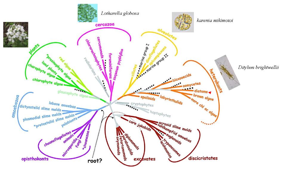
    [13] Baldauf SL (2008) An overview of the phylogeny and diversity of eukaryotes. J Syst Evol 46: 263-273.
    [14] Armbrust EV, Berges JA, Bowler C, et al. (2004) The genome of the diatom Thalassiosira pseudonana: ecology, evolution, and metabolism. Science 306: 79-86. doi: 10.1126/science.1101156
    [15] Lommer M, Specht M, Roy AS, et al. (2012) Genome and low-iron response of an oceanic diatom adapted to chronic iron limitation. Genome Biol 13: R66. doi: 10.1186/gb-2012-13-7-r66
    [16] Tanaka T, Maeda Y, Veluchamy A, et al. (2015) Oil Accumulation by the Oleaginous Diatom Fistulifera solaris as Revealed by the Genome and Transcriptome. Plant Cell 27: 162-176. doi: 10.1105/tpc.114.135194
    [17] Bowler C, De Martino A, Falciatore A (2010) Diatom cell division in an environmental context. Curr Opin Plant Biol 13: 623-630. doi: 10.1016/j.pbi.2010.09.014
    [18] Allen AE, Dupont CL, Obornik M, et al. (2011) Evolution and metabolic significance of the urea cycle in photosynthetic diatoms. Nature 473: 203-207. doi: 10.1038/nature10074
    [19] Tirichine L, Bowler C (2011) Decoding algal genomes: tracing back the history of photosynthetic life on Earth. Plant J 66: 45-57. doi: 10.1111/j.1365-313X.2011.04540.x
    [20] Maumus F, Allen AE, Mhiri C, et al. (2009) Potential impact of stress activated retrotransposons on genome evolution in a marine diatom. BMC Genomics 10: 624. doi: 10.1186/1471-2164-10-624
    [21] Lin X, Tirichine L, Bowler C (2012) Protocol: Chromatin immunoprecipitation (ChIP) methodology to investigate histone modifications in two model diatom species. Plant Methods 8: 48. doi: 10.1186/1746-4811-8-48
    [22] Tirichine L, Lin X, Thomas Y, et al. (2014) Histone extraction protocol from the two model diatoms Phaeodactylum tricornutum and Thalassiosira pseudonana. Mar Genomics 13: 21-25. doi: 10.1016/j.margen.2013.11.006
    [23] Veluchamy A, Lin X, Maumus F, et al. (2013) Insights into the role of DNA methylation in diatoms by genome-wide profiling in Phaeodactylum tricornutum. Nat Commun 4: 2091.
    [24] 
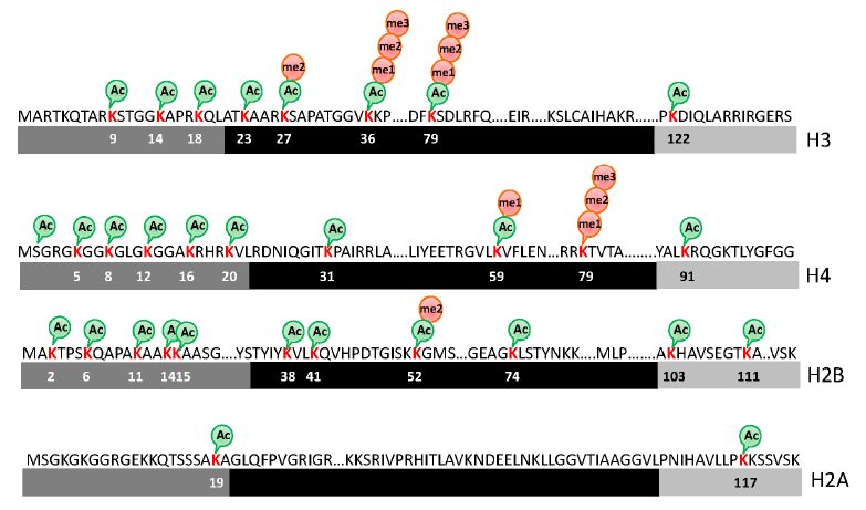
Veluchamy A, Rastogi A, Lin X, et al. (2015) An integrative analysis of post-translational histone modifications in the marine diatom Phaeodactylum tricornutum. Genome Biol 16: 102. doi: 10.1186/s13059-015-0671-8
    [25] Rogato A, Richard H, Sarazin A, et al. (2014) The diversity of small non-coding RNAs in the diatom Phaeodactylum tricornutum. BMC Genomics 15: 698. doi: 10.1186/1471-2164-15-698
    [26] Huang A, He L, Wang G (2011) Identification and characterization of microRNAs from Phaeodactylum tricornutum by high-throughput sequencing and bioinformatics analysis. BMC Genomics 12: 337. doi: 10.1186/1471-2164-12-337
    [27] Cock JM, Sterck L, Rouze P, et al. (2010) The Ectocarpus genome and the independent evolution of multicellularity in brown algae. Nature 465: 617-621. doi: 10.1038/nature09016
    [28] Suzuki MM, Bird A (2008) DNA methylation landscapes: provocative insights from epigenomics. Nat Rev Genet 9: 465-476.
    [29] Law JA, Jacobsen SE (2010) Establishing, maintaining and modifying DNA methylation patterns in plants and animals. Nat Rev Genet 11: 204-220. doi: 10.1038/nrg2719
    [30] Feng S, Cokus SJ, Zhang X, et al. (2010) Conservation and divergence of methylation patterning in plants and animals. Proc Natl Acad Sci U S A 107: 8689-8694. doi: 10.1073/pnas.1002720107
    [31] Zemach A, McDaniel IE, Silva P, et al. (2010) Genome-wide evolutionary analysis of eukaryotic DNA methylation. Science 328: 916-919. doi: 10.1126/science.1186366
    [32] Goll MG, Bestor TH (2005) Eukaryotic cytosine methyltransferases. Annu Rev Biochem 74: 481-514. doi: 10.1146/annurev.biochem.74.010904.153721
    [33] Huff JT, Zilberman D (2014) Dnmt1-independent CG methylation contributes to nucleosome positioning in diverse eukaryotes. Cell 156: 1286-1297. doi: 10.1016/j.cell.2014.01.029
    [34] Zhang X, Yazaki J, Sundaresan A, et al. (2006) Genome-wide high-resolution mapping and functional analysis of DNA methylation in arabidopsis. Cell 126: 1189-1201. doi: 10.1016/j.cell.2006.08.003
    [35] Molaro A, Hodges E, Fang F, et al. (2011) Sperm methylation profiles reveal features of epigenetic inheritance and evolution in primates. Cell 146: 1029-1041. doi: 10.1016/j.cell.2011.08.016
    [36] Zeng J, Konopka G, Hunt BG, et al. (2012) Divergent whole-genome methylation maps of human and chimpanzee brains reveal epigenetic basis of human regulatory evolution. Am J Hum Genet 91: 455-465. doi: 10.1016/j.ajhg.2012.07.024
    [37] Honeybee Genome Sequencing C (2006) Insights into social insects from the genome of the honeybee Apis mellifera. Nature 443: 931-949. doi: 10.1038/nature05260
    [38] Satou Y, Mineta K, Ogasawara M, et al. (2008) Improved genome assembly and evidence-based global gene model set for the chordate Ciona intestinalis: new insight into intron and operon populations. Genome Biol 9: R152. doi: 10.1186/gb-2008-9-10-r152
    [39] Goll MG, Kirpekar F, Maggert KA, et al. (2006) Methylation of tRNAAsp by the DNA methyltransferase homolog Dnmt2. Science 311: 395-398. doi: 10.1126/science.1120976
    [40] Ponger L, Li WH (2005) Evolutionary diversification of DNA methyltransferases in eukaryotic genomes. Mol Biol Evol 22: 1119-1128. doi: 10.1093/molbev/msi098
    [41] Maumus F, Rabinowicz P, Bowler C, et al. (2011) Stemming Epigenetics in Marine Stramenopiles. Current Genomics 12: 357-370. doi: 10.2174/138920211796429727
    [42] Bowler C, Vardi A, Allen AE (2010) Oceanographic and biogeochemical insights from diatom genomes. Ann Rev Mar Sci 2: 333-365. doi: 10.1146/annurev-marine-120308-081051
    [43] Zimmermann C, Guhl E, Graessmann A (1997) Mouse DNA methyltransferase (MTase) deletion mutants that retain the catalytic domain display neither de novo nor maintenance methylation activity in vivo. Biol Chem 378: 393-405.
    [44] Fatemi M, Hermann A, Pradhan S, et al. (2001) The activity of the murine DNA methyltransferase Dnmt1 is controlled by interaction of the catalytic domain with the N-terminal part of the enzyme leading to an allosteric activation of the enzyme after binding to methylated DNA. J Mol Biol 309: 1189-1199. doi: 10.1006/jmbi.2001.4709
    [45] Penterman J, Zilberman D, Huh JH, et al. (2007) DNA demethylation in the Arabidopsis genome. Proc Natl Acad Sci U S A 104: 6752-6757. doi: 10.1073/pnas.0701861104
    [46] Cokus SJ, Feng S, Zhang X, et al. (2008) Shotgun bisulphite sequencing of the Arabidopsis genome reveals DNA methylation patterning. Nature 452: 215-219. doi: 10.1038/nature06745
    [47] Hunt BG, Brisson JA, Yi SV, et al. Functional conservation of DNA methylation in the pea aphid and the honeybee. Genome Biol Evol 2: 719-728.
    [48] Foret S, Kucharski R, Pittelkow Y, et al. (2009) Epigenetic regulation of the honey bee transcriptome: unravelling the nature of methylated genes. BMC Genomics 10: 472. doi: 10.1186/1471-2164-10-472
    [49] Xiang H, Zhu J, Chen Q, et al. (2010) Single base-resolution methylome of the silkworm reveals a sparse epigenomic map. Nat Biotechnol 28: 516-520. doi: 10.1038/nbt.1626
    [50] Hellman A, Chess A (2007) Gene body-specific methylation on the active X chromosome. Science 315: 1141-1143. doi: 10.1126/science.1136352
    [51] Brenet F, Moh M, Funk P, et al. DNA methylation of the first exon is tightly linked to transcriptional silencing. PLoS One 6: e14524.
    [52] Maunakea AK, Nagarajan RP, Bilenky M, et al. (2010) Conserved role of intragenic DNA methylation in regulating alternative promoters. Nature 466: 253-257. doi: 10.1038/nature09165
    [53] Takuno S, Gaut BS (2012) Body-Methylated Genes in Arabidopsis thaliana Are Functionally Important and Evolve Slowly. Mol Biol Evol 29: 219-227. doi: 10.1093/molbev/msr188
    [54] Zilberman D, Gehring M, Tran RK, et al. (2007) Genome-wide analysis of Arabidopsis thaliana DNA methylation uncovers an interdependence between methylation and transcription. Nat Genet 39: 61-69. doi: 10.1038/ng1929
    [55] Lorincz MC, Dickerson DR, Schmitt M, et al. (2004) Intragenic DNA methylation alters chromatin structure and elongation efficiency in mammalian cells. Nat Struct Mol Biol 11: 1068-1075. doi: 10.1038/nsmb840
    [56] Luco RF, Pan Q, Tominaga K, et al. (2010) Regulation of alternative splicing by histone modifications. Science 327: 996-1000. doi: 10.1126/science.1184208
    [57] Lyko F, Foret S, Kucharski R, et al. (2010) The honey bee epigenomes: differential methylation of brain DNA in queens and workers. PLoS Biol 8: e1000506. doi: 10.1371/journal.pbio.1000506
    [58] Ammar R, Torti D, Tsui K, et al. (2012) Chromatin is an ancient innovation conserved between Archaea and Eukarya. Elife 1: e00078.
    [59] Nalabothula N, Xi L, Bhattacharyya S, et al. (2013) Archaeal nucleosome positioning in vivo and in vitro is directed by primary sequence motifs. BMC Genomics 14: 391. doi: 10.1186/1471-2164-14-391
    [60] Mersfelder EL, Parthun MR (2006) The tale beyond the tail: histone core domain modifications and the regulation of chromatin structure. Nucleic Acids Res 34: 2653-2662. doi: 10.1093/nar/gkl338
    [61] Cosgrove MS, Boeke JD, Wolberger C (2004) Regulated nucleosome mobility and the histone code. Nat Struct Mol Biol 11: 1037-1043. doi: 10.1038/nsmb851
    [62] Lermontova I, Schubert V, Fuchs J, et al. (2006) Loading of Arabidopsis centromeric histone CENH3 occurs mainly during G2 and requires the presence of the histone fold domain. Plant Cell 18: 2443-2451. doi: 10.1105/tpc.106.043174
    [63] Hashimoto H, Sonoda E, Takami Y, et al. (2007) Histone H1 variant, H1R is involved in DNA damage response. DNA Repair (Amst) 6: 1584-1595. doi: 10.1016/j.dnarep.2007.05.003
    [64] Maheswari U, Jabbari K, Petit JL, et al. (2010) Digital expression profiling of novel diatom transcripts provides insight into their biological functions. Genome Biol 11: R85. doi: 10.1186/gb-2010-11-8-r85
    [65] Bheda P, Swatkoski S, Fiedler KL, et al. (2012) Biotinylation of lysine method identifies acetylated histone H3 lysine 79 in Saccharomyces cerevisiae as a substrate for Sir2. Proc Natl Acad Sci U S A 109: E916-925. doi: 10.1073/pnas.1121471109
    [66] Zhang K, Sridhar VV, Zhu J, et al. (2007) Distinctive core histone post-translational modification patterns in Arabidopsis thaliana. PLoS One 2: e1210. doi: 10.1371/journal.pone.0001210
    [67] Tan M, Luo H, Lee S, et al. (2011) Identification of 67 histone marks and histone lysine crotonylation as a new type of histone modification. Cell 146: 1016-1028. doi: 10.1016/j.cell.2011.08.008
    [68] Sana J, Faltejskova P, Svoboda M, et al. (2012) Novel classes of non-coding RNAs and cancer. J Transl Med 10: 103. doi: 10.1186/1479-5876-10-103
    [69] Ulitsky I, Bartel DP (2013) lincRNAs: genomics, evolution, and mechanisms. Cell 154: 26-46. doi: 10.1016/j.cell.2013.06.020
    [70] Okazaki Y, Furuno M, Kasukawa T, et al. (2002) Analysis of the mouse transcriptome based on functional annotation of 60,770 full-length cDNAs. Nature 420: 563-573. doi: 10.1038/nature01266
    [71] Cabili MN, Trapnell C, Goff L, et al. (2011) Integrative annotation of human large intergenic noncoding RNAs reveals global properties and specific subclasses. Genes Dev 25: 1915-1927. doi: 10.1101/gad.17446611
    [72] Liu J, Jung C, Xu J, et al. (2012) Genome-wide analysis uncovers regulation of long intergenic noncoding RNAs in Arabidopsis. Plant Cell 24: 4333-4345. doi: 10.1105/tpc.112.102855
    [73] Pauli A, Valen E, Lin MF, et al. (2012) Systematic identification of long noncoding RNAs expressed during zebrafish embryogenesis. Genome Res 22: 577-591. doi: 10.1101/gr.133009.111
    [74] Cech TR, Steitz JA (2014) The noncoding RNA revolution-trashing old rules to forge new ones. Cell 157: 77-94. doi: 10.1016/j.cell.2014.03.008
    [75] Molnar A, Schwach F, Studholme DJ, et al. (2007) miRNAs control gene expression in the single-cell alga Chlamydomonas reinhardtii. Nature 447: 1126-1129. doi: 10.1038/nature05903
    [76] Zhao T, Li G, Mi S, et al. (2007) A complex system of small RNAs in the unicellular green alga Chlamydomonas reinhardtii. Genes Dev 21: 1190-1203. doi: 10.1101/gad.1543507
    [77] Lopez-Gomollon S, Beckers M, Rathjen T, et al. (2014) Global discovery and characterization of small non-coding RNAs in marine microalgae. BMC Genomics 15: 697. doi: 10.1186/1471-2164-15-697
    [78] Norden-Krichmar TM, Allen AE, Gaasterland T, et al. (2011) Characterization of the small RNA transcriptome of the diatom, Thalassiosira pseudonana. PLoS One 6: e22870. doi: 10.1371/journal.pone.0022870
    [79] 
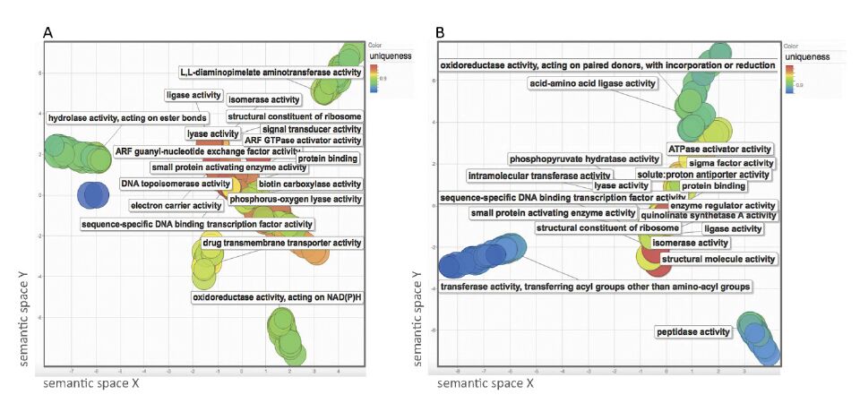
Supek F, Bosnjak M, Skunca N, et al. (2011) REVIGO summarizes and visualizes long lists of gene ontology terms. PLoS One 6: e21800. doi: 10.1371/journal.pone.0021800
  • This article has been cited by:

    1. Zhexin Fan, Bo Yang, R. Paul Ross, Catherine Stanton, Jianxin Zhao, Hao Zhang, Wei Chen, The prophylactic effects of different Lactobacilli on collagen-induced arthritis in rats, 2020, 11, 2042-6496, 3681, 10.1039/C9FO02556A
    2. Chunhong Liu, Li Lin, Weidong Cui, Lei Wang, Min Ai, Zhongwei Zhao, Xiaohan Ma, Shengyu Li, Lactiplantibacillus plantarum HG20 attenuates II type collagen-induced rheumatoid arthritis in rats via anti-inflammatory and inhibition of apoptosis, 2022, 132, 1365-2672, 2323, 10.1111/jam.15333
  • Reader Comments
  • © 2015 the Author(s), licensee AIMS Press. This is an open access article distributed under the terms of the Creative Commons Attribution License (http://creativecommons.org/licenses/by/4.0)
通讯作者: 陈斌, bchen63@163.com
  • 1. 

    沈阳化工大学材料科学与工程学院 沈阳 110142

  1. 本站搜索
  2. 百度学术搜索
  3. 万方数据库搜索
  4. CNKI搜索

Metrics

Article views(9382) PDF downloads(1631) Cited by(18)

Article outline

Figures and Tables

Figures(7)  /  Tables(2)

/

DownLoad:  Full-Size Img  PowerPoint
Return
Return

Catalog