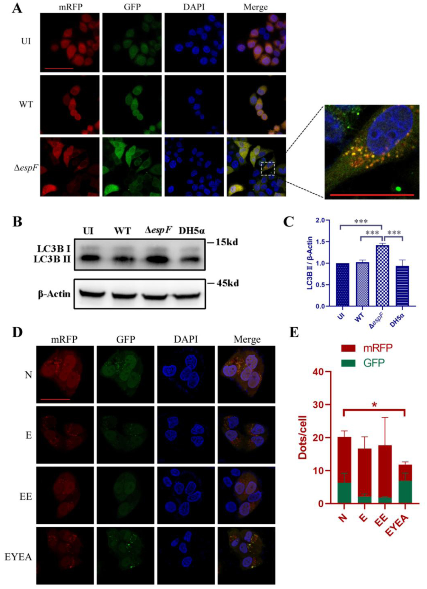
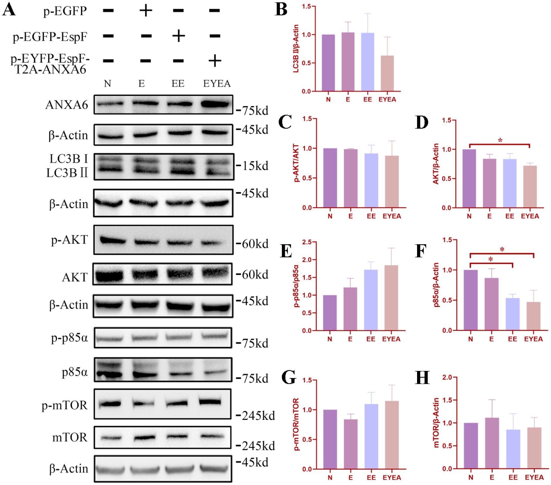
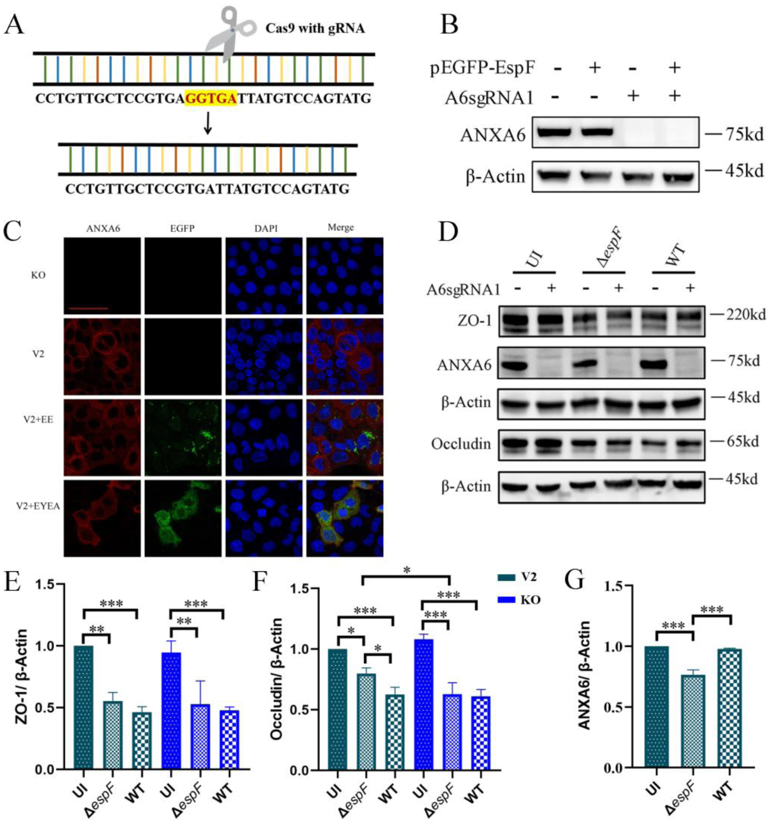
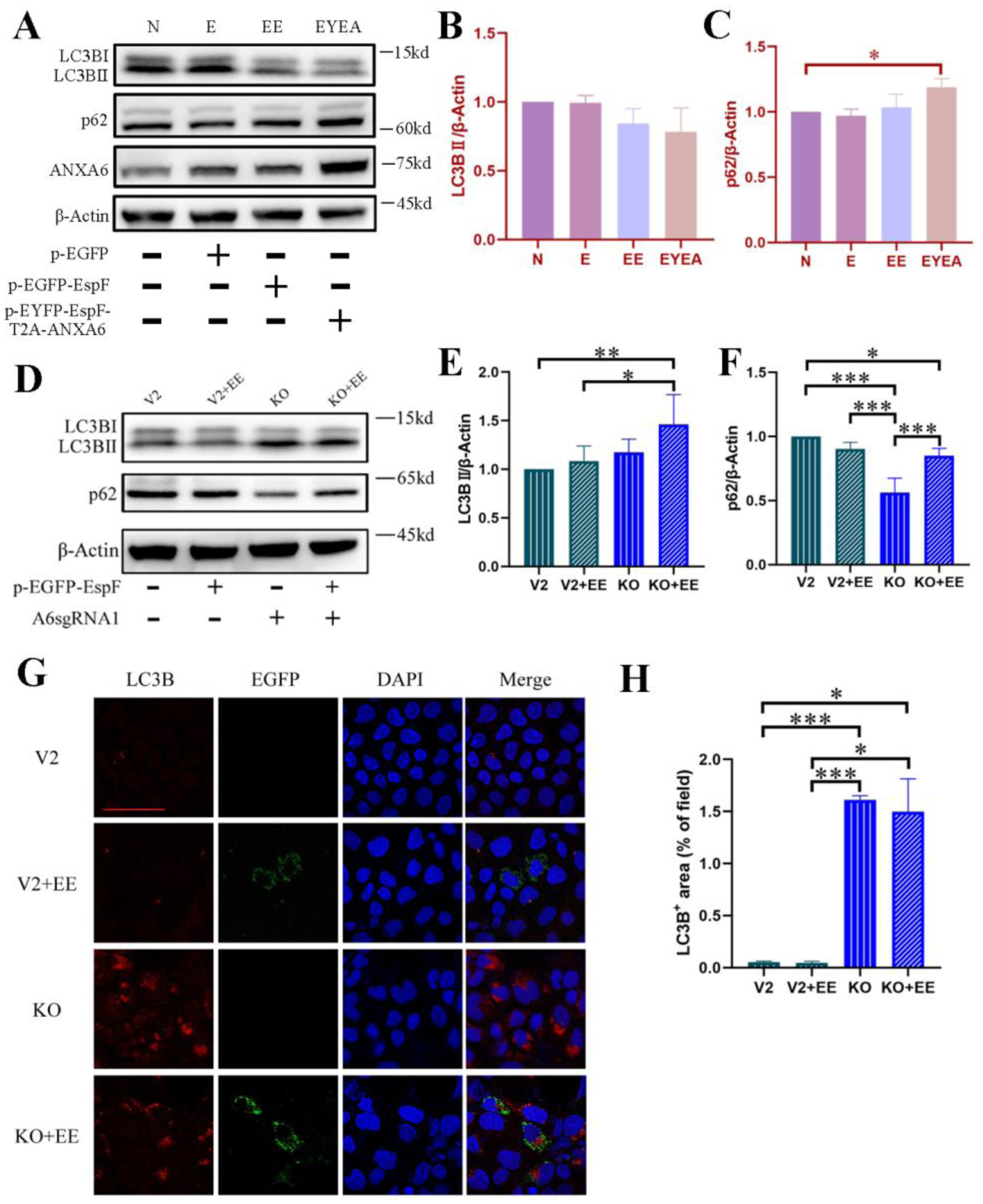
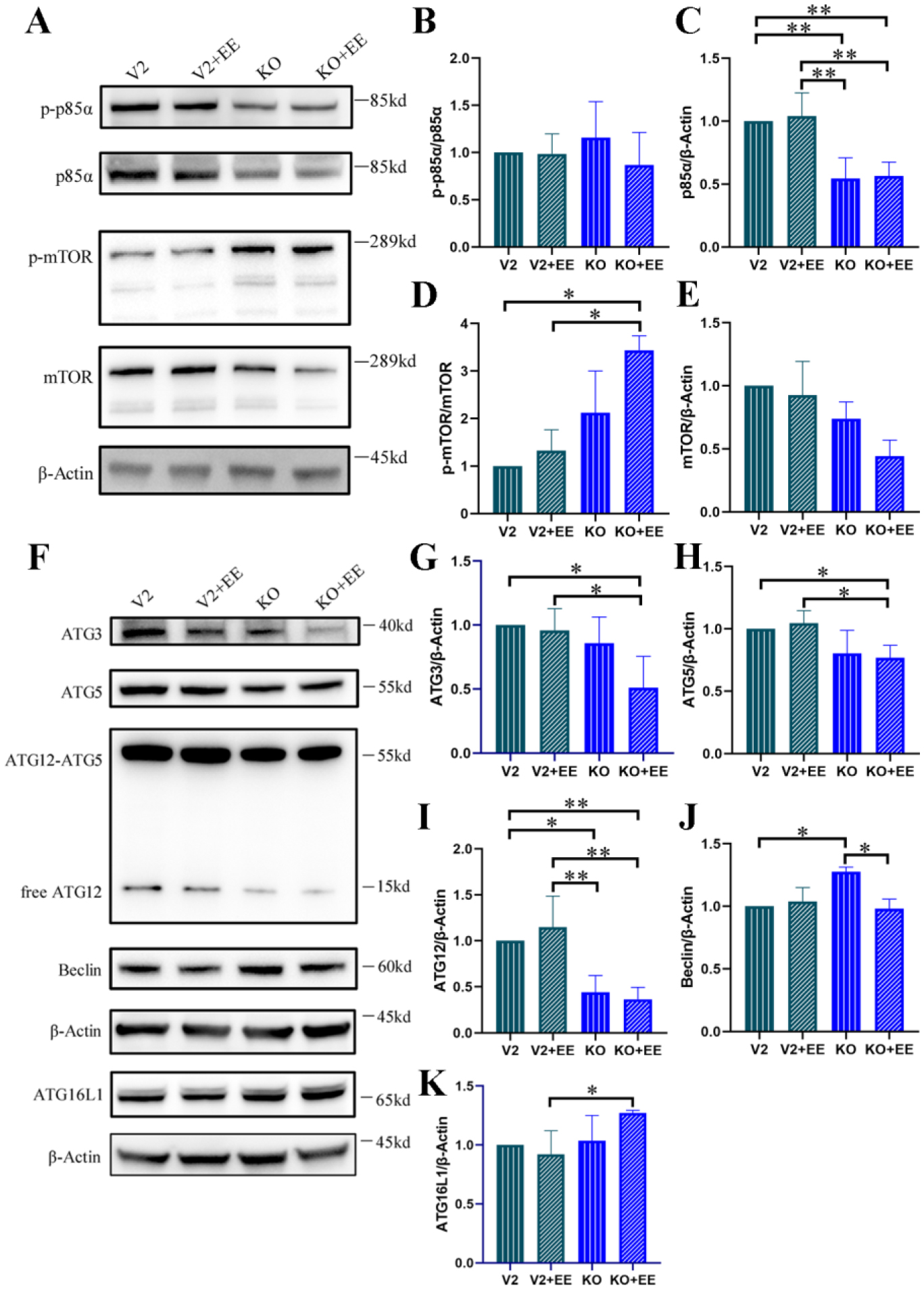
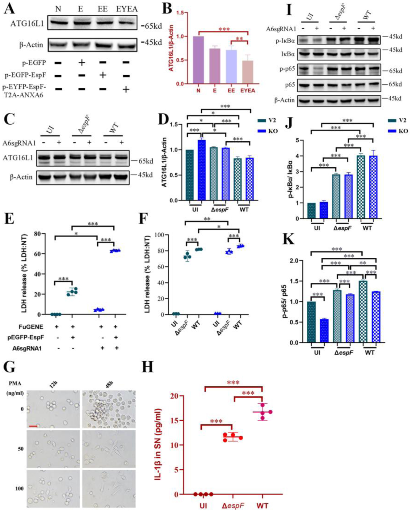
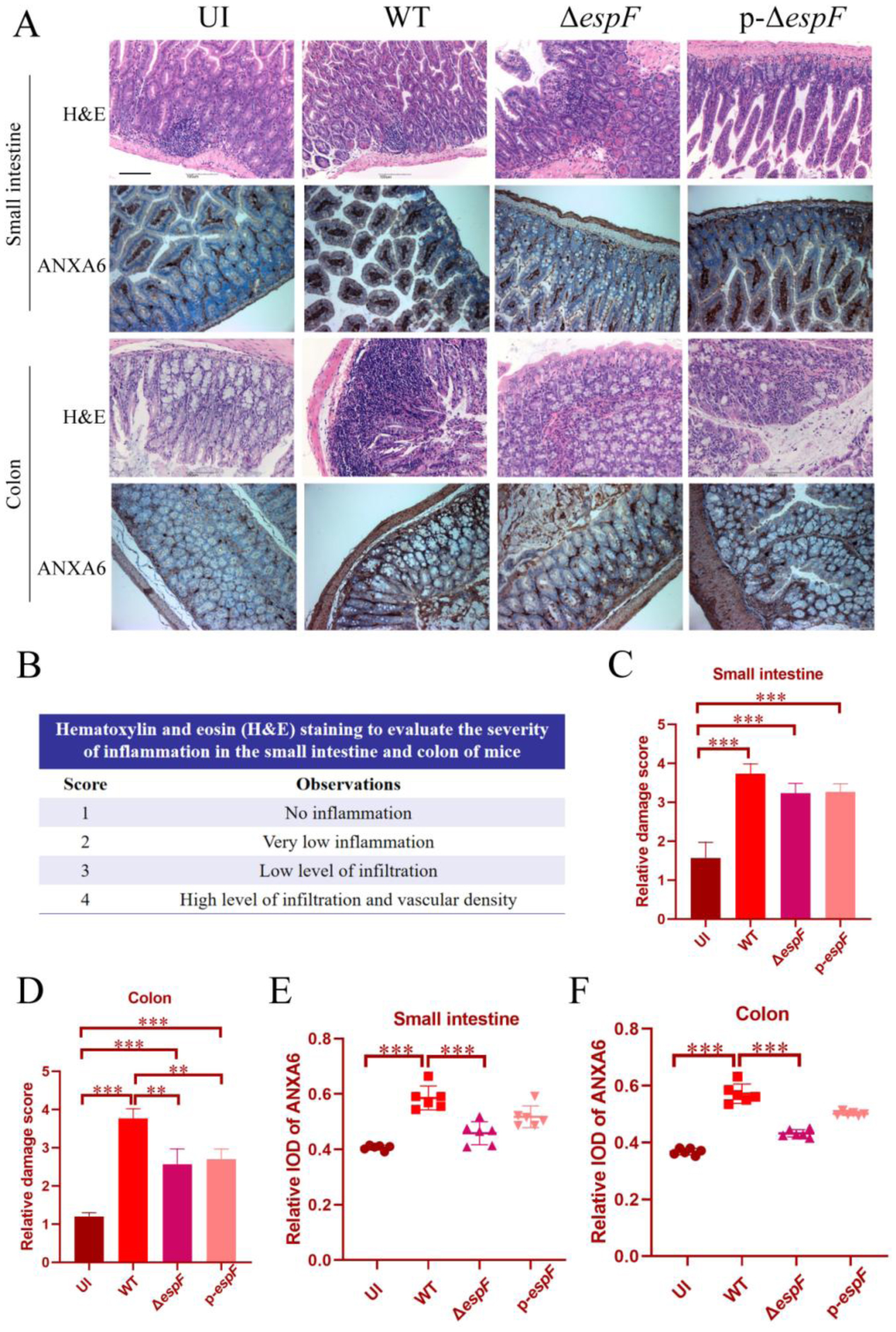
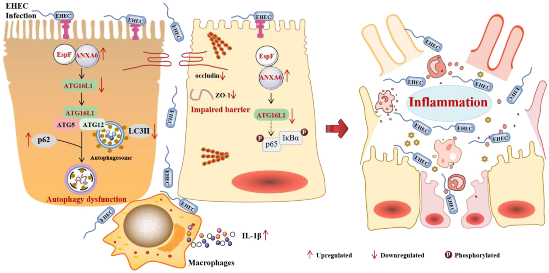
Autophagy is a critical host defense mechanism against pathogens; however, enterohemorrhagic Escherichia coli (EHEC) O157:H7 exploits it to establish infection. Here, we revealed how EHEC's effector EspF collaborates with host Annexin A6 (ANXA6) to suppress autophagy and drive inflammation. Our results showed that CRISPR/Cas9-mediated anxa6 knockout in intestinal epithelial cells reversed EHEC-induced autophagic inhibition, as evidenced by elevated LC3B-II levels and reduced p62 accumulation. Mechanistically, EspF stabilizes ANXA6 to disrupt PI3K/mTOR signaling and impair autophagosome formation, whereas ANXA6 suppresses the expression of ATG16L1, a key autophagy regulator. In this study, EHEC infection triggered IL-1β hypersecretion in macrophages, which was coupled with NF-κB pathway hyperactivation via IκBα/p65 phosphorylation. In vivo, EHEC infection regulated intestinal ANXA6 expression, correlating with mucosal inflammation and barrier dysfunction. Crucially, ANXA6/ATG16L1 axis disruption created a self-reinforcing cycle of impaired autophagy, bacterial persistence, and inflammatory escalation. Our findings identified ANXA6 as a context-dependent autophagy modulator and ATG16L1 as a novel EHEC target, providing mechanistic insights into EHEC pathogenesis.
Citation: Litai Xu, Min Gao, Yaoguo Wang, Bao Zhang, Wei Zhao, Weizhi Lu, Guanhua Cao, Chengsong Wan, Ying Hua. Enterohemorrhagic Escherichia coli targets Annexin A6 and ATG16L1 to inhibit autophagy and induce inflammation[J]. AIMS Microbiology, 2025, 11(4): 984-1006. doi: 10.3934/microbiol.2025044
Autophagy is a critical host defense mechanism against pathogens; however, enterohemorrhagic Escherichia coli (EHEC) O157:H7 exploits it to establish infection. Here, we revealed how EHEC's effector EspF collaborates with host Annexin A6 (ANXA6) to suppress autophagy and drive inflammation. Our results showed that CRISPR/Cas9-mediated anxa6 knockout in intestinal epithelial cells reversed EHEC-induced autophagic inhibition, as evidenced by elevated LC3B-II levels and reduced p62 accumulation. Mechanistically, EspF stabilizes ANXA6 to disrupt PI3K/mTOR signaling and impair autophagosome formation, whereas ANXA6 suppresses the expression of ATG16L1, a key autophagy regulator. In this study, EHEC infection triggered IL-1β hypersecretion in macrophages, which was coupled with NF-κB pathway hyperactivation via IκBα/p65 phosphorylation. In vivo, EHEC infection regulated intestinal ANXA6 expression, correlating with mucosal inflammation and barrier dysfunction. Crucially, ANXA6/ATG16L1 axis disruption created a self-reinforcing cycle of impaired autophagy, bacterial persistence, and inflammatory escalation. Our findings identified ANXA6 as a context-dependent autophagy modulator and ATG16L1 as a novel EHEC target, providing mechanistic insights into EHEC pathogenesis.

| [1] |
Liu S, Yao S, Yang H, et al. (2023) Autophagy: Regulator of cell death. Cell Death Dis 14: 1-17. https://doi.org/10.1038/s41419-023-06154-8 
|
| [2] |
Aparicio IM, Espino J, Bejarano I, et al. (2016) Autophagy-related proteins are functionally active in human spermatozoa and may be involved in the regulation of cell survival and motility. Sci Rep 6: 33647. https://doi.org/10.1038/srep33647
|
| [3] |
Losier TT, Akuma M, McKee-Muir OC, et al. (2019) AMPK promotes xenophagy through priming of autophagic kinases upon detection of bacterial outer membrane vesicles. Cell Rep 26: 2150-2165.e2155. https://doi.org/10.1016/j.celrep.2019.01.062
|
| [4] |
Mansilla Pareja ME, Colombo MI (2013) Autophagic clearance of bacterial pathogens: Molecular recognition of intracellular microorganisms. Front Cell Infect Microbiol 3: 54. https://doi.org/10.3389/fcimb.2013.00054
|
| [5] |
Gomes LC, Dikic I (2014) Autophagy in antimicrobial immunity. Mol Cell 54: 224-233. https://doi.org/10.1016/j.molcel.2014.03.009
|
| [6] |
Germic N, Frangez Z, Yousefi S, et al. (2019) Regulation of the innate immune system by autophagy: Monocytes, macrophages, dendritic cells and antigen presentation. Cell Death Differ 26: 715-727. https://doi.org/10.1038/s41418-019-0297-6
|
| [7] |
Ogawa M, Yoshimori T, Suzuki T, et al. (2005) Escape of intracellular shigella from autophagy. Science 307: 727-731. https://doi.org/10.1126/science.1106036
|
| [8] |
Zhuge X, Sun Y, Xue F, et al. (2018) A novel PhoP/PhoQ regulation pathway modulates the survival of extraintestinal pathogenic escherichia coli in macrophages. Front Immunol 9: 788. https://doi.org/10.3389/fimmu.2018.00788
|
| [9] |
Birmingham CL, Canadien V, Gouin E, et al. (2007) Listeria monocytogenes evades killing by autophagy during colonization of host cells. Autophagy 3: 442-451. https://doi.org/10.4161/auto.4450
|
| [10] |
Wu YW, Li F (2019) Bacterial interaction with host autophagy. Virulence 10: 352-362. https://doi.org/10.1080/21505594.2019.1602020
|
| [11] |
Rani A, Ravindran VB, Surapaneni A, et al. (2021) Review: Trends in point-of-care diagnosis for Escherichia coli O157:H7 in food and water. Int J Food Microbiol 349. https://doi.org/10.1016/j.ijfoodmicro.2021.109233
|
| [12] |
Tang B, Li Q, Zhao XH, et al. (2015) Shiga toxins induce autophagic cell death in intestinal epithelial cells via the endoplasmic reticulum stress pathway. Autophagy 11: 344-354. https://doi.org/10.1080/15548627.2015.1023682
|
| [13] |
Xue Y, Du M, Sheng H, et al. (2017) Escherichia coli O157:H7 suppresses host autophagy and promotes epithelial adhesion via tir-mediated and cAMP-independent activation of protein kinase A. Cell Death Discov 3: 17055. https://doi.org/10.1038/cddiscovery.2017.55
|
| [14] |
Li J, Guo S, Chai F, et al. (2021) Genetically incorporated crosslinkers reveal NleE attenuates host autophagy dependent on PSMD10. eLife 10: e69047. https://doi.org/10.7554/eLife.69047
|
| [15] |
Holmes A, Mühlen S, Roe AJ, et al. (2010) The EspF effector, a bacterial pathogen's swiss army knife. Infect Immun 78: 4445-4453. https://doi.org/10.1128/IAI.00635-10
|
| [16] |
Hua Y, Wu J, Fu M, et al. (2020) Enterohemorrhagic escherichia coli effector protein EspF interacts with host protein ANXA6 and triggers myosin light chain kinase (MLCK)-dependent tight junction dysregulation. Front Cell Dev Biol 8: 613061. https://doi.org/10.3389/fcell.2020.613061
|
| [17] |
Fu M, Liang S, Wu J, et al. (2021) An escherichia coli effector protein EspF may induce host DNA damage via interaction with SMC1. Front Microbiol 12: 682064. https://doi.org/10.3389/fmicb.2021.682064
|
| [18] |
Sun X, Shu Y, Xu M, et al. (2020) ANXA6 suppresses the tumorigenesis of cervical cancer through autophagy induction. Clin Transl Med 10: e208. https://doi.org/10.1002/ctm2.208
|
| [19] |
Enrich C, Rentero C, Grewal T (2017) Annexin A6 in the liver: From the endocytic compartment to cellular physiology. Biochim Biophys Acta Mol Cell Res 1864: 933-946. https://doi.org/10.1016/j.bbamcr.2016.10.017
|
| [20] |
Hua Y, Ju J, Wang X, et al. (2018) Screening for host proteins interacting with escherichia coli O157:H7 EspF using bimolecular fluorescence complementation. Future Microbiol 13: 37-58. https://doi.org/10.2217/fmb-2017-0087
|
| [21] |
Yu T, Guo F, Yu Y, et al. (2017) Fusobacterium nucleatum promotes chemoresistance to colorectal cancer by modulating autophagy. Cell 170: 548-563.e516. https://doi.org/10.1016/j.cell.2017.07.008
|
| [22] |
Schneider CA, Rasband WS, Eliceiri KW (2012) NIH image to imageJ: 25 years of image analysis. Nat Methods 9: 671-675. https://doi.org/10.1038/nmeth.2089
|
| [23] | Chen HZ, Liang S, Li X, et al. (2023) Knockout of human anxa6 gene in Caco-2 cells by CRISPR/Cas9 system. Acta Microbiol Sin 63: 1217-1229. |
| [24] |
Wang X, Du Y, Hua Y, et al. (2017) The EspF N-terminal of enterohemorrhagic escherichia coli O157:H7 EDL933w imparts stronger toxicity effects on HT-29 cells than the C-terminal. Front Cell Infect Microbiol 7: 410. https://doi.org/10.3389/fcimb.2017.00410
|
| [25] |
Yoshii SR, Mizushima N (2017) Monitoring and measuring autophagy. Int J Mol Sci 18: 1865. https://doi.org/10.3390/ijms18091865
|
| [26] |
van Breemen RB, Li Y (2005) Caco-2 cell permeability assays to measure drug absorption. Expert Opin Drug Metab Toxicol 1: 175-185. https://doi.org/10.1517/17425255.1.2.175
|
| [27] |
Gerke V, Creutz CE, Moss SE (2005) Annexins: Linking Ca2+ signalling to membrane dynamics. Nat Rev Mol Cell Biol 6: 449-461. https://doi.org/10.1038/nrm1661
|
| [28] |
Qi H, Liu S, Guo C, et al. (2015) Role of annexin A6 in cancer. Oncol Lett 10: 1947-1952. https://doi.org/10.3892/ol.2015.3498
|
| [29] |
Zheng Y, Qiu Y, Grace CRR, et al. (2019) A switch element in the autophagy E2 Atg3 mediates allosteric regulation across the lipidation cascade. Nat Commun 10: 3600. https://doi.org/10.1038/s41467-019-11435-y
|
| [30] |
Saitoh T, Fujita N, Jang MH, et al. (2008) Loss of the autophagy protein Atg16L1 enhances endotoxin-induced IL-1beta production. Nature 456: 264-268. https://doi.org/10.1038/nature07383
|
| [31] |
Deretic V, Levine B (2018) Autophagy balances inflammation in innate immunity. Autophagy 14: 243-251. https://doi.org/10.1080/15548627.2017.1402992
|
| [32] |
Cadwell K (2016) Crosstalk between autophagy and inflammatory signalling pathways: Balancing defence and homeostasis. Nat Rev Immunol 16: 661-675. https://doi.org/10.1038/nri.2016.100
|
| [33] |
Keller MD, Torres VJ, Cadwell K (2020) Autophagy and microbial pathogenesis. Cell Death Differ 27: 872-886. https://doi.org/10.1038/s41418-019-0481-8
|
| [34] |
Kimmey JM, Stallings CL (2016) Bacterial pathogens versus autophagy: Implications for therapeutic interventions. Trends Mol Med 22: 1060-1076. https://doi.org/10.1016/j.molmed.2016.10.008
|
| [35] |
Sharma S, Tiwari M, Tiwari V (2021) Therapeutic strategies against autophagic escape by pathogenic bacteria. Drug Discov Today 26: 704-712. https://doi.org/10.1016/j.drudis.2020.12.002
|
| [36] |
Lee M-S, Cherla RP, Jenson MH, et al. (2011) Shiga toxins induce autophagy leading to differential signalling pathways in toxin-sensitive and toxin-resistant human cells. Cell Microbiol 13: 1479-1496. https://doi.org/10.1111/j.1462-5822.2011.01634.x
|
| [37] |
Chen Q, Zheng W, Zhu L, et al. (2020) ANXA6 contributes to radioresistance by promoting autophagy via inhibiting the PI3K/AKT/mTOR signaling pathway in nasopharyngeal carcinoma. Front Cell Dev Biol 8: 232. https://doi.org/10.3389/fcell.2020.00232
|
| [38] |
Cao J, Wan S, Chen S, et al. (2023) ANXA6: A key molecular player in cancer progression and drug resistance. Discov Oncol 14: 53. https://doi.org/10.1007/s12672-023-00662-x
|
| [39] |
Komor MA, Bosch LJW, Coupé VMH, et al. (2020) Proteins in stool as biomarkers for non-invasive detection of colorectal adenomas with high risk of progression. J Pathol 250: 288-298. https://doi.org/10.1002/path.5369
|
| [40] |
Boye TL, Maeda K, Pezeshkian W, et al. (2017) Annexin A4 and A6 induce membrane curvature and constriction during cell membrane repair. Nat Commun 8: 1623. https://doi.org/10.1038/s41467-017-01743-6
|
| [41] |
Sorbara MT, Ellison LK, Ramjeet M, et al. (2013) The protein ATG16L1 suppresses inflammatory cytokines induced by the intracellular sensors Nod1 and Nod2 in an autophagy-independent manner. Immunity 39: 858-873. https://doi.org/10.1016/j.immuni.2013.10.013
|
| [42] |
Symington JW, Wang C, Twentyman J, et al. (2015) ATG16L1 deficiency in macrophages drives clearance of uropathogenic E. coli in an IL-1β-dependent manner. Mucosal Immunol 8: 1388-1399. https://doi.org/10.1038/mi.2015.7
|
| [43] |
Plantinga TS, Crisan TO, Oosting M, et al. (2011) Crohn's disease-associated ATG16L1 polymorphism modulates pro-inflammatory cytokine responses selectively upon activation of NOD2. Gut 60: 1229-1235. https://doi.org/10.1136/gut.2010.228908
|
| [44] |
Foerster EG, Mukherjee T, Cabral-Fernandes L, et al. (2022) How autophagy controls the intestinal epithelial barrier. Autophagy 18: 86-103. https://doi.org/10.1080/15548627.2021.1909406
|
Figures(8) / Tables(1)

Litai Xu, Min Gao, Yaoguo Wang, Bao Zhang, Wei Zhao, Weizhi Lu, Guanhua Cao, Chengsong Wan, Ying Hua. Enterohemorrhagic Escherichia coli targets Annexin A6 and ATG16L1 to inhibit autophagy and induce inflammation[J]. AIMS Microbiology, 2025, 11(4): 984-1006. doi: 10.3934/microbiol.2025044


 DownLoad: