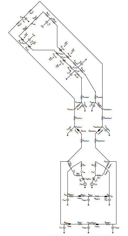
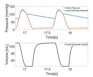
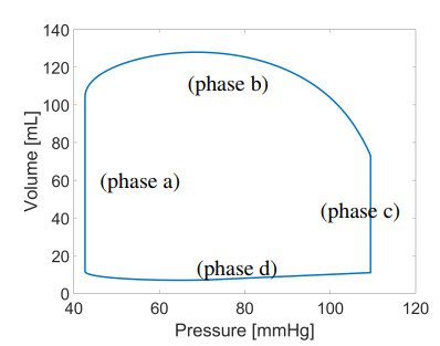
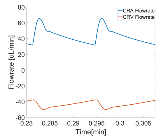
The cardiovascular and ocular systems are intricately connected, with hemodynamic interactions playing a crucial role in both physiological regulation and pathological conditions. However, existing models often treat these systems separately, thus limiting the understanding of their interdependence. In this study, we present the Eye2Heart model, which is a novel closed-loop mathematical framework that integrates cardiovascular and ocular dynamics. Using an electrical-hydraulic analogy, the model describes the interactions between the heart and retinal circulation through a nonlinear system of ordinary differential equations. The model is tested against clinical and experimental data, thus demonstrating its ability to reproduce key cardiovascular parameters (e.g., stroke volume, cardiac output) and ocular hemodynamics (e.g., retinal blood flow). Additionally, we explore in silico the effects of intraocular pressure and left ventricular compliance on both local ocular and global systemic circulation, thus revealing critical dependencies between cardiovascular and ocular health. The results highlight the model's potential for studying cardiovascular diseases with ocular manifestations and support emerging research in oculomics by providing a mechanistic basis to interpret ocular biomarkers within a systemic context. This paves the way for patient-specific data integration and broader applications in personalized medicine.
Citation: Lorenzo Sala, Mohamed Zaid, Faith Hughes, Marcela Szopos, Virginia H. Huxley, Alon Harris, Giovanna Guidoboni, Sergey Lapin. Eye2Heart: A reduced mathematical model bridging cardiovascular and ocular hemodynamics[J]. Mathematical Biosciences and Engineering, 2026, 23(2): 421-448. doi: 10.3934/mbe.2026017
The cardiovascular and ocular systems are intricately connected, with hemodynamic interactions playing a crucial role in both physiological regulation and pathological conditions. However, existing models often treat these systems separately, thus limiting the understanding of their interdependence. In this study, we present the Eye2Heart model, which is a novel closed-loop mathematical framework that integrates cardiovascular and ocular dynamics. Using an electrical-hydraulic analogy, the model describes the interactions between the heart and retinal circulation through a nonlinear system of ordinary differential equations. The model is tested against clinical and experimental data, thus demonstrating its ability to reproduce key cardiovascular parameters (e.g., stroke volume, cardiac output) and ocular hemodynamics (e.g., retinal blood flow). Additionally, we explore in silico the effects of intraocular pressure and left ventricular compliance on both local ocular and global systemic circulation, thus revealing critical dependencies between cardiovascular and ocular health. The results highlight the model's potential for studying cardiovascular diseases with ocular manifestations and support emerging research in oculomics by providing a mechanistic basis to interpret ocular biomarkers within a systemic context. This paves the way for patient-specific data integration and broader applications in personalized medicine.

| [1] |
J. Flammer, K. Konieczka, R. M. Bruno, A. Virdis, A. J. Flammer, S. Taddei, The eye and the heart, Eur. Heart J., 34 (2013), 1270–1278. https://doi.org/10.1093/eurheartj/eht023 doi: 10.1093/eurheartj/eht023

|
| [2] |
H. Hanssen, L. Streese, W. Vilser, Retinal vessel diameters and function in cardiovascular risk and disease, Prog. Retin. Eye Res., 91 (2022), 101095. https://doi.org/10.1016/j.preteyeres.2022.101095 doi: 10.1016/j.preteyeres.2022.101095
|
| [3] |
E. L. Michelson, J. Morganroth, C. W. Nichols, H. MacVaugh, Retinal arteriolar changes as an indicator of coronary artery disease, Arch. Intern. Med., 139 (1979), 1139–1141. https://doi.org/10.1001/archinte.1979.03630470051017 doi: 10.1001/archinte.1979.03630470051017
|
| [4] |
E. Tedeschi-Reiner, M. Strozzi, B. Skoric, Z. Reiner, Relation of atherosclerotic changes in retinal arteries to the extent of coronary artery disease, Am. J. Cardiol., 96 (2005), 1107–1109. https://doi.org/10.1016/j.amjcard.2005.05.070 doi: 10.1016/j.amjcard.2005.05.070
|
| [5] |
A. Abdin, A. D. Abdin, G. Merone, W. Aljundi, B. Haring, Y. A. Dail, et al., Cardio-ocular syndrome: Retinal microvascular changes in acutely decompensated heart failure, Eur. J. Heart Fail., 26 (2024), 2421–2430. https://doi.org/10.1002/ejhf.3474 doi: 10.1002/ejhf.3474
|
| [6] |
Z. Zhu, Y. Wang, Z. Qi, W. Hu, X. Zhang, S. K. Wagner, et al., Oculomics: Current concepts and evidence, Prog. Retin. Eye Res., 101350 (2025). https://doi.org/10.1016/j.preteyeres.2025.101350 doi: 10.1016/j.preteyeres.2025.101350
|
| [7] |
L. A. Ghenciu, M. Dima, E. R. Stoicescu, R. Iacob, C. Boru, O. A. Hațegan, Retinal imaging-based oculomics: Artificial intelligence as a tool in the diagnosis of cardiovascular and metabolic diseases, Biomedicines, 12 (2024), 2150. https://doi.org/10.3390/biomedicines12092150 doi: 10.3390/biomedicines12092150
|
| [8] |
W. Hu, Z. Lin, M. Clark, J. Henwood, X. Shang, R. Chen, et al., Real-world feasibility, accuracy and acceptability of automated retinal photography and AI-based cardiovascular disease risk assessment, npj Digit. Med., 8 (2025), 122. https://doi.org/10.1038/s41746-025-01436-1 doi: 10.1038/s41746-025-01436-1
|
| [9] |
E. Y. Chew, S. A. Burns, A. G. Abraham, M. F. Bakhoum, J. A. Beckman, T. Y. P. Chui, et al., Standardization and clinical applications of retinal imaging biomarkers for cardiovascular disease, Nat. Rev. Cardiol., 22 (2025), 47–63. https://doi.org/10.1038/s41569-024-01060-8 doi: 10.1038/s41569-024-01060-8
|
| [10] |
H. T. Caddy, L. J. Kelsey, L. P. Parker, D. J. Green, B. J. Doyle, Modelling large scale artery haemodynamics from the heart to the eye in response to simulated microgravity, npj Microgravity, 10 (2024), 7. https://doi.org/10.1038/s41526-024-00348-w doi: 10.1038/s41526-024-00348-w
|
| [11] |
G. Guidoboni, L. Sala, M. Enayati, R. Sacco, M. Szopos, J. M. Keller, et al., Cardiovascular function and Ballistocardiogram: A relationship interpreted via mathematical modeling, IEEE Trans. Biomed. Eng., 66 (2019), 2906–2917. https://doi.org/10.1109/tbme.2019.2897952 doi: 10.1109/tbme.2019.2897952
|
| [12] |
G. Guidoboni, A. Harris, S. Cassani, J. Arciero, B. Siesky, A. Amireskandari, et al., Intraocular pressure, blood pressure, and retinal blood flow autoregulation: A mathematical model to clarify their relationship and clinical relevance, Invest. Ophthalmol. Vis. Sci., 55 (2014), 4105–4118. https://doi.org/10.1167/iovs.13-13611 doi: 10.1167/iovs.13-13611
|
| [13] | A. C. Guyton, J. E. Hall, Cardiac output, venous return, and their regulation, In Guyton and Hall Textbook of Medical Physiology, 14th ed., Elsevier, Philadelphia (2021). https://doi.org/10.1016/b978-1-4160-5451-1.00020-7 |
| [14] | K. Kashiwagi, T. Tsumura, N. Iwasaki, S. Tsukahara, Blood flow of the ophthalmic artery in healthy individuals, Invest. Ophthalmol. Vis. Sci., 53 (2012), 6870–6876. |
| [15] |
A. C. Guyton, T. G. Coleman, H. J. Granger, Circulation: Overall regulation, Annu. Rev. Physiol., 34 (1972), 13–46. https://doi.org/10.1146/annurev.ph.34.030172.000305 doi: 10.1146/annurev.ph.34.030172.000305
|
| [16] |
G. Avanzolini, P. Barbini, A. Cappello, G. Cevenini, CADCS simulation of the closed-loop cardiovascular system, Int. J. Biomed. Comput., 22 (1988), 39–49. https://doi.org/10.1016/0020-7101(88)90006-2 doi: 10.1016/0020-7101(88)90006-2
|
| [17] |
G. T. Dorner, E. Polska, G. Garhöfer, C. Zawinka, B. Frank, L. Schmetterer, Calculation of the diameter of the central retinal artery from noninvasive measurements in humans, Curr. Eye Res., 25 (2002), 341–345. https://doi.org/10.1076/ceyr.25.6.341.14231 doi: 10.1076/ceyr.25.6.341.14231
|
| [18] |
L. F. Shampine, M. W. Reichelt, J. A. Kierzenka, Solving index-1 DAEs in MATLAB and Simulink, SIAM Rev., 41 (1999), 538–552. https://doi.org/10.1137/s003614459933425x doi: 10.1137/s003614459933425x
|
| [19] |
A. M. Maceira, S. K. Prasad, M. Khan, D. J. Pennell, Normalized left ventricular systolic and diastolic function by steady state free precession cardiovascular magnetic resonance, J. Cardiovasc. Magn. Reson., 8 (2006), 417–426. https://doi.org/10.1080/10976640600572889 doi: 10.1080/10976640600572889
|
| [20] |
A.M. Maceira, S. K. Prasad, M. Khan, D. J. Pennell, Reference right ventricular systolic and diastolic function normalized to age, gender and body surface area from steady-state free precession cardiovascular magnetic resonance, Eur. Heart J., 27 (2006), 2879–2888. https://doi.org/10.1093/eurheartj/ehl336 doi: 10.1093/eurheartj/ehl336
|
| [21] |
J. Sandstede, C. Lipke, M. Beer, S. Hofmann, T. Pabst, W. Kenn, et al., Age- and gender-specific differences in left and right ventricular cardiac function and mass determined by cine magnetic resonance imaging, Eur. Radiol., 10 (2000), 438–442. https://doi.org/10.1007/s003300050072 doi: 10.1007/s003300050072
|
| [22] |
M. S. Maurer, D. Burkhoff, L. P. Fried, J. Gottdiener, D. L. King, D. W. Kitzman, Ventricular Structure and Function in Hypertensive Participants With Heart Failure and a Normal Ejection Fraction, J. Am. Coll. Cardiol., 49 (2007), 972–981. https://doi.org/10.1016/j.jacc.2006.10.061 doi: 10.1016/j.jacc.2006.10.061
|
| [23] | Edwards Lifesciences, Normal hemodynamic parameters and laboratory values, 2014. |
| [24] |
M. M. Gruca, J. A. Slivnick, A. Singh, J. I. Cotella, V. Subashchandran, D. Prabhu, et al., Noninvasive assessment of left ventricular end-diastolic pressure using machine learning–derived phasic left atrial strain, Eur. Heart J. Cardiovasc. Imaging, 25 (2024), 18–26. https://doi.org/10.1093/ehjci/jead231 doi: 10.1093/ehjci/jead231
|
| [25] |
L. Manzi, L. Sperandeo, I. Forzano, D. S. Castiello, D. Florimonte, R. Paolillo, et al., Contemporary evidence and practice on right heart catheterization in patients with acute or chronic heart failure, Diagnostics, 14 (2024), 136. https://doi.org/10.3390/diagnostics14020136 doi: 10.3390/diagnostics14020136
|
| [26] |
G. de Simone, R. B. Devereux, S. R. Daniels, G. Mureddu, M. J. Roman, T. R. Kimball, et al., Stroke volume and cardiac output in normotensive children and adults, Circulation, 95 (1997), 1837–1843. https://doi.org/10.1161/01.cir.95.7.1837 doi: 10.1161/01.cir.95.7.1837
|
| [27] |
M. M. Redfield, S. J. Jacobsen, B. A. Borlaug, R. J. Rodeheffer, D. A. Kass, Age- and gender-related ventricular-vascular stiffening, Circulation, 112 (2005), 2254–2262. https://doi.org/10.1161/circulationaha.105.541078 doi: 10.1161/circulationaha.105.541078
|
| [28] |
I. Singh, R. K. F. Oliveira, P. M. Heerdt, R. Pari, D. M. Systrom, A. B. Waxman, Sex-related differences in dynamic right ventricular-pulmonary vascular coupling in heart failure with preserved ejection fraction, Chest, 159 (2021), 2402–2416. https://doi.org/10.1016/j.chest.2020.12.028 doi: 10.1016/j.chest.2020.12.028
|
| [29] |
H. D. Sesso, M. J. Stampfer, B. Rosner, C. H. Hennekens, J. M. Gaziano, J. E. Manson, et al., Systolic and diastolic blood pressure, pulse pressure, and mean arterial pressure as predictors of cardiovascular disease risk in men, Hypertension, 36 (2000), 801–807. https://doi.org/10.1161/01.hyp.36.5.801 doi: 10.1161/01.hyp.36.5.801
|
| [30] | J. E. Lock, Cardiac catheterization, In: J. F. Keane, J. E. Lock, D. C. Fyler (eds.), Nadas' Pediatric Cardiology, 2nd ed., WB Saunders, Philadelphia (2006), 213–250. https://doi.org/10.1016/b978-1-4160-2390-6.50019-2 |
| [31] | R. Klabunde, Cardiovascular physiology concepts, Lippincott Williams & Wilkins, 2011. |
| [32] |
C. E. Riva, G. T. Feke, B. Eberli, V. Benary, Bidirectional LDV system for absolute measurement of blood speed in retinal vessels, Appl. Opt., 18 (1979), 2301–2306. https://doi.org/10.1364/ao.18.002301 doi: 10.1364/ao.18.002301
|
| [33] |
A. Harris, K. Joos, M. Kay, D. Evans, R. Shetty, W. E. Sponsel, et al., Acute IOP elevation with scleral suction: Effects on retrobulbar haemodynamics, Br. J. Ophthalmol., 80 (1996), 1055–1059. https://doi.org/10.1136/bjo.80.12.1055 doi: 10.1136/bjo.80.12.1055
|
| [34] |
L. Julien, S. Bonnin, M. Paques, J.-M. Fullana, One-dimensional modeling of microvascular hemodynamics in the retina using multimodal imaging, Phys. Fluids, 35 (2023), 061901. https://doi.org/10.1063/5.0152499 doi: 10.1063/5.0152499
|
| [35] |
J. P. S. Garcia Jr., P. T. Garcia, R. B. Rosen, Retinal blood flow in the normal human eye using the Canon laser blood flowmeter, Ophthalm. Res., 34 (2002), 295–299. https://doi.org/10.1159/000065600 doi: 10.1159/000065600
|
| [36] |
G. T. Feke, C. E. Riva, Laser Doppler measurements of blood velocity in human retinal vessels, J. Opt. Soc. Am., 68 (1978), 526–531. https://doi.org/10.1364/josa.68.000526 doi: 10.1364/josa.68.000526
|
| [37] |
K. E. Lee, B. E. K. Klein, R. Klein, S. M. Meuer, Association of retinal vessel caliber to optic disc and cup diameters, Invest. Ophthalmol. Vis. Sci., 48 (2007), 63–67. https://doi.org/10.1167/iovs.05-1203 doi: 10.1167/iovs.05-1203
|
| [38] |
S. Hsu, J. C. Fang, B. A. Borlaug, Hemodynamics for the heart failure clinician: a state-of-the-art review, J. Card. Fail., 28 (2022), 133–148. https://doi.org/10.1016/j.cardfail.2021.07.012 doi: 10.1016/j.cardfail.2021.07.012
|
| [39] |
B. R. McClintic, J. I. McClintic, J. D. Bisognano, R. C. Block, The relationship between retinal microvascular abnormalities and coronary heart disease: A review, Am. J. Med., 123 (2010), 374. https://doi.org/10.1016/j.amjmed.2009.05.030 doi: 10.1016/j.amjmed.2009.05.030
|
| [40] |
M. Alnawaiseh, F. Eckardt, N. Mihailovic, G. Frommeyer, R. Diener, F. Rosenberger, et al., Ocular perfusion in patients with reduced left ventricular ejection fraction measured by optical coherence tomography angiography, Graefes Arch. Clin. Exp. Ophthalmol., 259 (2021), 3605–3611. https://doi.org/10.1007/s00417-021-05253-6 doi: 10.1007/s00417-021-05253-6
|
| [41] |
R. Rai, G. Guidoboni, C. K. Wikle, F. Topouzis, B. Siesky, A. V. Vercellin, et al., Retinal venous vulnerability in primary open angle glaucoma: The combined effects of intraocular pressure and blood pressure with application to the Thessaloniki eye study, La Matematica, (2024), 1–18. https://doi.org/10.1007/s44007-024-00144-8 doi: 10.1007/s44007-024-00144-8
|
| [42] |
M. Zaid, L. Sala, J. R. Ivey, D. L. Tharp, C. M. Mueller, P. K. Thorne, et al., Mechanism-driven modeling to aid non-invasive monitoring of cardiac function via ballistocardiography, Front. Med. Technol., 4 (2022), 788264. https://doi.org/10.3389/fmedt.2022.788264 doi: 10.3389/fmedt.2022.788264
|
| [43] |
L. Sala, K. Lyons, G. Guidoboni, A. Harris, M. Szopos, S. Lapin, Analysis of waveform parameters in the retinal vasculature via mathematical modeling and data analytics methods, La Matematica, 3 (2024), 1297–1319. https://doi.org/10.1007/s44007-024-00137-7 doi: 10.1007/s44007-024-00137-7
|
| [44] |
M. Zaid, L. Sala, L. Despins, D. Heise, M. Popescu, M. Skubic, et al., Cardiovascular sex-differences: Insights via physiology-based modeling and potential for noninvasive sensing via ballistocardiography, Front. Cardiovasc. Med., 10 (2023), 1215958. https://doi.org/10.3389/fcvm.2023.1215958 doi: 10.3389/fcvm.2023.1215958
|
| [45] |
C. Prud'homme, L. Sala, M. Szopos, Uncertainty propagation and sensitivity analysis: Results from the Ocular Mathematical Virtual Simulator, , Math. Biosci. Eng., 18 (2021), 2010–2032. https://doi.org/10.3934/mbe.2021105 doi: 10.3934/mbe.2021105
|
| [46] |
N. M. Marazzi, G. Guidoboni, M. Zaid, L. Sala, S. Ahmad, L. Despins, et al., Combining physiology-based modeling and evolutionary algorithms for personalized, noninvasive cardiovascular assessment based on electrocardiography and ballistocardiography, Front. Physiol., 12 (2022), 739035. https://doi.org/10.3389/fphys.2021.739035 doi: 10.3389/fphys.2021.739035
|
Figures(7) / Tables(8)

Lorenzo Sala, Mohamed Zaid, Faith Hughes, Marcela Szopos, Virginia H. Huxley, Alon Harris, Giovanna Guidoboni, Sergey Lapin. Eye2Heart: A reduced mathematical model bridging cardiovascular and ocular hemodynamics[J]. Mathematical Biosciences and Engineering, 2026, 23(2): 421-448. doi: 10.3934/mbe.2026017


 DownLoad: