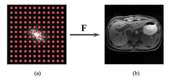
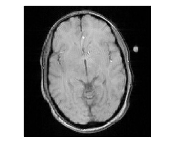
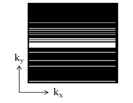
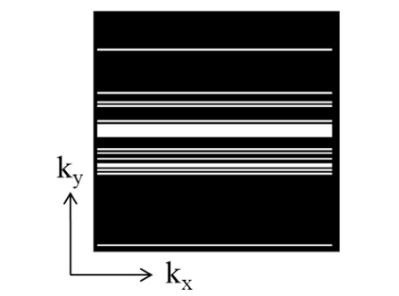
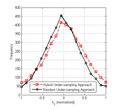
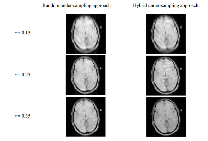
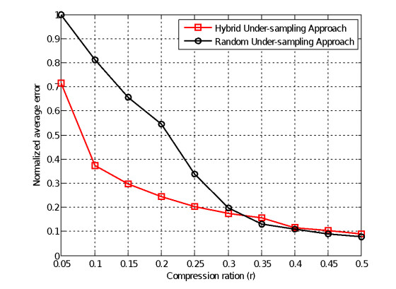
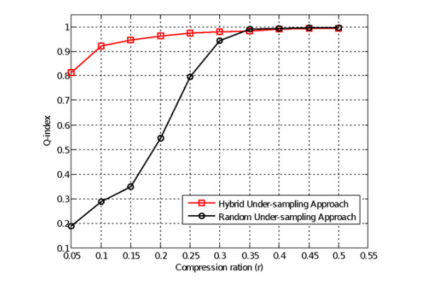
Compressive sampling (CS) has been commonly employed in the field of magnetic resonance imaging (MRI) to accurately reconstruct sparse and compressive signals. In a MR image, a large amount of encoded information focuses on the origin of the k-space. For the 2D Cartesian K-space MRI, under-sampling the frequency-encoding (kx) dimension does not affect to the acquisition time, thus, only the phase-encoding (ky) dimension can be exploited. In the traditional random under-sampling approach, it acquired Gaussian random measurements along the phaseencoding (ky) in the k-space. In this paper, we proposed a hybrid under-sampling approach; the number of measurements in (ky) is divided into two portions: 70% of the measurements are for random under-sampling and 30% are for definite under-sampling near the origin of the k-space. The numerical simulation consequences pointed out that, in the lower region of the under-sampling ratio r, both the average error and the universal image quality index of the appointed scheme are drastically improved up to 55 and 77% respectively as compared to the traditional scheme. For the first time, instead of using highly computational complexity of many advanced reconstruction techniques, a simple and efficient CS method based simulation is proposed for MRI reconstruction improvement. These findings are very useful for designing new MRI data acquisition approaches for reducing the imaging time of current MRI systems.
Citation: Anh Quang Tran, Tien-Anh Nguyen, Van Tu Duong, Quang-Huy Tran, Duc Nghia Tran, Duc-Tan Tran. MRI Simulation-based evaluation of an efficient under-sampling approach[J]. Mathematical Biosciences and Engineering, 2020, 17(4): 4048-4063. doi: 10.3934/mbe.2020224
Compressive sampling (CS) has been commonly employed in the field of magnetic resonance imaging (MRI) to accurately reconstruct sparse and compressive signals. In a MR image, a large amount of encoded information focuses on the origin of the k-space. For the 2D Cartesian K-space MRI, under-sampling the frequency-encoding (kx) dimension does not affect to the acquisition time, thus, only the phase-encoding (ky) dimension can be exploited. In the traditional random under-sampling approach, it acquired Gaussian random measurements along the phaseencoding (ky) in the k-space. In this paper, we proposed a hybrid under-sampling approach; the number of measurements in (ky) is divided into two portions: 70% of the measurements are for random under-sampling and 30% are for definite under-sampling near the origin of the k-space. The numerical simulation consequences pointed out that, in the lower region of the under-sampling ratio r, both the average error and the universal image quality index of the appointed scheme are drastically improved up to 55 and 77% respectively as compared to the traditional scheme. For the first time, instead of using highly computational complexity of many advanced reconstruction techniques, a simple and efficient CS method based simulation is proposed for MRI reconstruction improvement. These findings are very useful for designing new MRI data acquisition approaches for reducing the imaging time of current MRI systems.

| [1] |
P. C. Lauterbur, Image formation by induced local interactions: Examples employing nuclear magnetic resonance, Nature, 242 (1973), 190-191. doi: 10.1038/242296a0

|
| [2] | D. J. Larkman, R. G. Nunes, Parallel magnetic resonance imaging, Phys. Med. Biol., 52 (2007), R15-R55. |
| [3] |
K. P. Pruessmann, M. Weiger, M. B. Scheidegger, P. Boesiger, SENSE: Sensitivity encoding for fast MRI, Magnet. Reson. Med., 42 (1999), 952-962. doi: 10.1002/(SICI)1522-2594(199911)42:5<952::AID-MRM16>3.0.CO;2-S
|
| [4] | M. A. Griswold, P. M. Jakob, R. M. Heidemann, M. Nittka, V. Jellus, J. Wang, et al., Generalized autocalibrating partially parallel acquisitions (GRAPPA), Magnet. Reson. Med., 47 (2002), 1202-1210. |
| [5] | P. Kazmierczak, D. Theisen, K. Thierfelder, W. Sommer, M. Reiser, M. Notohamiprodjo, et al., Improved detection of hypervascular liver lesions with CAIPIRINHA-Dixon-TWIST-volume-interpolated breath-hold examination, Invest. Radiol., 50 (2014), 153-160. |
| [6] | W. A. Willinek, D. R. Hadizadeh, M. von Falkenhausen, H. Urbach, R. Hoogeveen, H. H. Schild, et al., 4D time-resolved MR angiography with keyhole (4D-TRAK): more than 60 times accelerated MRA using a combination of CENTRA, keyhole, and SENSE at 3.0T, J. Magn. Reson. Imaging, 27 (2008), 1455-1460. |
| [7] | J. H. Yoon, J. M. Lee, M. H. Yu, E. J. Kim, J. K. Han, Triple arterial phase MR imaging with gadoxetic acid using a combination of contrast enhanced time robust angiography, keyhole, and viewsharing techniques and two-dimensional parallel imaging in comparison with conventional single arterial phase, Korean. J. Radiol., 4 (2016), 522-532. |
| [8] |
T. A. Hope, M. Saranathan, I. Petkovska, B. A. Hargreaves, R. J. Herfkens, S. S. Vasanawala, Improvement of gadoxetate arterial phase capture with a high spatio-temporal resolution multiphase three-dimensional SPGR-Dixon sequence, J. Magn. Reson. Imaging, 38 (2013), 938-945. doi: 10.1002/jmri.24048
|
| [9] | D. L. Donoho, Compressed sensing, IEEE Trans. Inf. Theory, 52 (2006), 1289-1306. |
| [10] | F. Ong, R. Heckel, K. Ramchandran, A Fast and Robust Paradigm for Fourier Compressed Sensing Based on Coded Sampling, in ICASSP 2019-2019 IEEE International Conference on Acoustics, Speech and Signal Processing (ICASSP), 2019. |
| [11] |
Y. Li, R. Yang, Z. Zhang, Y. Wu, Chaotic-like k-space trajectory for compressed sensing MRI, J. Med. Imaging. Health. Inform., 5 (2015), 415-421. doi: 10.1166/jmihi.2015.1408
|
| [12] | D. V. Phong, N. Linh-Trung, T. D. Tan, H. V. Le, M. N. Do, Fast Image Acquisition in Magnetic Resonance Imaging by Chaotic Compressed Sensing, in 2011 IEEE International Symposium on Biomedical Imaging: From Nano to Macro, 2011. |
| [13] | T. Tran Duc, P. Dinh Van, C. Truong Minh, L. T. Nguyen, Accelerated Parallel Magnetic Resonance Imaging with Multi-Channel Chaotic Compressed Ssensing, in The 2010 International Conference on Advanced Technologies for Communications, 2010. |
| [14] |
M. Lustig, D. Donoho, J. M. Pauly, Sparse MRI: The application of compressed sensing for rapid MR imaging, Magnet. Reson. Med., 58 (2007), 1182-1195. doi: 10.1002/mrm.21391
|
| [15] |
G. Wang, Y. Bresler, V. Ntziachristos, Guest editorial compressive sensing for biomedical imaging, IEEE Trans. Med. Imaging, 30 (2011), 1013-1016. doi: 10.1109/TMI.2011.2145070
|
| [16] |
J. A. Tropp, A. C. Gilbert, Signal recovery from random measurements Vvia orthogonal matching pursuit, IEEE Trans. Inf. Theory, 53 (2007), 4655-4666. doi: 10.1109/TIT.2007.909108
|
| [17] |
E. J. Candes, J. Romberg, T. Tao, Robust uncertainty principles: Exact signal reconstruction from highly incomplete frequency information, IEEE Trans. Inf. Theory, 52 (2006), 489-509. doi: 10.1109/TIT.2005.862083
|
| [18] |
K. H. Jin, D. Lee, J. C. Ye, A general Fframework for compressed sensing and parallel MRI Uusing a filter based low-rank hankel matrix, IEEE Trans. Comput. Imaging, 2 (2016), 480-495. doi: 10.1109/TCI.2016.2601296
|
| [19] | G. Yang, S. Yu, H. Dong, G. Slabaugh, P. L. Dragotti, X. Ye, et al., DAGAN: Deep De-aliasing generative adversarial networks for fast compressed sensing MRI reconstruction, IEEE Trans. Med. Imaging, 37 (2018), 1310-1321. |
| [20] | S. Yu, H. Dong, G. Yang, G. Slabaugh, P. Dragotti, X. Ye, et al., Deep De-aliasing for fast compressive sensing MRI, arXiv: 1705.07137. |
| [21] | J. Schlemper, G. Yang, P. Ferreira, A. Scott, L. A. McGill, Z. Khalique, et al., Stochastic Deep Compressive Sensing for the Reconstruction of Diffusion Tensor Cardiac MRI, in Medical Image Computing and Computer Assisted Intervention, (2018), 295-303. |
| [22] | C. Wang, G. Papanastasiou, S. Tsaftaris, G. Yang, C. Gray, D. Newby, et al., TPSDicyc: Improved deformation invariant cross-domain medical image synthesis, in Machine Learning for Medical Image Reconstruction, Springer, Cham, (2019), 245-254. |
| [23] | J. Zhu, G. Yang, P. Lio, Lesion focused super resolution, in Medical Imaging 2019: Image Processing, SPIE, (2019), 109491L. |
| [24] | J. Zhu, G. Yang, P. Lio, How can we make GAN perform better in single medical image super-resolution? A lesion focused multi-scale approach, in 2019 IEEE 16th International Symposium on Biomedical Imaging (ISBI 2019), IEEE, 2019. |
| [25] | K. Thurnhofer-Hemsi, E. López-Rubio, E. Domínguez, R. M. Luque-Baena, N. Roé-Vellvé, Deep learning-based super-resolution of 3D magnetic resonance images by regularly spaced shifting, Neurocomputing, 398 (2020), 314-327. |
| [26] | M. Seitzer, G. Yang, J. Schlemper, O. Oktay, T. Würfl, V. Christlein, et al., Adversarial and Perceptual Refinement for Compressed Sensing MRI Reconstruction, MICCAI 2018, Springer International Publishing, (2018), 232-240. |
| [27] |
L. Feng, T. Benkert, K. T. Block, D. K. Sodickson, R. Otazo, H. Chandarana, Compressed sensing for body MRI, J. Magn. Reson. Imaging, 45 (2017), 966-987. doi: 10.1002/jmri.25547
|
| [28] | M. Sandilya, S. R. Nirmala, Compressed sensing trends in magnetic resonance imaging, Eng. Sci. Technol. Int. J., 20 (2017), 1342-1352. |
| [29] |
E. J. Candes, T. Tao, Decoding by linear programming, IEEE Trans. Inf. Theory, 51 (2005), 4203-4215. doi: 10.1109/TIT.2005.858979
|
| [30] |
E. Candès, J. Romberg, Sparsity and incoherence in compressive sampling, Inverse Probl., 23 (2007), 969-985. doi: 10.1088/0266-5611/23/3/008
|
| [31] |
J. P. Haldar, D. Hernando, Z. Liang, Compressed-sensing MRI with random encoding, IEEE Trans. Med. Imaging, 30 (2011), 893-903. doi: 10.1109/TMI.2010.2085084
|
| [32] | M. Lustig, D. L. Donoho, J. M. Santos, J. M. Pauly, Compressed sensing MRI, IEEE Signal Proc. Mag., 25 (2008), 72-82. |
| [33] |
J. H. Yoon, M. D. Nickel, J. M. Peeters, J. M. Lee, Rapid imaging: Recent advances in abdominal MRI for reducing acquisition time and its clinical applications, Korean J. Radiol., 20 (2019), 1597-1615. doi: 10.3348/kjr.2018.0931
|
| [34] |
F. Kong, Comparison of reconstruction algorithm for compressive sensing magnetic resonance imaging, Multimed. Tools. Appl., 77 (2018), 22617-22628. doi: 10.1007/s11042-017-4985-2
|
| [35] |
F. Wen, L. Pei, Y. Yang, W. Yu, P. Liu, Efficient and robust recovery of sparse signal and image using generalized nonconvex regularization, IEEE Trans. Comput. Imaging, 3 (2017), 566-579. doi: 10.1109/TCI.2017.2744626
|
| [36] |
W. Zhou, A. C. Bovik, A universal image quality index, IEEE Signal Proc. Lett., 9 (2002), 81-84. doi: 10.1109/97.995823
|
Figures(11) / Tables(1)

Anh Quang Tran, Tien-Anh Nguyen, Van Tu Duong, Quang-Huy Tran, Duc Nghia Tran, Duc-Tan Tran. MRI Simulation-based evaluation of an efficient under-sampling approach[J]. Mathematical Biosciences and Engineering, 2020, 17(4): 4048-4063. doi: 10.3934/mbe.2020224


 DownLoad: