


Citation: Álvaro Guzmán Aponte, María A Llano Ramírez, Yuliana Cadavid Mora, Juan F Santa Marín, Robison Buitrago Sierra. Cerium oxide nanoparticles for color removal of indigo carmine and methylene blue solutions[J]. AIMS Materials Science, 2020, 7(4): 468-485. doi: 10.3934/matersci.2020.4.468
| [1] | Ririn Cahyanti, Sumari Sumari, Fauziatul Fajaroh, Muhammad Roy Asrori, Yana Fajar Prakasa . Fe-TiO2/zeolite H-A photocatalyst for degradation of waste dye (methylene blue) under UV irradiation. AIMS Materials Science, 2023, 10(1): 40-54. doi: 10.3934/matersci.2023003 |
| [2] | Anne Heponiemi, Said Azalim, Tao Hu, Tuomas Vielma, Ulla Lassi . Efficient removal of bisphenol A from wastewaters: Catalytic wet air oxidation with Pt catalysts supported on Ce and Ce–Ti mixed oxides. AIMS Materials Science, 2019, 6(1): 25-44. doi: 10.3934/matersci.2019.1.25 |
| [3] | Carlos N. Kabengele, Giresse N. Kasiama, Etienne M. Ngoyi, Clement L. Inkoto, Juvenal M. Bete, Philippe B. Babady, Damien S. T. Tshibangu, Dorothée D. Tshilanda, Hercule M. Kalele, Pius T. Mpiana, Koto-Te-Nyiwa Ngbolua . Biogenic synthesis, characterization and effects of Mn-CuO composite nanocatalysts on Methylene blue photodegradation and Human erythrocytes. AIMS Materials Science, 2023, 10(2): 356-369. doi: 10.3934/matersci.2023019 |
| [4] | Muhammad Yakob, Hamdani Umar, Puji Wahyuningsih, Rachmad Almi Putra . Characterization of microstructural and optical CoFe2O4/SiO2 ferrite nanocomposite for photodegradation of methylene blue. AIMS Materials Science, 2019, 6(1): 45-51. doi: 10.3934/matersci.2019.1.45 |
| [5] | Khang Duy Vu Nguyen, Khoa Dang Nguyen Vo . Magnetite nanoparticles-TiO2 nanoparticles-graphene oxide nanocomposite: Synthesis, characterization and photocatalytic degradation for Rhodamine-B dye. AIMS Materials Science, 2020, 7(3): 288-301. doi: 10.3934/matersci.2020.3.288 |
| [6] | Mohamed Jaffer Sadiq Mohamed, Denthaje Krishna Bhat . Novel ZnWO4/RGO nanocomposite as high performance photocatalyst. AIMS Materials Science, 2017, 4(1): 158-171. doi: 10.3934/matersci.2017.1.158 |
| [7] | Denise Arrozarena Portilla, Arturo A. Velázquez López, Rosalva Mora Escobedo, Hernani Yee Madeira . Citrate coated iron oxide nanoparticles: Synthesis, characterization, and performance in protein adsorption. AIMS Materials Science, 2024, 11(5): 991-1012. doi: 10.3934/matersci.2024047 |
| [8] | Haiqing Yao, Fei Li, Jodie Lutkenhaus, Masaya Kotaki, Hung-Jue Sue . High-performance photocatalyst based on nanosized ZnO-reduced graphene oxide hybrid for removal of Rhodamine B under visible light irradiation. AIMS Materials Science, 2016, 3(4): 1410-1425. doi: 10.3934/matersci.2016.4.1410 |
| [9] | Kah Hon Leong, Azrina Abd Aziz, Yee Li Kang, Sheau Wei Goh, Kulhar Vijay Singh, Lan Ching Sim, Pichiah Saravanan . Synergized mechanistic and solar photocatalysis features of N-TiO2 functionalised activated carbon. AIMS Materials Science, 2017, 4(3): 800-813. doi: 10.3934/matersci.2017.3.800 |
| [10] | Amany Mahmoud, Ehssan Nassef, Hesham Salah, Yehia El-taweel . Use of hydrazide derivative of poly methylacrylate for the removal of cupric ions from solutions. AIMS Materials Science, 2020, 7(4): 420-430. doi: 10.3934/matersci.2020.4.420 |
Dyes are colored substances and they are solubilized during the application and impart color by selective absorption of light [1]. Dye molecules consist of a chromophore component (largely responsible for producing the color) and auxochrome component (as a supplement to the chromophore but also render the molecule soluble in water and improves its attachment toward the fibers) [2]. Yagub et al. [3] mentioned that dyes can be classified based on their particle charge upon dissolution in aqueous application. The dyes can be grouped into cationic (basic dyes), anionic (direct, acid and reactive) and non-ionic (dispersed).
Textile, cosmetic and paper industry use dyes as coloring agents. Five industries are known to be responsible for the presence of dye effluents in the environment: textile (54%), dyeing (21%), paper and pulp (10%), tannery and paint (8%) and dye manufacturers (7%) [4]. Therefore, the textile industry generates more than half of dye effluents, with a worldwide dye discharge estimated of 280,000 ton/year as mentioned by Jin et al. [5].
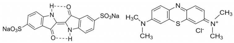
Indigo carmine (IC) and methylene blue (MB) are common dyes used as dyeing agents in the textile industry. They are anionic and cationic dyes, respectively [6,7,8,9]. Figure 1 shows the chemical structure of indigo carmine and methylene blue dyes.
The processes for color removal can be classified into physical, chemical and biological [2,4,12]; however, Hao et al. [12] have also mentioned the electrical process. Biological and physical methods are considered as the most efficient dye removal processes [2,4]. Murugan et al. [9] mentioned that most of methods adopted for removal of compounds (organic and inorganic) from the wastewater are expensive and not suitable for small scale industries. Moreover, photocatalysis (chemical treatment) is a high effective and low-cost process compared to other methods [9,13]. This method was applied by Frank and Bard [14] to degrade pollutants in aqueous solution and allowed the theorical foundation for photocatalytic oxidation technology in wastewater treatment. Furthermore, some authors have reported photocatalytic studies in this field [15,16,17,18]. It has been reported that the photocatalytic process involves three steps. Firstly, absorption of photons with energy larger than the band gap of a photocatalyst. Secondly, the generation, separation, migration or recombination of electron-hole pairs photo generated and finally, the redox reactions at the photocatalyst surface [9].
The photocatalytic activity of some semiconductors as titanium dioxide [15,16,19], zinc oxide [19,20,21], zirconium oxide [22], tin oxide [23], cadmium sulfide [24] and bismuth compounds [25,26], has been studied under light irradiation. CeO2 (ceria) is an n-type semiconductor with a wide bandgap [13] and remarkable features such as chemical stability, low-cost and low-toxicity [27,28]. It can be used in a wide range of different applications such as Solid Oxide Fuel Cells (SOFC) [13,29], oxidation of Volatile Organic Compounds (VOCs) [13,30], aerobic oxidation of alcohols [31], photodegradation of toluene gas [32,33], biomedical applications [13,34] and photocatalyst in wastewater treatment [13,32,35].
Cerium oxide has been used by several authors to degrade dyes. Sane et al. [27] reported > 95% removal of reactive dyes (reactive green-19, reactive orange-84 and reactive yellow-81) under visible light within 240 min by using CeO2 synthesized by precipitation. Zheng et al. [35] reported the adsorption capacity of CeO2 nanoparticles for Congo red (CR) is closely related to its morphology. In their work, the adsorption performance was attributed to the structure and presence of electrostatic interactions between the nanoparticles surface and dye molecules.
In addition, cerium oxide has been also used by diverse authors to degrade methylene blue. However, there is still a controversy in the literature about the effect of those particles in the dye degradation. Miyauchi et al. [36] and Kumar et al. [17] have mentioned that CeO2 thin films are inactive for photocatalytic degradation of methylene blue. Tuyen et al. [37] reported that CeO2 could not catalyze the photodegradation of MB. Majumder et al. [38] studied CeO2 nanoparticles with different morphologies as catalyst for degradation of MB at pH 3. They found that hexagonal and rectangular shapes had not a good catalytic performance even after 200 min of UV irradiation. However, CeO2 nanoparticles with square shape showed excellent photocatalytic performance with complete degradation of MB (4.6 μM) within 175 min.
However, other authors have reported the effectiveness of CeO2 NPs in the degradation of MB dye. Pouretedal and Kadkhodaie [39] studied the degradation efficiency of MB (20 mg/L) catalyzed by CeO2 nanoparticles (1.0 g/L) at different pH values, and reported 85% of dye degradation at pH 11 within 175 min. Zhang et al. [40] studied fly ash cenospheres (FACs)-supported CeO2 composite (CeO2/FACs) and obtained a promising catalyst for photocatalytic decolorization of MB. A color removal up to 60% after irradiation for 300 min was achieved. In a recent work, Murugan et al. [9] have mentioned than an enhanced photodegradation of MB can be found by using CeO2 nanoparticles doped with alkaline metal ion (Mg, Ca, Sr, Ba) compared to pure CeO2 nanoparticles. Yang et al. [28] mentioned that, calcination temperature for synthesis of CeO2 nanofibers catalyst has a positive effect for the photocatalytic performance. They reported that the photodegradation rate of MB increases from 67% to 98% for CeO2 nanofibers obtained at 500 and 800 ℃, respectively.
Moreover, based on reported literature, cerium oxide has been used to degrade several dyes, but limited work has been done and focused on the degradation of indigo carmine solutions by using of CeO2. Indigo carmine is one of the oldest and most important dyes used as dyeing agent of clothes (blue jeans) and other blue denim products [41,42,43,44,45,46,47]. To the best of the authors knowledge the work of Liyanage et al. [48] is the only article available in the literature related to degradation of indigo carmine by using cerium oxide. They found that yttrium-doped ceria nanorods displayed a better photocatalytic activity for the degradation of indigo carmine dye solution at room temperature under UV irradiation, compared to the pure ceria.
Summarizing, only a few of authors have studied the removal of color from indigo carmine solutions using cerium oxide and the materials used in that paper are not nanoparticles but nanorods. In addition, as shown in Table 1, several references were carefully reviewed and there is no consensus in the literature to conclude that cerium oxide particles are useful to degrade dye solutions. Accordingly, the aim of this article is to determine the ability of ceria nanoparticles synthesized via sol-gel method to remove and/or degrade methylene blue and indigo carmine, by modifying the pH of the dye solution. Moreover, the data reported in this article provides useful information related to the use of cerium oxide to degrade methylene blue since there is not agreement in the literature.
Cerium oxide nanoparticles were prepared via sol-gel method, following the procedure mentioned by He et al. [49] with some modifications. The chemical reagents used were cerium (III) nitrate hexahydrate (Ce(NO3)3·6H2O, ≥ 98.5%, Chemí) as cerium oxide precursor, urea (CO(NH2)2, ≥ 99.5%, Carlo Erba) and polyvinylpyrrolidone ((C6H9NO)n, ≥ 95.00%, M.W. 58,000, Alfa Aesar) as surfactant. The desired concentration of the reagents was dissolved into deionized water (conductivity lower than 0.4 μS) in a beaker to produce a clear solution. The solution was stirred at 300 rpm (magnetic stirrer) during 15 min and subsequently, the temperature of the solution was increased from room temperature to 90 ℃ and it was kept for up to 3 h. The pH was controlled between 8.2–8.5 (by adding dropwise ammonia with a burette) for about 4 h. A color variation phenomenon was observed, and it was correlated with chemical changes as mentioned by He et al. [49]. All mentioned chemical reagents were analytical grade and they were used without any further purification.
The solvents were eliminated from the solution by rotary evaporation at 85 rpm and 85 ℃ during 4 h. Nanoparticles (3 g) were added in 40 mL of deionized water (DI) or ethanol (EtOH). Later, the nanoparticles were separated by sonication and centrifugation at 10,000 rpm during 30 min/cycle to remove traces of ammonia and cerium nitrate. The resulting precipitates were drained and dried in an oven (Binder KB 115) at 80 ℃ overnight. The resulting particles were calcined at 500 ℃ in a tube furnace (Nabertherm P330) for 2 h.
The structural properties of CeO2 NPs were determined by X-ray diffraction (XRD) with a PANalytical X’Pert PRO diffractometer. The samples were scanned in the range of 2θ = 20–80° at a scanning speed of 0.02 °/s, using Cu Kα radiation at 45 kV and 40 mA. The crystallite size was calculated by using Scherrer’s formula [50] and the most intense peak of the diffractogram (111) was used for this calculation. The surface chemistry of CeO2 NPs was analyzed by using an X-ray photoelectron spectroscopy (XPS, SPECS) with a PHOIBOS 150 1D-DLD analyzer and monochromatic Al Kα radiation (13 kV, 1487 eV and 100 W). The calibration and correction of binding energy were accomplished by assuming the binding energy of the adventitious carbon (C 1s) to 284.6 eV. The relative concentration of the cations Ce3+ and Ce4+ were calculated as [51]:
| [Ce3+]=Av0+Av′+Au0+Au′Av0+Av′+Au0+Au′+Av+Av″+Av‴+Au+Au″+Au‴ | (1) |
| [Ce4+]=1−[Ce3+]=Av+Av″+Av‴+Au+Au″+Au‴Av0+Av′+Au0+Au′+Av+Av″+Av‴+Au+Au″+Au‴ | (2) |
Where Ai is the integrated area of peak “i”, u and v indicate the spin-orbit coupling states of 3d3/2 and 3d5/2, respectively. v0, v’, u0 and u’ are characteristic peaks of Ce3+; while v, v’’, v’’’, u, u’’, and u’’’ are characteristic of Ce4+.
The morphology of the nanoparticles was studied by using Field-Emission Scanning Electron Microscopy (FEG-SEM) (JEOL JSM-7100 F). The particles were measured using several SEM images and the size distribution of the nanoparticles was determined by using ImageJ free software. In addition, an elemental analysis of nanoparticles was performed using Energy-Dispersive Spectrometry (EDS) coupled to the SEM.
The presence of functional groups in ceria nanoparticles was investigated by using Fourier Transform Infrared Spectroscopy (FTIR) in a Shimadzu IR Tracer-100 spectrometer with wave number range 4000–400 cm−1 and resolution 4 cm−1. The samples were prepared by using conventional KBr. Zeta potential of nanoparticles was measured by using a Zetasizer Nanoplus 3HD instrument and the absorption spectra was determined on a UV-vis Agilent 8453 spectrophotometer, in the range of wavelengths between 200–900 nm. For both tests (Z potential and UV) a suspension with a concentration around of 1000 ppm was previously prepared and sonicated for 15 min.
Photocatalytic activity of CeO2 nanoparticles was studied for Indigo Carmine (IC) and Methylene Blue (MB) dye solutions (30 ppm in all cases). During color removal tests, 60 mg of ceria nanoparticles were added to 100 mL of dye solution. In this study, the concentration of NPs was fixed at 600 ppm since it is a medium value reported in the literature by other authors [16,35]. The pH of the dye solution is an important parameter during the adsorption process because it can alter the surface charge of the adsorbent and the dyes ionization degree [35]. Accordingly, the effect of the dye solution pH (2.5, 8.0 and 10.0) on the IC and MB degradation by CeO2 NPs was studied. The pH values were adjusted using 0.1 M NaOH and 0.1 M HCl. Moreover, the degradation of IC dye was carried out in absence of ceria nanoparticles under UV irradiation (photolysis) at the unmodified pH of the solution (pH = 5.6).
During the degradation tests, the solution was controlled, kept at room temperature (298 ± 1 K) and magnetically stirred (300 rpm). Prior to irradiation, the nanoparticles were added to the dye solution to reach an adsorption-desorption equilibrium (60 min under dark and previous UV irradiation). A lamp with 15 W (Lumek) was used as a UV-C source to trigger the photocatalytic reaction. This UV source covers the wavelength range of 100–280 nm, in accordance to suitable radiation < 420 nm to transfer electrons from the valance band to the conduction band mentioned by Pouretedal and Kadkhodaie [39].
The distance between the UV source and the beaker containing the solution was controlled to 10 cm [9,17]. Aliquots were taken at pre-set time intervals during irradiation and ceria nanoparticles were separated by centrifugation at 10,000 rpm for 15 min. The concentration of remaining IC and MB in the supernatant was monitored by measuring the absorbance at λmax 610 and 663 nm, respectively, by UV-vis spectroscopy (Agilent 8453 spectrophotometer). The color removal of dye solution was calculated by the equation reported by Ameen et al. [52], as follows:
| colorremoval(%)=((C0−C)C0)x100=((A0−A)A0)x100 | (3) |
Where C0 and C refers to initial and variable concentrations of dye solution, respectively. A0 represents the initial absorbance and A at pre-set time.
The XRD patterns of synthesized CeO2 nanoparticles can be observed in Figure 2. The CeO2 nanoparticles exhibited diffraction peaks at 28.54°, 33.07°, 47.55°, 56.22°, 59.05°, 69.26°, 76.75° and 79.03° attributed to cerium oxide phase. The particles have an ordered structure as indicated by the sharp diffraction peaks. The lattice parameter (a0=5.41 ˙A) is similar to reported for CeO2 (a0=5.41 ˙A) in the standard data (ICSD 072155), and its corresponding interplanar distance (d) and crystallite size (D) are 0.31 nm and 13.50 nm, respectively. These values are close to those reported by Andreescu et al. [53], Phoka et al. [54] and Murugan et al. [9]. No other phases were detected indicating the CeO2 NPs are chemically pure.
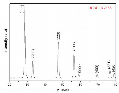
Figure 3 shows the FTIR spectra of the CeO2 nanoparticles. Different signals related to the presence of functional groups can be observed from the spectra. Two bands at 461 and 420 cm−1 corresponding to the Ce-O stretching vibration can be observed. This in agreement with the reports by Calvache-Muñoz et al. [55] and Miri and Sarani [56]. The band at 959 cm−1 could be ascribed to the vibrational stretching mode of H2O [55].
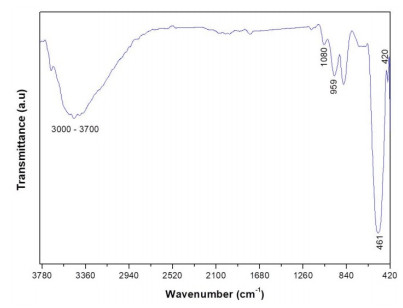
The band at ≈ 1080 cm−1 has been assigned to ν(Ce-O-Ce) vibration [56]. A broad band in the range of 3000–3700 cm−1 is associated to the O-H stretching vibration coming from absorbed water and/or hydroxyl groups [55,56,57].
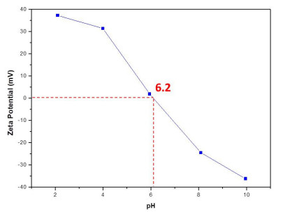
The point of zero charge (pzc) of CeO2 NPs was found at 6.2 (Figure 4). This result is similar to the value reported by Zheng et al. [35] which was 6.7. The pzc can be related with the adsorption capacity of the nanoparticles [35,58]. Consequently, higher adsorption capacity could be expected at lower pH of the solution (pH < pHpzc) because the electrostatic attraction between negative dye molecules and positive charged CeO2 NPs surface [35,58].
Kar et al. [34], Emsley [59], Singh et al. [60] and Dutta et al. [61] have reported that cerium can exist either in Ce(Ⅲ) or in Ce(Ⅳ) oxidation states, being Ce(Ⅲ) usually colorless and Ce(Ⅳ) turning from yellow to red in color. Accordingly, an approximation to the chemical state of Ce in the particles can be achieved from their UV absorption spectra. In this work the particles had a reddish color as seen in Figure 5.
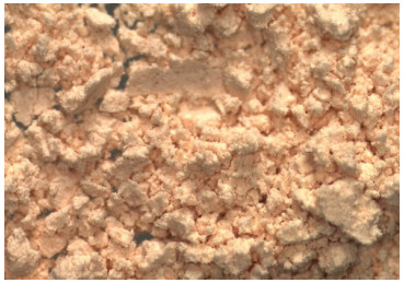
Figure 6 shows the UV-vis absorption spectra of CeO2 NPs as a function of time. The main absorption peak was observed with a shift from 311 nm (day 1) to around 327 nm (day 15). From the day 12 the main absorption peak remained stable (327 nm). Accordingly to Emsley [59], the characteristic absorption peak in the range between 300–400 nm corresponds to Ce(IV) state. This absorption peak is very close to the value (333 nm) reported by Miri and Sarani [56].
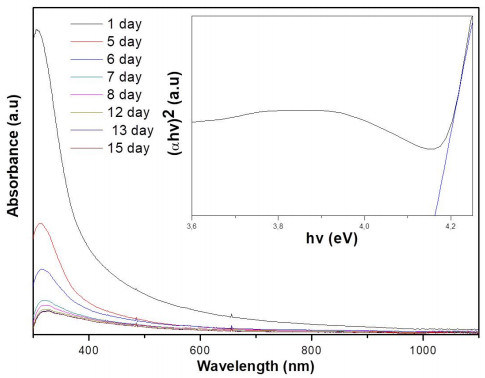
The indirect band gap energy (Eg) was calculated as the inset found with the Tauc-plot at the fifteenth day (Figure 6). The band gap energy (Eg) was determined by using the Tauc equation [62]:
| (αhv)2=A(hv−Eg) | (4) |
Where, α is the optical absorption coefficient, Eg is the direct band gap, h is the Planck constant, v is the frequency (hv = the photon energy) and A is a constant. The Eg was calculated by plotting (αhv)2 versus hv by extrapolating of the linear part of the curve to the (αhv)2 = 0. The observed band gap energy for CeO2 NPs at the fifteenth day was 4.16 eV. This bandgap value was higher than the value reported by Phoka et al. [54], Atla et al. [57], Mishra et al. [63] (Eg = 2.78–3.44 eV), but lower than reported by Gogoi and Sarma [62] (Eg = 4.91 eV). This increase in band gap value may be due to the charge transition of Ce ion (Ce(IV)) as mentioned by Phoka et al. [54].
The XPS spectra for the CeO2 nanoparticles of the Ce 3d and O 1s peaks are shown in Figure 7.
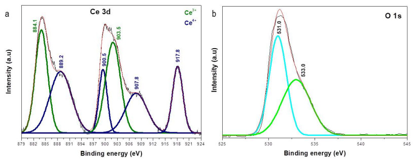
Deshpande et al. [51], Tuyen et al. [37], Kumar et al. [17] and Majumder et al. [38] reported the mix of valence states (Ce3+ and Ce4+) in CeO2 nanoparticles and CeO2 thin films. In this work, six characteristics peaks (Figure 7a) of Ce3+ at 884.1 and 903.5 eV, and of Ce4+ at 889.2,900.5,907.8 and 917.8 eV were found. The position of the peaks is according to the other XPS spectra reported in the literature [17,37,51]. These binding energies corresponded with v’, u’, v’’, u, u’’ and u’’’, respectively.
The relative concentration of Ce3+ and Ce4+ was calculated from the deconvoluted curves shown in Figure 7a. The calculations showed that CeO2 NPs have more Ce4+ (56.16%) than Ce3+ (43.84%). Truffault et al. [64] and Majumder et al. [38] have reported that a high relative concentration of Ce3+ and chemisorbed oxygen improved the photocatalytic activity of CeO2.
The XPS profile of O 1s (Figure 7b), evidenced two characteristic peaks attributed to lattice oxygen and chemisorbed oxygen (~ 531 and ~ 533 eV, respectively), as mentioned by Majumder et al. [38], the chemisorbed oxygen is directly proportional to the oxygen vacancies. From the deconvolution the calculated amount of lattice oxygen was 51.77% and chemisorbed oxygen 48.23%.
SEM images of synthesized ceria nanoparticles are shown in Figure 8. From this image, it could be observed that the CeO2 NPs exhibit a semi-spherical morphology and considerable agglomeration (Figure 8a, b). This phenomenon has been previously reported. He et al. [49] mentioned in their work that ceria particles tend to form agammaegation.
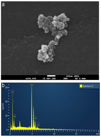
The EDS spectra confirmed the existence of Ce and O in NPs (Figure 8b). The atomic percentage of each element were 66.42 ± 3.34% and 33.58 ± 3.34% of O and Ce, respectively. This result supports the hypothesis of particles with a stoichiometry near to CeO2. Consequently, the presence of Ce (IV) can be inferred by analyzing together the results from the XPS and EDS.
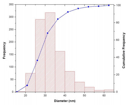
The particle size distribution for CeO2 (Figure 9) showed that the mean nanoparticle size was 33.3 ± 7.3 nm. Besides, the D10, D50 and D90 were 25.2 nm, 31.9 nm and 42.5 nm respectively.
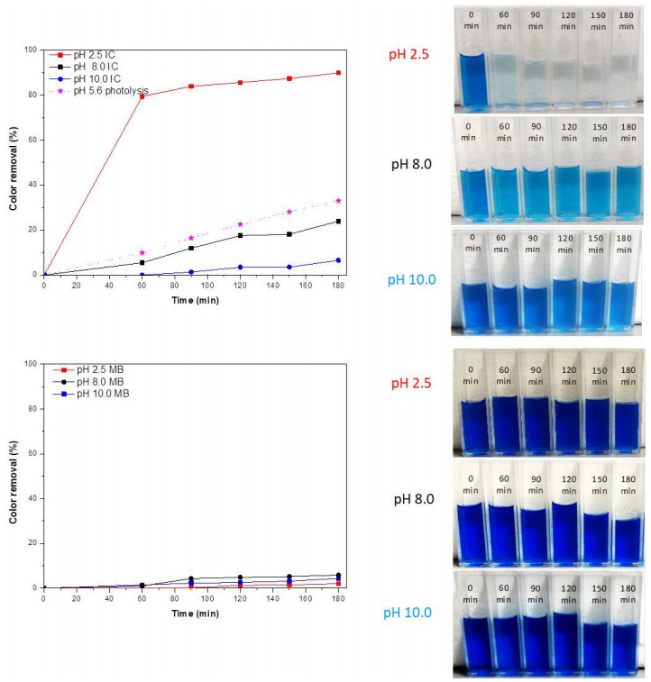
The color removal of IC and MB by CeO2 NPs can be observed in Figure 10. All samples were followed by 180 min and photography of the different solutions are included for the visual inspection. In photolysis conditions, the removal of the IC molecules occurred at a slow rate (33%) within 180 min. Compared with IC solution (pH 2.5), the color removal of the IC (pH 8.0 and 10.0) and MB (pH 2.5, 8.0 and 10.0) solutions were lower.
Some authors have mentioned the heterogeneous photocatalytic oxidation organic pollutants can be explained into five independent steps as follows: (1) transfer of the dye molecules from the liquid phase to the surface of CeO2; (2) adsorption of the dye molecules on the surface of CeO2; (3) the adsorbed dye pollutants react with reactive species in the adsorbed phase, where oxidation and reduction reactions occur once being excited; (4) desorption of the products from CeO2 surface; and (5) removal of the products form the interface region [18,65,66]. Ji et al. [58] also mentioned that the adsorption capacity on photocatalyst of dyes is a key factor for the degradation rate in photocatalytic system. For this reason, to explain the enhanced photocatalytic activity of CeO2 NPs, the adsorption of IC and MB on CeO2 NPs was examined at different pH.
Hao et al. [12] and Vautier et al. [41] have mentioned that photocatalytic oxidation is the main cause for color removal through degradation of chromophore structure. This can be explained by means of two possible mechanisms: a reductive [8] and an oxidative paths [67], that include the cleavage of the chromophore molecule bond. Generally speaking, the photocatalytic degradation of dyes and aromatic pollutants is caused by the degradation of the benzene rings and heteropolyaromatic linkage [39,41,65]. Some authors have shown that the nanoparticles contribute to oxidative cleavage of indigo carmine and consequently, the by-products would include carboxylic acids [10].
The highest color removal was 90%, 24% and 7% (at pH 2.5, 8.0 and 10.0 within 180 min, respectively) for IC, and 2%, 6% and 4% for MB (at pH 2.5, 8.0 and 10.0 within 180 min, respectively). In this case, CeO2 NPs exhibited a poor catalytic performance for IC even after 180 min under UV irradiation at pH 8.0 and 10.0, since the photocatalytic reaction take place in the third step (adsorbed phase). However, these results indicate the ceria NPs could be used as catalyst to photocatalytic degradation of IC dye at pH 2.5, enhancing the color removal about 2.72 times compared to photolysis conditions.
This behavior can be attributed to the electrostatic attraction between dye molecules and charged ceria NPs surface according to the results of Ji et al. [58] and Zheng et al. [35]. In aqueous solution, anionic dyes carry a net negative charge due to the presence of sulphonate groups (SO3-), while cationic dyes carry a net positive charge due to the presence of protonated amine or sulfur containing groups [67,68,69], and thereby, adsorption of anions is favored at pH < pHpzc, while the adsorption of cations is favored at pH > pHpzc. Therefore, a higher color removal capacity could be attained for IC at lower pH of solution (pH = 2.5 < pHpzc = 6.2) because the electrostatic attraction between negative dye molecules and positive charged ceria nanoparticles surface. Hence, a lower color removal capacity was attained for IC at higher pH of solution (pH = 8.0 or 10.0 > pHpzc = 6.2) even compared to photolysis conditions, because the electrostatic repulsion between negative dye molecules and negative charged ceria nanoparticles surface.
Is important to highlight that some authors have mentioned the CeO2 can be easily regenerated by a heat treatment (673 K in air for 2 h) and it can be reused even after four cycles, demonstrating good stability and reusability [35]. Majumder et al. [38] found that the photocatalytic efficiency of the CeO2 remains almost the same after three consecutive cycles which indicates its high stability.
A similar behavior was observed for MB at pH 2.5 (electrostatic repulsion); however, for MB at pH 10.0 greater color removal capacity was expected, but it was inhibited because hydroxyl ions compete with dye molecules in the adsorption process on the ceria nanoparticles surface, as mentioned by Pouretedal and Kadkhodaie [39]. Previous studies have indicated that there is still a controversy about the effect of using CeO2 in the MB dye degradation as shown in Table 1.
| Dye | Degradation (%) | Comment | Reference |
| Methylene blue | ≈95.0 | Dye concentration: 5 mg/L, catalyzed by CeO2 NPs obtained by precipitation and calcined at 600 ℃ (1 g/L) at pH 11 within 125 min | Pouretedal and Kadkhodaie [39] |
| Methylene blue | ≈60.0 | Dye concentration: 10 mg/L, catalyzed by CeO2 films coated on FACs (fly ash cenospheres) (4 g/L) within 300 min. | Zhang et al. [40] |
| ≈45.0 | Dye concentration: 10 mg/L, catalyzed by CeO2 NPs (0.6 g/L) within 300 min. | ||
| Methylene blue | ≈80.0 | Dye concentration: 10 mg/L, catalyzed by metal ion (barium) doped CeO2 (50 ppm) within 120 min. | Murugan et al. [9] |
| ≈30.0 | Dye concentration: 10 mg/L, catalyzed by CeO2 nanoparticles (50 ppm) within 120 min. | ||
| Methylene blue | 98.0 | Dye concentration: 30 mg/L, catalyzed by CeO2 nanofibers obtained at 800 ℃ (200 ppm) within 60 min. | Yang et al. [28] |
| Methylene blue | ≈100.0 | Dye concentration: 4.6 μM, catalyzed by CeO2 nanoparticles with square shape (4.6 μM) at pH 3 within 175 min. | Majumder et al. [38] |
| Methylene blue | Inability to decompose | Dye concentration: 1 × 10⁻³ mol/L, catalyzed by CeO2 films within 60 min. | Miyauchi et al. [36] |
| Methylene blue | ≈32.0 | Dye concentration: 5 mL (50 mM), and 10 mL of 30% H2O2 (50 mM) catalyzed by CeO2 (0.1 g) at room temperature within 60 min. | Gogoi and Sarma [62] |
| Methylene blue | ≈10.0 | Dye concentration: 15 mg/L, catalyzed by CeO2 (0.8 g/L) at neutral pH within 150 min. | Tuyen et al. [37] |
| Methylene blue | 18.9 | Dye concentration: 3.2 mg/L, catalyzed by CeO2 film (0.5 M) within 24 h. | Kumar et al. [17] |
| 2.1 | Dye concentration: 3.2 mg/L, catalyzed by CeO2 film (0.15 M) within 24 h. | ||
| Methylene blue | Poor photocatalytic performance (The peak intensity at 663 nm without change significantly) | Dye concentration: 4.6 μM, catalyzed by CeO2 nanoparticles with hexagonal and rectangular shape (4.6 μM) at pH 3 within 200 min. | Majumder et al. [38] |
| Methylene blue | 2, 6 and 4, respectively | Dye concentration: 30 mg/L, catalyzed by CeO2 nanoparticles (0.6 g/L) at pH 2.5, 8.0 and 10.0 respectively, within 120 min. | This work |
 DownLoad:
CSV
DownLoad:
CSV
This research confirmed that CeO2 NPs exhibits a poor catalytic performance even after 120 min under UV irradiation, in accordance with Miyauchi et al. [36], Tuyen et al. [37], Kumar et al. [17] and Majumder et al. [38]. However, as mentioned by Majumder et al. [38], the degradation of MB could be increased because of higher photocatalytic activity of CeO2 NPs (containing higher percentage of Ce3+ ions and oxygen vacancies that decrease the band gap). Thus, CeO2 NPs may be more easily photoactivated and suitable for removal of cationic dye effluents.
Based on the experimental results of this research, the following conclusions are drawn:
➢ Ceria nanoparticles photocatalyst was synthesized by sol-gel method and they were used for color removal indigo carmine and methylene blue dye solutions at different pH under UV irradiation.
➢ The color removal performance was strongly affected to the electrostatic attraction between dye molecules and the charge at the surface of ceria nanoparticles.
➢ CeO2 NPs exhibit more color removal for IC dye (≈ 90%, 24% and 7% at pH 2.5, 8.0 and 10.0 within 180 min, respectively) compared to MB dye (≈ 2%, 6% and 4% at pH 2.5, 8.0 and 10.0 within 180 min, respectively).
The authors would like to thank the Grupo de Investigación en Materiales Avanzados y Energía MATyER (Advanced materials and energy research group (MATyER)) of Instituto Tecnológico Metropolitano de Medellín (Metropolitan Technological Institute from Medellín) and the Centro Administrativo de Ciencia, Tecnología e Innovación (Department of Science, Technology and Innovation; COLCIENCIAS-Colombia) and its program of Postdoctoral research stay No. 811 for all their support. The authors would also like to acknowledge J.A.C Cornelio for proving support for XPS measurements.
All authors declare no conflicts of interest in this paper.
| [1] | Gürses A, Açıkyıldız M, Güneş K, et al. (2016) Dyes and pigments: their structure and properties. Dyes Pigments 2016: 13-29. |
| [2] |
Adegoke KA, Bello OS (2015) Dye sequestration using agricultural wastes as adsorbents. Water Resour Ind 12: 8-24. doi: 10.1016/j.wri.2015.09.002

|
| [3] |
Yagub MT, Sen TK, Afroze S, et al. (2014) Dye and its removal from aqueous solution by adsorption: A review. Adv Colloid Interface Sci 209: 172-184. doi: 10.1016/j.cis.2014.04.002
|
| [4] |
Katheresan V, Kansedo J, Lau SY (2018) Efficiency of various recent wastewater dye removal methods: A review. J Environ Chem Eng 6: 4676-4697. doi: 10.1016/j.jece.2018.06.060
|
| [5] |
Jin XC, Liu GQ, Xu ZH, et al. (2007) Decolorization of a dye industry effluent by Aspergillus fumigatus XC6. Appl Microbiol Biot 74: 239-243. doi: 10.1007/s00253-006-0658-1
|
| [6] |
Ammar S, Abdelhedi R, Flox C, et al. (2006) Electrochemical degradation of the dye indigo carmine at boron-doped diamond anode for wastewaters remediation. Environ Chem Lett 4: 229-233. doi: 10.1007/s10311-006-0053-2
|
| [7] | Quintero L, Cardona S (2010) Technologies for the decolorization of dyes: Indigo and indigo carmine. Dyna 77: 371-386. |
| [8] |
Li HX, Xu B, Tang L, et al. (2015) Reductive decolorization of indigo carmine dye with Bacillus sp. MZS10. Int Biodeter Biodegr 103: 30-37. doi: 10.1016/j.ibiod.2015.04.007
|
| [9] |
Murugan R, Kashinath L, Subash R, et al. (2018) Pure and alkaline metal ion (Mg, Ca, Sr, Ba) doped cerium oxide nanostructures for photo degradation of methylene blue. Mater Res Bull 97: 319-325. doi: 10.1016/j.materresbull.2017.09.026
|
| [10] |
Chacón-Patiño ML, Blanco-Tirado C, Hinestroza JP, et al. (2013) Biocomposite of nanostructured MnO2 and fique fibers for efficient dye degradation. Green Chem 15: 2920-2928. doi: 10.1039/c3gc40911b
|
| [11] |
de Oliveira Brito SM, Andrade HMC, Soares LF, et al. (2010) Brazil nut shells as a new biosorbent to remove methylene blue and indigo carmine from aqueous solutions. J Hazard Mater 174: 84-92. doi: 10.1016/j.jhazmat.2009.09.020
|
| [12] |
Hao OJ, Kim H, Chiang PC (2000) Decolorization of wastewater. Crit Rev Env Sci Tec 30: 449-505. doi: 10.1080/10643380091184237
|
| [13] |
Montini T, Melchionna M, Monai M, et al. (2016) Fundamentals and catalytic applications of CeO2-based materials. Chem Rev 116: 5987-6041. doi: 10.1021/acs.chemrev.5b00603
|
| [14] |
Frank SN, Bard AJ (1977) Heterogeneous photocatalytic oxidation of cyanide ion in aqueous solutions at titanium dioxide powder. J Am Chem Soc 99: 303-304. doi: 10.1021/ja00443a081
|
| [15] |
Cho IH, Zoh KD (2007) Photocatalytic degradation of azo dye (Reactive Red 120) in TiO2/UV system: Optimization and modeling using a response surface methodology (RSM) based on the central composite design. Dyes Pigments 75: 533-543. doi: 10.1016/j.dyepig.2006.06.041
|
| [16] | Bansal P, Sud D (2013) Photocatalytic degradation of commercial dye, CI Reactive Red 35 in aqueous suspension: Degradation pathway and identification of intermediates by LC/MS. J Mol Catal A-Chem 374: 66-72. |
| [17] |
Kumar V, Chen W, Zhang X, et al. (2019) Properties and performance of photocatalytic CeO2, TiO2, and CeO2-TiO2 layered thin films. Ceram Int 45: 22085-22094. doi: 10.1016/j.ceramint.2019.07.225
|
| [18] |
Ma R, Zhang S, Wen T, et al. (2019) A critical review on visible-light-response CeO2-based photocatalysts with enhanced photooxidation of organic pollutants. Catal Today 335: 20-30. doi: 10.1016/j.cattod.2018.11.016
|
| [19] |
Neppolian B, Sakthivel S, Arabindoo B, et al. (1998) Photocatalytic degradation of textile dye commonly used in cotton fabrics. Stud Surf Sci Catal 113: 329-335. doi: 10.1016/S0167-2991(98)80304-2
|
| [20] |
He Z, Li Y, Zhang Q, et al. (2010) Capillary microchannel-based microreactors with highly durable ZnO/TiO2 nanorod arrays for rapid, high efficiency and continuous-flow photocatalysis. Appl Catal B-Environ 93: 376-382. doi: 10.1016/j.apcatb.2009.10.011
|
| [21] | Nagaraja R, Kottam N, Girija CR, et al. (2012) Photocatalytic degradation of Rhodamine B dye under UV/solar light using ZnO nanopowder synthesized by solution combustion route. Powder Technol 215: 91-97. |
| [22] |
Moradi M, Ghanbari F, Manshouri M, et al. (2016) Photocatalytic degradation of azo dye using nano-ZrO2/UV/Persulfate: Response surface modeling and optimization. Korean J Chem Eng 33: 539-546. doi: 10.1007/s11814-015-0160-5
|
| [23] |
Pascariu P, Airinei A, Olaru N, et al. (2016) Photocatalytic degradation of Rhodamine B dye using ZnO-SnO2 electrospun ceramic nanofibers. Ceram Int 42: 6775-6781. doi: 10.1016/j.ceramint.2016.01.054
|
| [24] |
Fard NE, Fazaeli R (2016) A novel kinetic approach for photocatalytic degradation of azo dye with CdS and Ag/CdS nanoparticles fixed on a cement bed in a continuous‐flow photoreactor. Int J Chem Kinet 48: 691-701. doi: 10.1002/kin.21025
|
| [25] |
Sun L, Xiang L, Zhao X, et al. (2015) Enhanced visible-light photocatalytic activity of BiOI/BiOCl heterojunctions: key role of crystal facet combination. ACS Catal 5: 3540-3551. doi: 10.1021/cs501631n
|
| [26] |
Huang H, Xiao K, He Y, et al. (2016) In situ assembly of BiOI@Bi12O17Cl2 pn junction: charge induced unique front-lateral surfaces coupling heterostructure with high exposure of BiOI {001} active facets for robust and nonselective photocatalysis. Appl Catal B-Environ 199: 75-86. doi: 10.1016/j.apcatb.2016.06.020
|
| [27] |
Sane PK, Tambat S, Sontakke S, et al. (2018) Visible light removal of reactive dyes using CeO2 synthesized by precipitation. J Environ Chem Eng 6: 4476-4489. doi: 10.1016/j.jece.2018.06.046
|
| [28] |
Yang X, Liu Y, Li J, et al. (2019) Effects of calcination temperature on morphology and structure of CeO2 nanofibers and their photocatalytic activity. Mater Lett 241: 76-79. doi: 10.1016/j.matlet.2019.01.006
|
| [29] |
Ray C, Pal T (2017) Recent advances of metal-metal oxide nanocomposites and their tailored nanostructures in numerous catalytic applications. J Mater Chem A 5: 9465-9487. doi: 10.1039/C7TA02116J
|
| [30] |
Liu F, Zuo S, Xia X, et al. (2013) Generalized and high temperature synthesis of a series of crystalline mesoporous metal oxides based nanocomposites with enhanced catalytic activities for benzene combustion. J Mater Chem A 1: 4089-4096. doi: 10.1039/c3ta01505j
|
| [31] |
Wang H, Kong W, Zhu W, et al. (2014) One-step synthesis of Pd nanoparticles functionalized crystalline nanoporous CeO2 and their application for solvent-free and aerobic oxidation of alcohols. Catal Commun 50: 87-91. doi: 10.1016/j.catcom.2014.03.010
|
| [32] | Di Paola A, García-López E, Marcí G, et al. (2012) A survey of photocatalytic materials for environmental remediation. J Hazard Mater 211: 3-29. |
| [33] |
Koli VB, Kim JS (2019) Photocatalytic oxidation for removal of gases toluene by TiO2-CeO2 nanocomposites under UV light irradiation. Mater Sci Semicond Process 94: 70-79. doi: 10.1016/j.mssp.2019.01.032
|
| [34] | Kar S, Patel C, Santra S (2009) Direct room temperature synthesis of valence state engineered ultra-small ceria nanoparticles: investigation on the role of ethylenediamine as a capping agent. J Phys Chem C 113: 4862-4867. |
| [35] |
Zheng NC, Wang Z, Long JY, et al. (2018) Shape-dependent adsorption of CeO2 nanostructures for superior organic dye removal. J Colloid Interf Sci 525: 225-233. doi: 10.1016/j.jcis.2018.03.087
|
| [36] |
Miyauchi M, Nakajima A, Watanabe T, et al. (2002) Photocatalysis and photoinduced hydrophilicity of various metal oxide thin films. Chem Mater 14: 2812-2816. doi: 10.1021/cm020076p
|
| [37] |
Tuyen LTT, Quang DA, Tam Toan TT, et al. (2018) Synthesis of CeO2/TiO2 nanotubes and heterogeneous photocatalytic degradation of methylene blue. J Environ Chem Eng 6: 5999-6011. doi: 10.1016/j.jece.2018.09.022
|
| [38] |
Majumder D, Chakraborty I, Mandal K, et al. (2019) Facet-dependent photodegradation of methylene blue using pristine CeO2 nanostructures. ACS Omega 4: 4243-4251. doi: 10.1021/acsomega.8b03298
|
| [39] |
Pouretedal HR, Kadkhodaie A (2010) Synthetic CeO2 nanoparticle catalysis of methylene blue photodegradation: kinetics and mechanism. Chinese J Catal 31: 1328-1334. doi: 10.1016/S1872-2067(10)60121-0
|
| [40] |
Zhang J, Wang B, Cui H, et al. (2014) Synthesis of CeO2/fly ash cenospheres composites as novel photocatalysts by modified pyrolysis process. J Rare Earth 32: 1120-1125. doi: 10.1016/S1002-0721(14)60192-7
|
| [41] |
Vautier M, Guillard C, Herrmann JM (2001) Photocatalytic degradation of dyes in water: case study of indigo and of indigo carmine. J Catal 201: 46-59. doi: 10.1006/jcat.2001.3232
|
| [42] |
Roessler A, Crettenand D, Dossenbach O, et al. (2002) Direct electrochemical reduction of indigo. Electrochim Acta 47: 1989-1995. doi: 10.1016/S0013-4686(02)00028-2
|
| [43] |
Gemeay AH, Mansour IA, El-Sharkawy RG, et al. (2003) Kinetics and mechanism of the heterogeneous catalyzed oxidative degradation of indigo carmine. J Mol Catal A-Chem 193: 109-120. doi: 10.1016/S1381-1169(02)00477-6
|
| [44] |
Othman I, Mohamed RM, Ibrahem FM (2007) Study of photocatalytic oxidation of indigo carmine dye on Mn-supported TiO2. J Photochem Photobiol A 189: 80-85. doi: 10.1016/j.jphotochem.2007.01.010
|
| [45] |
Prado AGS, Bolzon LB, Pedroso CP, et al. (2008) Nb2O5 as efficient and recyclable photocatalyst for indigo carmine degradation. Appl Catal B-Environ 82: 219-224. doi: 10.1016/j.apcatb.2008.01.024
|
| [46] | Oliveira AS, Saggioro EM, Barbosa NR, et al. (2011) Surface photocatalysis: A study of the thickness of TiO2 layers on the photocatalytic decomposition of soluble indigo blue dye. Rev Chim 62: 462-468. |
| [47] |
Maruyama SA, Tavares SR, Leitão AA, et al. (2016) Intercalation of indigo carmine anions into zinc hydroxide salt: A novel alternative blue pigment. Dyes Pigments 128: 158-164. doi: 10.1016/j.dyepig.2016.01.022
|
| [48] |
Liyanage AD, Perera SD, Tan K, et al. (2014) Synthesis, characterization, and photocatalytic activity of Y-doped CeO2 nanorods. ACS Catal 4: 577-584. doi: 10.1021/cs400889y
|
| [49] |
He HW, Wu XQ, Ren W, et al. (2012) Synthesis of crystalline cerium dioxide hydrosol by a sol-gel method. Ceram Int 38: S501-S504. doi: 10.1016/j.ceramint.2011.05.063
|
| [50] | Cullity BD (1956) Diffraction I: The directions of diffracted beams, Elements of X-ray Diffraction, Boston: Addison-Wesley Publishing Company, 89-102. |
| [51] |
Deshpande S, Patil S, Kuchibhatla SV, et al. (2005) Size dependency variation in lattice parameter and valency states in nanocrystalline cerium oxide. Appl Phys Lett 87: 133113. doi: 10.1063/1.2061873
|
| [52] |
Ameen S, Shaheer Akhtar M, Seo HK, et al. (2014) Solution-processed CeO2/TiO2 nanocomposite as potent visible light photocatalyst for the degradation of bromophenol dye. Chem Eng J 247: 193-198. doi: 10.1016/j.cej.2014.02.104
|
| [53] |
Andreescu D, Matijević E, Goia DV (2006) Formation of uniform colloidal ceria in polyol. Colloid Surface A 291: 93-100. doi: 10.1016/j.colsurfa.2006.05.006
|
| [54] |
Phoka S, Laokul P, Swatsitang E, et al. (2009) Synthesis, structural and optical properties of CeO2 nanoparticles synthesized by a simple polyvinyl pyrrolidone (PVP) solution route. Mater Chem Phys 115: 423-428. doi: 10.1016/j.matchemphys.2008.12.031
|
| [55] |
Calvache-Muñoz J, Prado FA, Rodríguez-Páez JE (2017) Cerium oxide nanoparticles: Synthesis, characterization and tentative mechanism of particle formation. Colloid Surface A 529: 146-159. doi: 10.1016/j.colsurfa.2017.05.059
|
| [56] |
Miri A, Sarani M (2018) Biosynthesis, characterization and cytotoxic activity of CeO2 nanoparticles. Ceram Int 44: 12642-12647. doi: 10.1016/j.ceramint.2018.04.063
|
| [57] |
Atla SB, Chen YJ, Chen CY, et al. (2014) Characterization of CeO2 crystals synthesized with different amino acids. Mater Charact 98: 202-208. doi: 10.1016/j.matchar.2014.10.022
|
| [58] |
Ji P, Zhang J, Chen F, et al. (2009) Study of adsorption and degradation of acid orange 7 on the surface of CeO2 under visible light irradiation. Appl Catal B-Environ 85: 148-154. doi: 10.1016/j.apcatb.2008.07.004
|
| [59] | Emsley J (2011) Cerium, Nature's Building Blocks: an AZ Guide to the Elements, Oxford: Oxford University Press, 120-125. |
| [60] |
Singh S, Dosani T, Karakoti AS, et al. (2011) A phosphate-dependent shift in redox state of cerium oxide nanoparticles and its effects on catalytic properties. Biomaterials 32: 6745-6753. doi: 10.1016/j.biomaterials.2011.05.073
|
| [61] |
Dutta D, Mukherjee R, Patra M, et al. (2016) Green synthesized cerium oxide nanoparticle: A prospective drug against oxidative harm. Colloid Surface B 147: 45-53. doi: 10.1016/j.colsurfb.2016.07.045
|
| [62] |
Gogoi A, Sarma KC (2017) Synthesis of the novel β-cyclodextrin supported CeO2 nanoparticles for the catalytic degradation of methylene blue in aqueous suspension. Mater Chem Phys 194: 327-336. doi: 10.1016/j.matchemphys.2017.04.003
|
| [63] |
Mishra S, Soren S, Debnath AK, et al. (2018) Rapid microwave-Hydrothermal synthesis of CeO2 nanoparticles for simultaneous adsorption/photodegradation of organic dyes under visible light. Optik 169: 125-136. doi: 10.1016/j.ijleo.2018.05.045
|
| [64] |
Truffault L, Ta MT, Devers T, et al. (2010) Application of nanostructured Ca doped CeO2 for ultraviolet filtration. Mater Res Bull 45: 527-535. doi: 10.1016/j.materresbull.2010.02.008
|
| [65] |
Herrmann JM (1999) Heterogeneous photocatalysis: Fundamentals and applications to the removal of various types of aqueous pollutants. Catal Today 53: 115-129. doi: 10.1016/S0920-5861(99)00107-8
|
| [66] |
Ong CB, Ng LY, Mohammad AW (2018) A review of ZnO nanoparticles as solar photocatalysts: Synthesis, mechanisms and applications. Renew Sustain Energy Rev 81: 536-551. doi: 10.1016/j.rser.2017.08.020
|
| [67] | Cuervo Blanco T, Sierra Ávila CA, Zea Ramírez HR (2016) Nanostructured MnO2 catalyst in E. crassipes (water hyacinth) for indigo carmine degradation. Rev Colomb Quim 45: 30. |
| [68] |
Aldegs Y, Elbarghouthi M, Elsheikh A, et al. (2008) Effect of solution pH, ionic strength, and temperature on adsorption behavior of reactive dyes on activated carbon. Dyes Pigments 77: 16-23. doi: 10.1016/j.dyepig.2007.03.001
|
| [69] |
Mora SL, Cadavid Y, Cadena Ch EM, et al. (2018) Plantain fibers obtained from pseudostems residues for efficient color degradation of indigo carmine dye. Ind Crops Prod 126: 302-308. doi: 10.1016/j.indcrop.2018.10.030
|
| 1. | Wedad A. Al-Onazi, Mohamed H. H. Ali, Synthesis and characterization of cerium oxide hybrid with chitosan nanoparticles for enhancing the photodegradation of Congo Red dye, 2021, 0957-4522, 10.1007/s10854-021-05832-7 | |
| 2. | Arshad Iqbal, Arham S. Ahmed, Nafees Ahmad, Adil Shafi, Tanveer Ahamad, Mohammad Zain Khan, Seema Srivastava, Biogenic synthesis of CeO2 nanoparticles and its potential application as an efficient photocatalyst for the degradation of toxic amido black dye, 2021, 16, 22151532, 100505, 10.1016/j.enmm.2021.100505 | |
| 3. | Nurfina Yudasari, Wildan P. Tresna, Iyon T. Sugiarto, Yuyun Irmawati, Andi Suhandi, , 2022, 2708, 0094-243X, 050002, 10.1063/5.0106465 | |
| 4. | Sundas Ali, F. Akbar Jan, Rahat Ullah, Naimat Ullah, Kinetic and Thermodynamic Study of the Photo Catalytic Degradation of Methylene Blue (MB) in Aqueous Solution Using Cadmium Sulphide (CdS) Nanocatalysts, 2022, 5, 2522-5758, 293, 10.1007/s42250-022-00327-2 | |
| 5. | Anum Shahzadi, Sawaira Moeen, Aryan Dilawar Khan, Ali Haider, Junaid Haider, Anwar Ul-Hamid, Walid Nabgan, Iram Shahzadi, Muhammad Ikram, Ali Al-Shanini, La-Doped CeO2 Quantum Dots: Novel Dye Degrader, Antibacterial Activity, and In Silico Molecular Docking Analysis, 2023, 8, 2470-1343, 8605, 10.1021/acsomega.2c07753 | |
| 6. | Muhammad Danish, Hooriya Ayub, Zeshan Ali Sandhu, Aaqiba Shoaib, Sadia Akram, Jawayria Najeeb, Sumaira Naeem, Synthesis of cerium oxide/cadmium sulfide nanocomposites using inverse microemulsion methodology for photocatalytic degradation of methylene blue, 2021, 11, 2190-5509, 2503, 10.1007/s13204-021-02027-8 | |
| 7. | K.B. Kusuma, M. Manju, C.R. Ravikumar, N Raghavendra, M.A. Shilpa Amulya, H.P. Nagaswarupa, H.C. Ananda Murthy, M.R. Anil Kumar, T.R. Shashi Shekhar, Photocatalytic degradation of Methylene Blue and electrochemical sensing of paracetamol using Cerium oxide nanoparticles synthesized via sonochemical route, 2022, 11, 26665239, 100304, 10.1016/j.apsadv.2022.100304 | |
| 8. | Truong Thi Phuong Nguyet Xuan Trinh, Dinh Ngoc Trinh, Duong Chi Cuong, Nguyen Duy Hai, Le Minh Huong, Doan Ba Thinh, Huynh Nhut Hoa, Che Quang Cong, Nguyen Thanh Hoai Nam, Hoang An, Ta Dang Khoa, Vo Nguyen Dai Viet, Mai Thanh Phong, Nguyen Huu Hieu, Optimization of crystal violet photodegradation and investigation of the antibacterial performance by silver-doped titanium dioxide/graphene aerogel nanocomposite, 2023, 02728842, 10.1016/j.ceramint.2023.03.147 | |
| 9. | Indumati D Yadav, Aleem Ansari, Dineshkumar Yadav, Shivram S Garje, Facile synthesis of CeO2 nanoparticles and their applications in photodegradation of methylene blue and as supercapacitor electrode material, 2023, 46, 0973-7669, 10.1007/s12034-023-02921-7 | |
| 10. | Ararso N. Wagassa, Enyew A. Zereffa, Tofik A. Shifa, Amit Bansiwal, Breakthrough Study of CO2 Adsorption and Regeneration Performance of Mn‐ and Ce‐Doped Ni–Al Layered Double Hydroxides Derived Mixed Oxides in Packed‐Bed Column, 2024, 8, 2366-7486, 10.1002/adsu.202300558 | |
| 11. | S. C. Asha, B. Mahesh, C. R. Ravikumar, N. A. Chamaraja, H. C. Ananda Murthy, Green synthesis of calcium oxide nanoparticles using Ocimum sanctum leaf extracts: photocatalytic and electrochemical sensor applications, 2024, 35, 0957-4522, 10.1007/s10854-024-13374-x | |
| 12. | Falak Naz, Khalid Saeed, Fabrication of Reduced Band Gap Copper Oxide Photocatalysts with Enhanced Dye Degradation under Visible Light, 2024, 1059-9495, 10.1007/s11665-024-10243-w | |
| 13. | Büşra Erden, Gamze Katırcıoğlu Sınmaz, Meryem Aksu, N. Pınar Tanattı, Muhammed Has, Comparative Study on Removal of Acid Violet 90 dye by using Catalytic Ozonation Processes with n.CeO2, n.ZnO and n.CeO2/n.ZnO Nanocatalysts and Kinetic Examination, 2024, 235, 0049-6979, 10.1007/s11270-024-07397-7 | |
| 14. | Manasa Manjunatha, Hari Mahalingam, Upcycling of waste EPS beads to immobilized codoped TiO2 photocatalysts for ciprofloxacin degradation and E. coli disinfection under sunlight, 2023, 13, 2045-2322, 10.1038/s41598-023-41705-1 | |
| 15. | Muhammad Ikram, Rizwan Karim, Ali Haider, Anum Shahzadi, Mohammed M. Algaradah, Anwar Ul-Hamid, Walid Nabgan, Ahmed M. Fouda, Salamat Ali, Anomalous catalytic and antibacterial activity confirmed by molecular docking analysis of silver and polyacrylic acid doped CeO2 nanostructures, 2023, 13, 2046-2069, 26149, 10.1039/D3RA04760A | |
| 16. | S. Thanka Rajan, B. Subramanian, A. Arockiarajan, Synergistic performance of biomedical textiles incorporated with cerium oxide carbon nanocomposites for the antibacterial and sunlight-driven photocatalytic activity of self-cleaning, 2024, 298, 00092509, 120390, 10.1016/j.ces.2024.120390 | |
| 17. | H. V. Harini, H. P. Nagaswarupa, Ramachandra Naik, A. Naveen Kumar, C. R. Ravikumar, N. Basavaraju, A Benign Approach for Sunlight and UV Driven Photocatalytic Applications of Green Synthesized Novel Calcium Magnesium Aluminate Nanoparticles for Acid Red-88 Dye Degradation, 2024, 7, 2522-5758, 1107, 10.1007/s42250-023-00774-5 | |
| 18. | Tutuk Djoko Kusworo, Dani Puji Utomo, Andri Cahyo Kumoro, Mohd Hafiz Dzarfan Othman, Tonni Agustiono Kurniawan, Construction of CeO2-decorated carboxyl functionalized graphene oxide as a durable and efficient photocatalyst for the ciprofloxacin elimination in wastewater, 2024, 18761070, 105550, 10.1016/j.jtice.2024.105550 | |
| 19. | Ana Claudia B. Queiróz, Adriana P. B. Santos, Thaiza S. Queiroz, Aline E. B. Lima, Rejane Maria P. da Silva, Renato A. Antunes, Geraldo E. Luz, Anne Gabriella D. Santos, Vinícius P. S. Caldeira, Ciprofloxacin Photodegradation by CeO2 Nanostructures with Different Morphologies, 2023, 234, 0049-6979, 10.1007/s11270-023-06424-3 | |
| 20. | Ramachandra Naik, A. Naveen Kumar, Yashwanth V. Naik, N. Basavaraju, G.V. Ashok Reddy, H.P. Nagaswarupa, Bandar Ali Al-Asbahi, Nipa Roy, Sang Woo Joo, Green combustion synthesis of multifunctional inorganic ZnO: Co2+ (1–11 mol%) nanoparticles: Electrochemical and photocatalytic applications, 2024, 563, 00201693, 121924, 10.1016/j.ica.2024.121924 | |
| 21. | Lubna Jaber, Alaa Abushawish, Yehia Manawi, Abdallah Shanableh, Muataz Ali Atieh, Mathias Ulbricht, Ismail W. Almanassra, Successful preparation of CeO2-modified 2D boron nitride for enhanced dye and humic acid separation with ultrafiltration membranes, 2024, 68, 22147144, 106464, 10.1016/j.jwpe.2024.106464 | |
| 22. | Izabela Wojtczak, Weronika Brzozowska, Viorica Railean, Zhanar Bekissanova, Grzegorz Trykowski, Myroslav Sprynskyy, Diatomaceous Biosilica Doped with Heteroepitaxially Growing Ag/AgCl/CeO2 Composite Nanoparticles: Synthesis, Characterisation and Antibacterial Application, 2024, 35, 1040-7278, 429, 10.1007/s10876-023-02492-x | |
| 23. | Poojashri Ravindra Naik, Vinod Alurdoddi Rajashekara, Sudeep Mudhulu, Manjunatha Channegowda, Batch studies on uranium uptake by CeO2 nanoparticles from its aqueous solution, 2024, 163, 13877003, 112292, 10.1016/j.inoche.2024.112292 | |
| 24. | M. Mylarappa, S. Chandruvasan, K. S. Harisha, G. Krishnamurthy, Multifloral honey incorporated CeO 2 nanoparticle for optical, sensors, dye removal, and antioxidant studies , 2024, 2, 2836-1466, 10.1080/28361466.2024.2379391 | |
| 25. | Dani Puji Utomo, Tutuk Djoko Kusworo, Andri Cahyo Kumoro, Mohd Hafiz Dzarfan Othman, Developing a versatile and resilient PVDF/CeO2@GO-COOH photocatalytic membrane for efficient treatment of antibiotic-contaminated wastewater, 2023, 56, 22147144, 104353, 10.1016/j.jwpe.2023.104353 | |
| 26. | Ararso Nagari Wagassa, Tofik Ahmed Shifa, Amit Bansiwal, Enyew Amare Zereffa, Kinetics, isotherm, mechanism, and recyclability of novel nano-sized Ce4+-doped Ni–Al layered double hydroxide for defluoridation of aqueous solutions, 2023, 30, 1614-7499, 119084, 10.1007/s11356-023-30723-1 | |
| 27. | Rajashree P. Mishra, Madoori Mrinalini, Niharika Kumar, Sweta Bastia, Yatendra S. Chaudhary, Efficient Photocatalytic CO2 Reduction with High Selectivity for Ethanol by Synergistically Coupled MXene-Ceria and the Charge Carrier Dynamics, 2023, 39, 0743-7463, 14189, 10.1021/acs.langmuir.3c01064 | |
| 28. | Ararso Nagari Wagassa, Amit Bansiwal, Tofik Ahmed Shifa, Enyew Amare Zereffa, Ce4+-Substituted Ni–Al mixed oxide: fluoride adsorption performance and reusability, 2024, 14, 2046-2069, 1229, 10.1039/D3RA07690C | |
| 29. | Vijay Dubey, Ketan D. Parikh, Ravirajsinh J. Jadav, Devarshi H. Vyas, B. Avinash, C. R. Ravikumar, A. Naveen Kumar, H. Seshagiri Rao, Deepak Kumar, Suresh Ghotekar, Eco-Friendly Fabrication of Cr2V4O13 Nanoparticles: A Promising Material for Photocatalysis, Electrochemical Sensing, and Supercapacitor Applications, 2025, 36, 1040-7278, 10.1007/s10876-024-02720-y | |
| 30. | Laouedj Nadjia, Elaziouti Abdelkader, Design, synthesis and characterization of ceria: assessment of crystallite size and intrinsic strain using XRD profile analysis and its photocatalytic applications, 2024, 1735-207X, 10.1007/s13738-024-03149-w | |
| 31. | Keerthana Vedhantham, K. Karthik, P. Keerthi, Karthikeyan Ramalingam, Biogenic Novel Z- scheme Ag-CeO2/MgAl-LDH composite for enhanced photocatalytic dye degradation, 2025, 02540584, 130539, 10.1016/j.matchemphys.2025.130539 |
Figures(10) / Tables(1)

Álvaro Guzmán Aponte, María A Llano Ramírez, Yuliana Cadavid Mora, Juan F Santa Marín, Robison Buitrago Sierra. Cerium oxide nanoparticles for color removal of indigo carmine and methylene blue solutions[J]. AIMS Materials Science, 2020, 7(4): 468-485. doi: 10.3934/matersci.2020.4.468

| Dye | Degradation (%) | Comment | Reference |
| Methylene blue | ≈95.0 | Dye concentration: 5 mg/L, catalyzed by CeO2 NPs obtained by precipitation and calcined at 600 ℃ (1 g/L) at pH 11 within 125 min | Pouretedal and Kadkhodaie [39] |
| Methylene blue | ≈60.0 | Dye concentration: 10 mg/L, catalyzed by CeO2 films coated on FACs (fly ash cenospheres) (4 g/L) within 300 min. | Zhang et al. [40] |
| ≈45.0 | Dye concentration: 10 mg/L, catalyzed by CeO2 NPs (0.6 g/L) within 300 min. | ||
| Methylene blue | ≈80.0 | Dye concentration: 10 mg/L, catalyzed by metal ion (barium) doped CeO2 (50 ppm) within 120 min. | Murugan et al. [9] |
| ≈30.0 | Dye concentration: 10 mg/L, catalyzed by CeO2 nanoparticles (50 ppm) within 120 min. | ||
| Methylene blue | 98.0 | Dye concentration: 30 mg/L, catalyzed by CeO2 nanofibers obtained at 800 ℃ (200 ppm) within 60 min. | Yang et al. [28] |
| Methylene blue | ≈100.0 | Dye concentration: 4.6 μM, catalyzed by CeO2 nanoparticles with square shape (4.6 μM) at pH 3 within 175 min. | Majumder et al. [38] |
| Methylene blue | Inability to decompose | Dye concentration: 1 × 10⁻³ mol/L, catalyzed by CeO2 films within 60 min. | Miyauchi et al. [36] |
| Methylene blue | ≈32.0 | Dye concentration: 5 mL (50 mM), and 10 mL of 30% H2O2 (50 mM) catalyzed by CeO2 (0.1 g) at room temperature within 60 min. | Gogoi and Sarma [62] |
| Methylene blue | ≈10.0 | Dye concentration: 15 mg/L, catalyzed by CeO2 (0.8 g/L) at neutral pH within 150 min. | Tuyen et al. [37] |
| Methylene blue | 18.9 | Dye concentration: 3.2 mg/L, catalyzed by CeO2 film (0.5 M) within 24 h. | Kumar et al. [17] |
| 2.1 | Dye concentration: 3.2 mg/L, catalyzed by CeO2 film (0.15 M) within 24 h. | ||
| Methylene blue | Poor photocatalytic performance (The peak intensity at 663 nm without change significantly) | Dye concentration: 4.6 μM, catalyzed by CeO2 nanoparticles with hexagonal and rectangular shape (4.6 μM) at pH 3 within 200 min. | Majumder et al. [38] |
| Methylene blue | 2, 6 and 4, respectively | Dye concentration: 30 mg/L, catalyzed by CeO2 nanoparticles (0.6 g/L) at pH 2.5, 8.0 and 10.0 respectively, within 120 min. | This work |
DownLoad:
CSV
| Dye | Degradation (%) | Comment | Reference |
| Methylene blue | ≈95.0 | Dye concentration: 5 mg/L, catalyzed by CeO2 NPs obtained by precipitation and calcined at 600 ℃ (1 g/L) at pH 11 within 125 min | Pouretedal and Kadkhodaie [39] |
| Methylene blue | ≈60.0 | Dye concentration: 10 mg/L, catalyzed by CeO2 films coated on FACs (fly ash cenospheres) (4 g/L) within 300 min. | Zhang et al. [40] |
| ≈45.0 | Dye concentration: 10 mg/L, catalyzed by CeO2 NPs (0.6 g/L) within 300 min. | ||
| Methylene blue | ≈80.0 | Dye concentration: 10 mg/L, catalyzed by metal ion (barium) doped CeO2 (50 ppm) within 120 min. | Murugan et al. [9] |
| ≈30.0 | Dye concentration: 10 mg/L, catalyzed by CeO2 nanoparticles (50 ppm) within 120 min. | ||
| Methylene blue | 98.0 | Dye concentration: 30 mg/L, catalyzed by CeO2 nanofibers obtained at 800 ℃ (200 ppm) within 60 min. | Yang et al. [28] |
| Methylene blue | ≈100.0 | Dye concentration: 4.6 μM, catalyzed by CeO2 nanoparticles with square shape (4.6 μM) at pH 3 within 175 min. | Majumder et al. [38] |
| Methylene blue | Inability to decompose | Dye concentration: 1 × 10⁻³ mol/L, catalyzed by CeO2 films within 60 min. | Miyauchi et al. [36] |
| Methylene blue | ≈32.0 | Dye concentration: 5 mL (50 mM), and 10 mL of 30% H2O2 (50 mM) catalyzed by CeO2 (0.1 g) at room temperature within 60 min. | Gogoi and Sarma [62] |
| Methylene blue | ≈10.0 | Dye concentration: 15 mg/L, catalyzed by CeO2 (0.8 g/L) at neutral pH within 150 min. | Tuyen et al. [37] |
| Methylene blue | 18.9 | Dye concentration: 3.2 mg/L, catalyzed by CeO2 film (0.5 M) within 24 h. | Kumar et al. [17] |
| 2.1 | Dye concentration: 3.2 mg/L, catalyzed by CeO2 film (0.15 M) within 24 h. | ||
| Methylene blue | Poor photocatalytic performance (The peak intensity at 663 nm without change significantly) | Dye concentration: 4.6 μM, catalyzed by CeO2 nanoparticles with hexagonal and rectangular shape (4.6 μM) at pH 3 within 200 min. | Majumder et al. [38] |
| Methylene blue | 2, 6 and 4, respectively | Dye concentration: 30 mg/L, catalyzed by CeO2 nanoparticles (0.6 g/L) at pH 2.5, 8.0 and 10.0 respectively, within 120 min. | This work |
