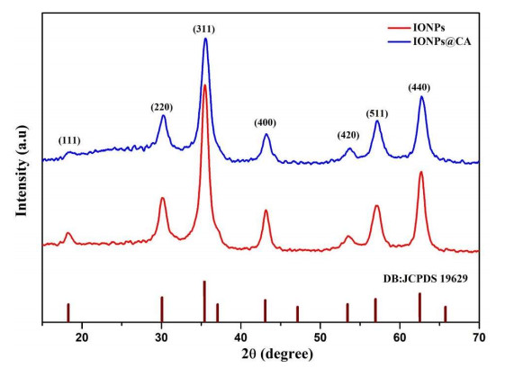
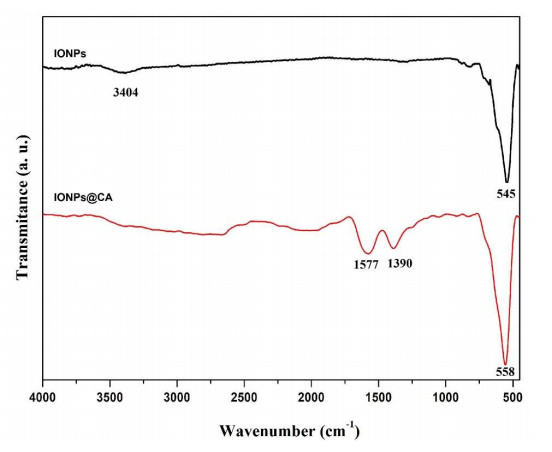
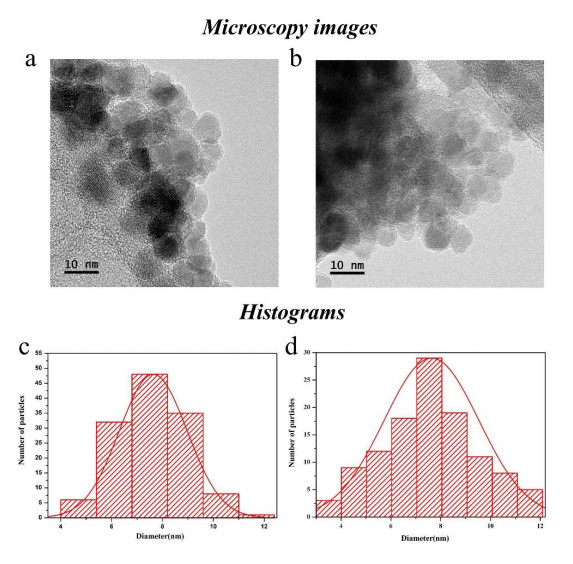
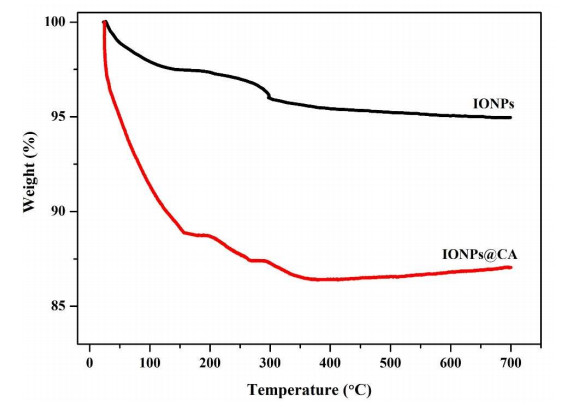
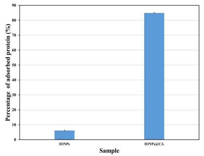
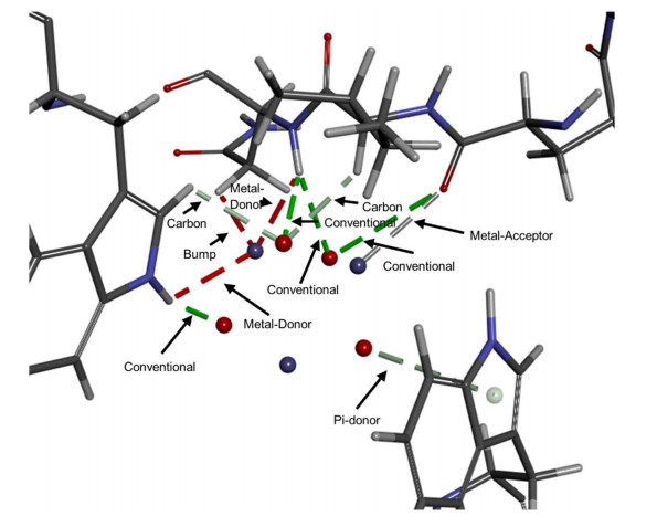
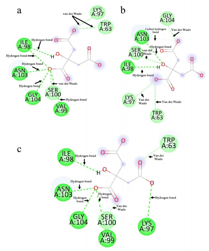
Magnetic nanoparticles (MNPs) are extensively utilized in biomedicine as part of controlled drug release systems, hyperthermia, and magnetic resonance imaging. Surface modification of MNPs not only enhances their stability and biocompatibility but also increases affinity with certain molecules, allowing them to be used in protein separation and adsorption processes. This article reports the synthesis and characterization of iron oxide MNPs functionalized with citric acid (IONPs@CA) to evaluate their performance in protein adsorption. The nanoparticles were characterized using various techniques such as transmission electron microscopy (TEM), X-ray diffraction (XRD), dynamic light scattering (DLS), thermogravimetric analysis (TGA), and Fourier-transform infrared spectroscopy (FT-IR). The percentage of lysozyme (Lyz) adsorbed by IONPs@CA was 84.9%, while the IONPs sample only adsorbed 5.9%. In silico evaluation results showed some repulsion bonds obtained in Lyz-IONPs and hydrogen bonds, carbon-hydrogen bonds, and van der Waals interactions in Lyz-IONPs@CA. These results may be novel since no previous research was found specifying this type of interaction between lysozyme and IONPs and/or IONPs@CA. The maximum adsorption efficiency obtained for the coated nanoparticles was 88.3%.
Citation: Denise Arrozarena Portilla, Arturo A. Velázquez López, Rosalva Mora Escobedo, Hernani Yee Madeira. Citrate coated iron oxide nanoparticles: Synthesis, characterization, and performance in protein adsorption[J]. AIMS Materials Science, 2024, 11(5): 991-1012. doi: 10.3934/matersci.2024047
Magnetic nanoparticles (MNPs) are extensively utilized in biomedicine as part of controlled drug release systems, hyperthermia, and magnetic resonance imaging. Surface modification of MNPs not only enhances their stability and biocompatibility but also increases affinity with certain molecules, allowing them to be used in protein separation and adsorption processes. This article reports the synthesis and characterization of iron oxide MNPs functionalized with citric acid (IONPs@CA) to evaluate their performance in protein adsorption. The nanoparticles were characterized using various techniques such as transmission electron microscopy (TEM), X-ray diffraction (XRD), dynamic light scattering (DLS), thermogravimetric analysis (TGA), and Fourier-transform infrared spectroscopy (FT-IR). The percentage of lysozyme (Lyz) adsorbed by IONPs@CA was 84.9%, while the IONPs sample only adsorbed 5.9%. In silico evaluation results showed some repulsion bonds obtained in Lyz-IONPs and hydrogen bonds, carbon-hydrogen bonds, and van der Waals interactions in Lyz-IONPs@CA. These results may be novel since no previous research was found specifying this type of interaction between lysozyme and IONPs and/or IONPs@CA. The maximum adsorption efficiency obtained for the coated nanoparticles was 88.3%.

| [1] |
Song YF, Feng LX, Basuray S, et al. (2023) Hemoglobin-BSA separation and purification by internally staged ultrafiltration. Sep Purif Technol 312: 123363. https://doi.org/10.1016/j.seppur.2023.123363 doi: 10.1016/j.seppur.2023.123363

|
| [2] |
Ayan K, Ganar K, Deshpande S, et al. (2023) Continuous counter-current electrophoretic separation of oleosomes and proteins from oilseeds. Food Hydrocolloid 144: 109053. https://doi.org/10.1016/j.foodhyd.2023.109053 doi: 10.1016/j.foodhyd.2023.109053
|
| [3] |
Gómez-García R, Campos DA, Aguilar C, et al. (2021) Biological protein precipitation: A green process for the extraction of cucumisin from melon (Cucumis melo L. inodorus) by-products. Food Hydrocolloid 116: 106650. https://doi.org/10.1016/j.foodhyd.2021.106650 doi: 10.1016/j.foodhyd.2021.106650
|
| [4] |
Kanoh S, Shiraki K, Wada M, et al. (2023) Chromatographic purification of histidine-tagged proteins using zirconia particles modified with phosphate groups. J Chromatogr A 1703: 464112. https://doi.org/10.1016/j.chroma.2023.464112 doi: 10.1016/j.chroma.2023.464112
|
| [5] |
Rodriguez EL, Poddar S, Iftekhar S, et al. (2020) Affinity chromatography: A review of trends and developments over the past 50 years. J Chromatogr B 1157: 122332. https://doi.org/10.1016/j.jchromb.2020.122332 doi: 10.1016/j.jchromb.2020.122332
|
| [6] |
Mahmoudi GM, Saraygord-Afshari N, Farsimadan M, et al. (2020) Opportunities and challenges of the tag-assisted protein purification techniques: Applications in the pharmaceutical industry. Biotechnol Adv 45: 107653. https://doi.org/10.1016/j.biotechadv.2020.107653 doi: 10.1016/j.biotechadv.2020.107653
|
| [7] |
Chu W, Prodromou R, Day KN, et al. (2021) Peptides and pseudopeptide ligands: A powerful toolbox for the affinity purification of current and next-generation biotherapeutics. J Chromatogr A 1635: 461632. https://doi.org/10.1016/j.chroma.2020.461632 doi: 10.1016/j.chroma.2020.461632
|
| [8] |
Huang H, Dong X, Sun Y, et al. (2023) Biomimetic affinity chromatography for antibody purification: Host cell protein binding and impurity removal. J Chromatogr A 1707: 464305. https://doi.org/10.1016/j.chroma.2023.464305 doi: 10.1016/j.chroma.2023.464305
|
| [9] |
Hamedani NS, Happich FL, Klein EM, et al. (2022) Aptamer loaded superparamagnetic beads for selective capturing and gentle release of activated protein C. Sci Rep 12: 7091. https://doi.org/10.1038/s41598-022-11198-5 doi: 10.1038/s41598-022-11198-5
|
| [10] | Perret G, Boschetti E (2020) Aptamer-based affinity chromatography for protein extraction and purification, In: Urmann K, Walter JG, Advances in Biochemical Engineering/Biotechnology, Cham: Springer. https://doi.org/10.1007/10_2019_106 |
| [11] |
Yıldırım D, Kip Ç, Tsogtbaatar K, et al. (2020) Microfluidic immobilized metal affinity chromatography based on Ti(Ⅳ)-decorated silica microspheres for purification of phosphoproteins. J Chromatogr B 1140: 122010. https://doi.org/10.1016/j.jchromb.2020.122010 doi: 10.1016/j.jchromb.2020.122010
|
| [12] |
Marini T, Gallina DA, Nabeshima EH, et al. (2022) Development of probiotic yoghurts with high protein content by ultrafiltration. NFS J 29: 16–25. https://doi.org/10.1016/j.nfs.2022.09.003 doi: 10.1016/j.nfs.2022.09.003
|
| [13] |
Çambay KF, Koçer İ, Kip Ç, et al. (2023) Ni(Ⅱ) functionalized polyhedral oligomeric silsesquioxane based capillary monolith for purification of histidine-tagged proteins by immobilized metal affinity micro-chromatography. J Chromatogr B 1225: 123759. https://doi.org/10.1016/j.jchromb.2023.123759 doi: 10.1016/j.jchromb.2023.123759
|
| [14] |
Choi HJ, Cheong DE, Yoo SK, et al. (2023) One-step metal affinity purification of recombinant hFGF19 without using tags. Protein Expr Purif 201: 106186. https://doi.org/10.1016/j.pep.2022.106186 doi: 10.1016/j.pep.2022.106186
|
| [15] |
Hoffman DL (1989) Purification and large-scale preparation of antithrombin Ⅲ. Am J Med 87: 23–26. https://doi.org/10.1016/0002-9343(89)80527-3 doi: 10.1016/0002-9343(89)80527-3
|
| [16] |
N'cho JS, Fofana I, Hadjadj Y, et al. (2016) Review of physicochemical-based diagnostic techniques for assessing insulation condition in aged transformers. Energies 9: 367. https://doi.org/10.3390/en9050367 doi: 10.3390/en9050367
|
| [17] |
Ali AH (2022) High-performance liquid chromatography (HPLC): A review. Ann Adv Chem 6: 010–020. https://doi.org/10.29328/journal.aac.1001026 doi: 10.29328/journal.aac.1001026
|
| [18] |
Wang J, Han Q, Wang K, et al. (2023) Recent advances in development of functional magnetic adsorbents for selective separation of proteins/peptides. Talanta 253: 123919. https://doi.org/10.1016/j.talanta.2022.123919 doi: 10.1016/j.talanta.2022.123919
|
| [19] |
Eivazzadeh-Keihan R, Bahreinizad H, Amiri Z, et al. (2021) Functionalized magnetic nanoparticles for the separation and purification of proteins and peptides. TrAC Trends Anal Chem 141: 116291. https://doi.org/10.1016/j.trac.2021.116291 doi: 10.1016/j.trac.2021.116291
|
| [20] |
Thomas SL, Thacker JB, Schug KA, et al. (2021) Sample preparation and fractionation techniques for intact proteins for mass spectrometric analysis. J Sep Sci 44: 211–246. https://doi.org/10.1002/jssc.202000936 doi: 10.1002/jssc.202000936
|
| [21] |
Cai L, Gao Y, Chu Y, et al. (2023) Green synthesis of silica-coated magnetic nanocarriers for simultaneous purification-immobilization of β-1, 3-xylanase. Int J Biol Macromol 233: 123223. https://doi.org/10.1016/j.ijbiomac.2023.123223 doi: 10.1016/j.ijbiomac.2023.123223
|
| [22] |
Wahajuddin, Arora S (2012) Superparamagnetic iron oxide nanoparticles: Magnetic nanoplatforms as drug carriers. Int J Nanomedicine 7: 3445–3471. https://doi.org/10.2147/IJN.S30320 doi: 10.2147/IJN.S30320
|
| [23] |
Ghutepatil P, Khot VM, Salunkhe AB, et al. (2022) Design of monodispersed PVP functionalized biocompatible manganese ferrite nanoparticles for hyperthermia application. Mater Today Proc 62: 5341–5346. https://doi.org/10.1016/j.matpr.2022.03.417 doi: 10.1016/j.matpr.2022.03.417
|
| [24] |
Valdeperez D, Wutke N, Ackermann LM, et al. (2022) Colloidal stability of polymer coated zwitterionic Au nanoparticles in biological media. Inorg Chim Acta 534: 120820. https://doi.org/10.1016/j.ica.2022.120820 doi: 10.1016/j.ica.2022.120820
|
| [25] |
Amatya R, Lee D, Sultana M, et al. (2023) Albumin-coated copper nanoparticles for photothermal cancer therapy: Synthesis and in vitro characterization. Heliyon 9: 17732. https://doi.org/10.1016/j.heliyon.2023.e17732 doi: 10.1016/j.heliyon.2023.e17732
|
| [26] |
Turrina C, Schoenen M, Milani D, et al. (2023) Application of magnetic iron oxide nanoparticles: Thrombotic activity, imaging and cytocompatibility of silica-coated and carboxymethyl dextran-coated particles. Colloids Surf B Biointerfaces 228: 113428. https://doi.org/10.1016/j.colsurfb.2023.113428 doi: 10.1016/j.colsurfb.2023.113428
|
| [27] |
Zeng K, Sun EJ, Liu ZW, et al. (2020) Synthesis of magnetic nanoparticles with an IDA or TED modified surface for purification and immobilization of poly-histidine tagged proteins. RSC Adv 10: 11524–11534. https://doi.org/10.1039/C9RA10473A doi: 10.1039/C9RA10473A
|
| [28] |
Lodhi MS, Shaheen A, Khan MT, et al. (2022) A novel method of affinity purification and characterization of polygalacturonase of Aspergillus flavus by galacturonic acid engineered magnetic nanoparticle. Food Chem 372: 131317. https://doi.org/10.1016/j.foodchem.2021.131317 doi: 10.1016/j.foodchem.2021.131317
|
| [29] |
Tavakoli Z, Rasekh B, Yazdian F, et al. (2019) One-step separation of the recombinant protein by using the amine-functionalized magnetic mesoporous silica nanoparticles; an efficient and facile approach. Int J Biol Macromol 135: 600–608. https://doi.org/10.1016/j.ijbiomac.2019.05.114 doi: 10.1016/j.ijbiomac.2019.05.114
|
| [30] |
Medina-Espinosa T, Asimbaya C, Galeas S, et al. (2021) Adsorptive removal of chromium (Ⅵ) from synthetic waters using magnetic lignocellulosic composites. IOP Conf Ser Earth Environ Sci 897: 012020. https://doi.org/10.1088/1755-1315/897/1/012020 doi: 10.1088/1755-1315/897/1/012020
|
| [31] |
Liu J, Dai C, Hu Y, et al. (2018) Aqueous aggregation behavior of citric acid coated magnetite nanoparticles: Effects of pH, cations, anions, and humic acid. Environ Res 161: 49–60. https://doi.org/10.1016/j.envres.2017.10.045 doi: 10.1016/j.envres.2017.10.045
|
| [32] |
Bradford MM (1976) A rapid and sensitive method for the quantitation of microgram quantities of protein utilizing the principle of protein-dye binding. Anal Biochem 72: 248–254. https://doi.org/10.1016/0003-2697(76)90527-3 doi: 10.1016/0003-2697(76)90527-3
|
| [33] |
Rahman Z, Dong YL, Ren C, et al. (2012) Protein adsorption on citrate modified magnetic nanoparticles. J Nanosci Nanotechnol 12: 2598–2606. https://doi.org/10.1166/jnn.2012.5751 doi: 10.1166/jnn.2012.5751
|
| [34] |
Yang X, Zhang H, Cheng S, et al. (2022) Optimization of the adsorption and removal of Sb(ⅲ) by MIL-53(Fe)/GO using response surface methodology. RSC Adv 12: 4101–4112. https://doi.org/10.1039/d1ra08169a doi: 10.1039/d1ra08169a
|
| [35] |
Al-Madhagi H, Yazbik V, Abdelwahed W, et al. (2023) Magnetite nanoparticle co-precipitation synthesis, characterization, and applications: Mini review. BioNanoScience 13: 853–859. https://doi.org/10.1007/s12668-023-01113-1 doi: 10.1007/s12668-023-01113-1
|
| [36] |
Dubey M, Challagulla N, Wadhwa S, et al. (2021) Ultrasound assisted synthesis of magnetic Fe3O4/ɑ-MnO2 nanocomposite for photodegradation of organic dye. Colloids Surf A 609: 125720. https://doi.org/10.1016/j.colsurfa.2020.125720 doi: 10.1016/j.colsurfa.2020.125720
|
| [37] |
González-Martínez DA, González G, Escalante-Bermúdez C, et al. (2023) Efficient capture of recombinant SARS-CoV-2 receptor-binding domain (RBD) with citrate-coated magnetic iron oxide nanoparticles. Nanoscale 15: 7854–7869. https://doi.org/10.1039/d3nr01109g doi: 10.1039/d3nr01109g
|
| [38] |
Kristianto H, Reynaldi E, Prasetyo S, et al. (2020) Adsorbed leucaena protein on citrate modified Fe3O4 nanoparticles: Synthesis, characterization, and its application as magnetic coagulant. Sustain Environ Res 30: 32. https://doi.org/10.1186/s42834-020-00074-4 doi: 10.1186/s42834-020-00074-4
|
| [39] |
Andronenko SI, Nikolaev AM, Suharzhevsky SM, et al. (2023) Phase composition and magnetic properties of nanoparticles with magnetite-maghemite structure. Ceramics 6: 1623–1631. https://doi.org/10.3390/ceramics6030099 doi: 10.3390/ceramics6030099
|
| [40] |
Petcharoen K, Sirivat A (2012) Synthesis and characterization of magnetite nanoparticles via the chemical co-precipitation method. Mater Sci Eng B 177: 421–427. https://doi.org/10.1016/j.mseb.2012.01.003 doi: 10.1016/j.mseb.2012.01.003
|
| [41] |
González-Martínez E, Gómez A, González-Martínez DA, et al. (2021) Chitosan-coated magnetic nanoparticles; exploring their potentialities for DNA and Cu(Ⅱ) recovery. Inorg Nano-Met Chem 51: 1098–1107. https://doi.org/10.1080/24701556.2020.1814335 doi: 10.1080/24701556.2020.1814335
|
| [42] |
Augusto-Jimenez YE, González-Montoya M, Naranjo-Feliciano D, et al. (2021) Antioxidant activity of bioactive peptide fractions from germinated soybeans conjugated to Fe3O4 nanoparticles by the Ugi multicomponent reaction. Molecules 26: 5726. https://doi.org/10.3390/molecules26195726 doi: 10.3390/molecules26195726
|
| [43] |
Peternele WS, Monge-Fuentes V, Fascineli ML, et al. (2014) Experimental investigation of the coprecipitation method: An approach to obtain magnetite and maghemite nanoparticles with improved properties. J Nanomater 2014: 682985. https://doi.org/10.1155/2014/682985 doi: 10.1155/2014/682985
|
| [44] |
Kim W, Suh CY, Cho SW, et al. (2012) A new method for the identification and quantification of magnetite-maghemite mixture using conventional X-ray diffraction technique. Talanta 94: 348–352. https://doi.org/10.1016/j.talanta.2012.03.001 doi: 10.1016/j.talanta.2012.03.001
|
| [45] |
Freire TM, Dutra LMU, Queiroz DC, et al. (2016) Fast ultrasound assisted synthesis of chitosan-based magnetite nanocomposites as a modified electrode sensor. Carbohydr Polym 151: 760–769. https://doi.org/10.1016/j.carbpol.2016.05.095 doi: 10.1016/j.carbpol.2016.05.095
|
| [46] |
Velásquez AA, Urquijo JP (2021) Synthesis and characterization of magnetite-maghemite nanoparticles in presence of polyethylene glycol obtained by mechanical milling. Mater Sci Eng B 263: 114873. https://doi.org/10.1016/j.mseb.2020.114873 doi: 10.1016/j.mseb.2020.114873
|
| [47] |
Qureashi A, Pandith AH, Bashir A, et al. (2021) Citrate coated magnetite: A complete magneto dielectric, electrochemical and DFT study for detection and removal of heavy metal ions. Surf Interfaces 23: 101004. https://doi.org/10.1016/j.surfin.2021.101004 doi: 10.1016/j.surfin.2021.101004
|
| [48] |
Shabatina TI, Vernaya OI, Shabatin VP, et al. (2020) Magnetic nanoparticles for biomedical purposes: Modern trends and prospects. Magnetochemistry 6: 30. https://doi.org/10.3390/magnetochemistry6030030 doi: 10.3390/magnetochemistry6030030
|
| [49] |
Dolai J, Mandal K, Jana NR, et al. (2021) Nanoparticle size effects in biomedical applications. ACS Appl Nano Mater 4: 6471–6496. https://doi.org/10.1021/acsanm.1c00987 doi: 10.1021/acsanm.1c00987
|
| [50] |
Li L, Mak KY, Leung CW, et al. (2013) Effect of synthesis conditions on the properties of citric-acid coated iron oxide nanoparticles. Microelectron Eng 110: 329–334. https://doi.org/10.1016/j.mee.2013.02.045 doi: 10.1016/j.mee.2013.02.045
|
| [51] |
Atrei A, Mahdizadeh FF, Baratto MC, et al. (2021) Effect of citrate on the size and the magnetic properties of primary Fe3O4 nanoparticles and their aggregates. Appl Sci 11: 6974. https://doi.org/10.3390/app11156974 doi: 10.3390/app11156974
|
| [52] |
Chesnel K, Trevino M, Cai Y, et al. (2014) Particle size effects on the magnetic behaviour of 5 to 11 nm Fe3O4 nanoparticles coated with oleic acid. J Phys Conf Ser 521: 012004. https://doi.org/10.1088/1742-6596/521/1/012004 doi: 10.1088/1742-6596/521/1/012004
|
| [53] |
Maleki H, Simchi A, Imani M, et al. (2012) Size-controlled synthesis of superparamagnetic iron oxide nanoparticles and their surface coating by gold for biomedical applications. J Magn Magn Mater 324: 3997–4005. https://doi.org/10.1016/j.jmmm.2012.06.045 doi: 10.1016/j.jmmm.2012.06.045
|
| [54] |
Surpi A, Shelyakova T, Murgia M, et al. (2023) Versatile magnetic configuration for the control and manipulation of superparamagnetic nanoparticles. Sci Rep 13: 5301. https://doi.org/10.1038/s41598-023-32299-9 doi: 10.1038/s41598-023-32299-9
|
| [55] |
Hah HY, Gray S, Johnson CE, et al. (2021) Mössbauer spectroscopy of superparamagnetic Fe3O4 nanoparticles. J Magn Magn Mater 539: 168382. https://doi.org/10.1016/j.jmmm.2021.168382 doi: 10.1016/j.jmmm.2021.168382
|
| [56] |
Araujo JF, Tahir, Arsalani S, et al. (2020) Novel scanning magnetic microscopy method for the characterization of magnetic nanoparticles. J Magn Magn Mater 499: 166300. https://doi.org/10.1016/j.jmmm.2019.166300 doi: 10.1016/j.jmmm.2019.166300
|
| [57] |
Rezanezhad A, Hajalilou A, Eslami F, et al. (2021) Superparamagnetic magnetite nanoparticles for cancer cells treatment via magnetic hyperthermia: Effect of natural capping agent, particle size and concentration. J Mater Sci Mater Electron 32: 24026–24040. https://doi.org/10.1007/s10854-021-06865-8 doi: 10.1007/s10854-021-06865-8
|
| [58] |
Abdolrahimi M, Vasilakaki M, Slimani S, et al. (2021) Magnetism of nanoparticles: Effect of the organic coating. Nanomaterials 11: 1787. https://doi.org/10.3390/nano11071787 doi: 10.3390/nano11071787
|
| [59] |
Guduri BR, Luyt AS (2008) Structure and mechanical properties of polycarbonate modified clay nanocomposites. J Nanosci Nanotechnol 8: 1880–1885. https://doi.org/10.1166/jnn.2008.18253 doi: 10.1166/jnn.2008.18253
|
| [60] |
Omelyanchik A, Gomes da Silva F, Gomide G, et al. (2021) Effect of citric acid on the morpho-structural and magnetic properties of ultrasmall iron oxide nanoparticles. J Alloys Compd 883: 160779. https://doi.org/10.1016/j.jallcom.2021.160779 doi: 10.1016/j.jallcom.2021.160779
|
| [61] |
Blanco-Andujar C, Ortega D, Southern P, et al. (2015) High performance multi-core iron oxide nanoparticles for magnetic hyperthermia: Microwave synthesis, and the role of core-to-core interactions. Nanoscale 7: 1768–1775. https://doi.org/10.1039/c4nr06239f doi: 10.1039/c4nr06239f
|
| [62] |
Walkey CD, Olsen JB, Guo H, et al. (2012) Nanoparticle size and surface chemistry determine serum protein adsorption and macrophage uptake. J Am Chem Soc 134: 2139–2147. https://doi.org/10.1021/ja2084338 doi: 10.1021/ja2084338
|
| [63] |
Ibrahim MA, Jaafar MZ, Yusof MA, et al. (2023) Influence of size and surface charge on the adsorption behaviour of silicon dioxide nanoparticles on sand particles. Colloids Surf A 674: 131943. https://doi.org/10.1016/j.colsurfa.2023.131943 doi: 10.1016/j.colsurfa.2023.131943
|
| [64] |
Dongargaonkar AA, Clogston JD (2018) Quantitation of surface coating on nanoparticles using thermogravimetric analysis. Methods Mol Biol 1682: 57–63. https://doi.org/10.1007/978-1-4939-7352-1_6 doi: 10.1007/978-1-4939-7352-1_6
|
| [65] |
Malik LA, Bashir A, Ahmad N, et al. (2020) Exploring metal ion adsorption and antifungal properties of carbon-coated magnetite composite. ChemistrySelect 5: 3208–3216. https://doi.org/10.1002/slct.201904830 doi: 10.1002/slct.201904830
|
| [66] |
Hancock ML, Yokel RA, Beck MJ, et al. (2021) The characterization of purified citrate-coated cerium oxide nanoparticles prepared via hydrothermal synthesis. Appl Surf Sci 535: 147681. https://doi.org/10.1016/j.apsusc.2020.147681 doi: 10.1016/j.apsusc.2020.147681
|
| [67] |
Vassallo M, Martella D, Barrera G, et al. (2023) Improvement of hyperthermia properties of iron oxide nanoparticles by surface coating. ACS Omega 8: 2143–2154. https://doi.org/10.1021/acsomega.2c06244 doi: 10.1021/acsomega.2c06244
|
| [68] |
Jia ZX, Li JT, Gao L, et al. (2023) Dynamic light scattering: A powerful tool for in situ nanoparticle sizing. Colloids Interfaces 7: 15. https://doi.org/10.3390/colloids7010015 doi: 10.3390/colloids7010015
|
| [69] |
Jummes B, Sganzerla WG, Gonçalves da Rosa G, et al. (2020) Antioxidant and antimicrobial poly-ε-caprolactone nanoparticles loaded with Cymbopogon martinii essential oil. Biocatal Agric Biotechnol 23: 101499. https://doi.org/10.1016/j.bcab.2020.101499 doi: 10.1016/j.bcab.2020.101499
|
| [70] | Joseph E, Singhvi G (2019) Chapter 4—Multifunctional nanocrystals for cancer therapy: A potential nanocarrier, In: Grumezescu AM, Nanomaterials for Drug Delivery and Therapy, Norwich: William Andrew Publishing, 91–116. https://doi.org/10.1016/b978-0-12-816505-8.00007-2 |
| [71] |
Dheyab MA, Aziz AA, Jameel MS, et al. (2020) Simple rapid stabilization method through citric acid modification for magnetite nanoparticles. Sci Rep 10: 10793. https://doi.org/10.1038/s41598-020-67869-8 doi: 10.1038/s41598-020-67869-8
|
| [72] |
Stein R, Friedrich B, Mühlberger M, et al. (2020) Synthesis and characterization of citrate-stabilized gold-coated superparamagnetic iron oxide nanoparticles for biomedical applications. Molecules 25: 4425. https://doi.org/10.3390/molecules25194425 doi: 10.3390/molecules25194425
|
| [73] |
Raji Z, Karim A, Karam A, et al. (2023) A review on the heavy metal adsorption capacity of dietary fibers derived from agro-based wastes: Opportunities and challenges for practical applications in the food industry. Trends Food Sci Technol 137: 74–91. https://doi.org/10.1016/j.tifs.2023.05.004 doi: 10.1016/j.tifs.2023.05.004
|
| [74] |
Alderton G, Fevold HL (1946) Direct crystallization of lysozyme from egg white and some crystalline salts of lysozyme. J Biol Chem 164: 1–5. https://doi.org/10.1016/S0021-9258(18)43040-2 doi: 10.1016/S0021-9258(18)43040-2
|
| [75] |
Abdelsattar AS, Dawoud A, Helal MA (2021) Interaction of nanoparticles with biological macromolecules: A review of molecular docking studies. Nanotoxicology 15: 66–95. https://doi.org/10.1080/17435390.2020.1842537 doi: 10.1080/17435390.2020.1842537
|
| [76] |
Maulydia NB, Tallei TE, Ginting B, et al. (2022) Analysis of flavonoid compounds of Orange (Citrus sp.) peel as anti-main protease of SARS-CoV-2: A molecular docking study. IOP Conf Ser Earth Environ Sci 951: 012078. https://doi.org/10.1088/1755-1315/951/1/012078 doi: 10.1088/1755-1315/951/1/012078
|
| [77] |
Weinhold F, Klein RA (2014) Anti-electrostatic hydrogen bonds. Angew Chem Int Ed 53: 11214–11217. https://doi.org/10.1002/anie.201405812 doi: 10.1002/anie.201405812
|
| [78] |
Vaidyanathan R, Murugan SS, Ravichandran K, et al. (2023) Molecular docking approach on the binding stability of derivatives of phenolic acids (DPAs) with human serum albumin (HSA): Hydrogen-bonding versus hydrophobic interactions or combined influences? JCIS Open 12: 100096. https://doi.org/10.1016/j.jciso.2023.100096 doi: 10.1016/j.jciso.2023.100096
|
| [79] |
Pinzi L, Rastelli G (2019) Molecular docking: Shifting paradigms in drug discovery. Int J Mol Sci 20: 4331. https://doi.org/10.3390/ijms20184331 doi: 10.3390/ijms20184331
|
| [80] |
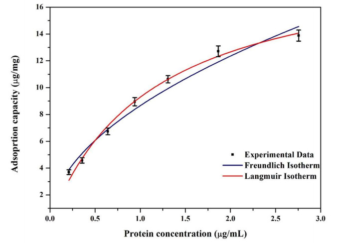
Langmuir I (1916) The constitution and fundamental properties of solids and liquids. Part Ⅰ. Solids. J Am Chem Soc 38: 2221–2295. https://doi.org/10.1021/ja02268a002 doi: 10.1021/ja02268a002
|
| [81] | Singh AK (2016) Chapter 8—Nanoparticle ecotoxicology, In: Singh AK, Engineered Nanoparticles, Boston: Academic Press, 343–350. http://dx.doi.org/10.1016/b978-0-12-801406-6.00008-x |
| [82] |
Cho M, Mahmoodi Z, Shetty P, et al. (2024) Protein adsorption on solid surfaces: Data mining, database, molecular surface-derived properties, and semiempirical relationships. ACS Appl Mater Interfaces 16: 28290–28306. https://doi.org/10.1021/acsami.4c06759 doi: 10.1021/acsami.4c06759
|
| [83] |
Latour RA (2015) The Langmuir isotherm: A commonly applied but misleading approach for the analysis of protein adsorption behavior. J Biomed Mater Res A 103: 949–958. https://doi.org/10.1002/jbm.a.35235 doi: 10.1002/jbm.a.35235
|
| [84] |
Rahdar S, Rahdar A, Ahmadi S, et al. (2019) Adsorption of bovine serum albumin (BSA) by bare magnetite nanoparticles with surface oxidative impurities that prevent aggregation. Can J Chem 97: 577–583. https://doi.org/10.1139/cjc-2019-0008 doi: 10.1139/cjc-2019-0008
|
| [85] |
Zhang J, Huang L, Zheng J, et al. (2020) SiO2-assisted synthesis of Fe3O4@SiO2@C-Ni nanochains for effective catalysis and protein adsorption. J Magn Magn Mater 497: 166011. https://doi.org/10.1016/j.jmmm.2019.166011 doi: 10.1016/j.jmmm.2019.166011
|
| [86] |
Maleki MS, Moradi O, Tahmasebi S (2017) Adsorption of albumin by gold nanoparticles: Equilibrium and thermodynamics studies. Arab J Chem 7: 1104–1109. https://doi.org/10.1016/j.arabjc.2012.09.003 doi: 10.1016/j.arabjc.2012.09.003
|
| [87] |
Wang G, Hou H, Wang S, et al. (2017) Exploring the interaction of silver nanoparticles with lysozyme: Binding behaviors and kinetics. Colloids Surf B Biointerfaces 1: 138–145. https://doi.org/10.1016/j.colsurfb.2017.05.071 doi: 10.1016/j.colsurfb.2017.05.071
|
| [88] |
Saptarshi SR, Duschl A, Lopata AL (2013) Interaction of nanoparticles with proteins: Relation to bio-reactivity of the nanoparticle. J Nanobiotechnol 11: 26. https://doi.org/10.1186/1477-3155-11-26 doi: 10.1186/1477-3155-11-26
|
| [89] |
Rabe M, Verdes D, Seeger S (2011) Understanding protein adsorption phenomena at solid surfaces. Adv Colloid Interface Sci 162: 87–106. https://doi.org/10.1016/j.cis.2010.12.007 doi: 10.1016/j.cis.2010.12.007
|
| [90] |
Esmaeilnejad-Ahranjani P, Maboudi SA, Arpanaei A (2023) Effect of the structure of magnetic nanocomposite adsorbents on the lysozyme separation efficiency. ACS Appl Bio Mater 6: 191–202. https://doi.org/10.1016/j.mtcomm.2023.105632 doi: 10.1016/j.mtcomm.2023.105632
|
| [91] |
Pota G, Gallucci N, Cavasso D, et al. (2023) Controlling the adsorption of β-glucosidase onto wrinkled SiO2 nanoparticles to boost the yield of immobilization of an efficient biocatalyst. Langmuir 39: 1482–1494. https://doi.org/10.1021/acs.langmuir.2c02861 doi: 10.1021/acs.langmuir.2c02861
|
 matersci-11-05-047-Supplementary.pdf matersci-11-05-047-Supplementary.pdf |
 |
Figures(8) / Tables(4)

Denise Arrozarena Portilla, Arturo A. Velázquez López, Rosalva Mora Escobedo, Hernani Yee Madeira. Citrate coated iron oxide nanoparticles: Synthesis, characterization, and performance in protein adsorption[J]. AIMS Materials Science, 2024, 11(5): 991-1012. doi: 10.3934/matersci.2024047


 DownLoad: