Loading [Contrib]/a11y/accessibility-menu.js
Review Topical Sections

Tissue-engineered products capable of enhancing wound healing

  • In a general wound healing process, foreign bodies and tissue detritus are broken down and then a new tissue is produced. However, the new tissue formation sometimes fails to proceed under the impaired conditions. The endogenous levels of growth factors are reduced in some chronic wounds. To improve these wound conditions, several types of tissue-engineered products composed of cell, growth factor, and/or biomaterial have been developed. This review includes the practical design of tissue-engineered products capable of enhancing wound healing. The first product is a cultured dermal substitute composed of cell and biomaterial. The cultured dermal substitute is prepared by seeding allogeneic fibroblasts into a hyaluronic acid and collagen spongy sheet. Although allogeneic fibroblasts are rejected gradually in immune system, they are able to release some types of growth factors, and thereby regenerate a damaged tissue. The second products are a wound dressing and a skin care product composed of growth factor and biomaterial. These products are prepared by freeze-drying an aqueous solution of hyaluronic acid and collagen containing epidermal growth factor. Epidermal growth factor is beneficial for wound healing because of its effects on proliferation of keratinocytes and fibroblasts, thus capable of facilitating granulation tissue formation and re-epithelialization. In addition, epidermal growth factor can stimulate fibroblasts to synthesize an increased amount of vascular endothelial growth factor and hepatocyte growth factor that are essential for angiogenesis. The third product is an anti-adhesive product composed of growth factor and biomaterial. This product is prepared by freeze-drying an aqueous solution of hyaluronic acid and collagen containing epidermal growth factor. Wound dressings are applied to external wounds, while anti-adhesive products are applied to internal wounds. However, both products should be designed to have a common function that facilitates wound healing.

    Citation: Misato Kuroyanagi, Yoshimitsu Kuroyanagi. Tissue-engineered products capable of enhancing wound healing[J]. AIMS Materials Science, 2017, 4(3): 561-581. doi: 10.3934/matersci.2017.3.561

    Related Papers:

    [1] Sukhwinder K. Bhullar, Harpal S. Buttar . Perspectives on nanofiber dressings for the localized delivery of botanical remedies in wound healing. AIMS Materials Science, 2017, 4(2): 370-382. doi: 10.3934/matersci.2017.2.370
    [2] Raffaele Conte, Anna Di Salle, Francesco Riccitiello, Orsolina Petillo, Gianfranco Peluso, Anna Calarco . Biodegradable polymers in dental tissue engineering and regeneration. AIMS Materials Science, 2018, 5(6): 1073-1101. doi: 10.3934/matersci.2018.6.1073
    [3] Dorel Feldman . Poly(vinyl alcohol) nanocomposites: Recent contributions to engineering and medicine. AIMS Materials Science, 2021, 8(1): 119-129. doi: 10.3934/matersci.2021008
    [4] Elena Kossovich . Theoretical study of chitosan-graphene and other chitosan-based nanocomposites stability. AIMS Materials Science, 2017, 4(2): 317-327. doi: 10.3934/matersci.2017.2.317
    [5] Chiara Emma Campiglio, Chiara Marcolin, Lorenza Draghi . Electrospun ECM macromolecules as biomimetic scaffold for regenerative medicine: challenges for preserving conformation and bioactivity. AIMS Materials Science, 2017, 4(3): 638-669. doi: 10.3934/matersci.2017.3.638
    [6] Huilin Yang, Amy M. Peterson . Inkjet printed drug-releasing polyelectrolyte multilayers for wound dressings. AIMS Materials Science, 2017, 4(2): 452-469. doi: 10.3934/matersci.2017.2.452
    [7] Vincenzo Guarino, Tania Caputo, Rosaria Altobelli, Luigi Ambrosio . Degradation properties and metabolic activity of alginate and chitosan polyelectrolytes for drug delivery and tissue engineering applications. AIMS Materials Science, 2015, 2(4): 497-502. doi: 10.3934/matersci.2015.4.497
    [8] Osmar Alejandro Chanes-Cuevas, Adriana Perez-Soria, Iriczalli Cruz-Maya, Vincenzo Guarino, Marco Antonio Alvarez-Perez . Macro-, micro- and mesoporous materials for tissue engineering applications. AIMS Materials Science, 2018, 5(6): 1124-1140. doi: 10.3934/matersci.2018.6.1124
    [9] Rudolf Kawalla, Anett Stöcker, Ulrich Prahl, Xuefei Wei, Jens Dierdorf, Gerhard Hirt, Martin Heller, Stefan Roggenbuck, Sandra Korte-Kerzel, Hannes Alois Weiss, Philipp Tröber, Lucas Böhm, Wolfram Volk, Nora Leuning, Kay Hameyer . Low-loss FeSi sheet for energy-efficient electrical drives. AIMS Materials Science, 2018, 5(6): 1184-1198. doi: 10.3934/matersci.2018.6.1184
    [10] Martin O. Steinhauser . Multiscale modeling, coarse-graining and shock wave computer simulations in materials science. AIMS Materials Science, 2017, 4(6): 1319-1357. doi: 10.3934/matersci.2017.6.1319
  • In a general wound healing process, foreign bodies and tissue detritus are broken down and then a new tissue is produced. However, the new tissue formation sometimes fails to proceed under the impaired conditions. The endogenous levels of growth factors are reduced in some chronic wounds. To improve these wound conditions, several types of tissue-engineered products composed of cell, growth factor, and/or biomaterial have been developed. This review includes the practical design of tissue-engineered products capable of enhancing wound healing. The first product is a cultured dermal substitute composed of cell and biomaterial. The cultured dermal substitute is prepared by seeding allogeneic fibroblasts into a hyaluronic acid and collagen spongy sheet. Although allogeneic fibroblasts are rejected gradually in immune system, they are able to release some types of growth factors, and thereby regenerate a damaged tissue. The second products are a wound dressing and a skin care product composed of growth factor and biomaterial. These products are prepared by freeze-drying an aqueous solution of hyaluronic acid and collagen containing epidermal growth factor. Epidermal growth factor is beneficial for wound healing because of its effects on proliferation of keratinocytes and fibroblasts, thus capable of facilitating granulation tissue formation and re-epithelialization. In addition, epidermal growth factor can stimulate fibroblasts to synthesize an increased amount of vascular endothelial growth factor and hepatocyte growth factor that are essential for angiogenesis. The third product is an anti-adhesive product composed of growth factor and biomaterial. This product is prepared by freeze-drying an aqueous solution of hyaluronic acid and collagen containing epidermal growth factor. Wound dressings are applied to external wounds, while anti-adhesive products are applied to internal wounds. However, both products should be designed to have a common function that facilitates wound healing.


    Abbreviations

    EGF   epidermal growth factor

    VEGF   vascular endothelial growth factor

    HGF   hepatocyte growth factor

    bFGF   basic fibroblast growth factor

    TGF   transforming growth factor

    ECM    extracellular matrix

    CDS   cultured dermal substitute

    HA   hyarulonic acid

    Col   collagen

    Arg   arginin

    VC   vitamin C

    PGA   poly-γ-glutamic acid

    GC   glucosyl ceramide


    1. Introduction

    The creation of tissue-engineered product needs the basic knowledge on wound healing, especially the functions of cell, growth factor, and biomaterial in wound healing process. Tissue response to injury has been divided into three overlapping phases: inflammation, proliferation, and remodeling (Figure 1). Immediately following injury, platelets aggregate and release coagulation factors and growth factors that are essential for a normal wound healing process to proceed. During the inflammation phase for a period of 2 weeks, neutrophils, monocytes, and macrophages are able to kill and digest bacteria and detritus of damaged tissues. Macrophages also serve as an important source of growth factors that are essential for wound healing. During the proliferation phase, endothelial cells and fibroblasts are predominant cell types. The neovascularization process requires migration and proliferation of endothelial cells in order to form new functioning blood vessels. The granulation process requires migration and proliferation of fibroblasts. At this stage, fibroblasts release stimulatory and inhibitory cytokines, thereby regulating the synthesis and deposition of ECM components such as fibronectin, laminin, glycosaminoglycans, and collagen. The epithelialization process requires migration, proliferation, and differentiation of keratinocytes found in the residual epidermis and skin appendages. This process is facilitated by growth factors secreted from fibroblasts and keratinocytes. During the remodeling phase, collagen degradation is regulated by a complex counterbalance of stimulatory and inhibitory cytokines [1].

    Figure 1. Normal wound healing response through a series of overlapping phases: inflammation, proliferation, and remodeling. Several cells at coagulation phase and following phases release growth factors that are essential for a normal wound healing process to proceed [1].

    The normal wound healing process is a coordinated and predictable series of cellular and biochemical events. Various cytokines are present in acute wound fluid. These cytokines are essentia for a normal wound healing to proceed. However, some chronic wounds fail to proceed through an orderly and timely healing process, because certain pathophysiologic conditions and metabolic factors involved in these chronic wounds can alter cellular functions. These chronic wounds result from deficiency of growth factors. Such a deficiency may be partly the result of elevated levels of proteinases that degrade growth factors and ECM components at the wound site [2,3,4,5,6]. To improve these wound conditions, several types of tissue-engineered products have been developed.

    In general, the prime constituents of the tissue-engineered products are cell, growth factor, and biomaterial (often referred to as scaffold). In the first approach, the procedure entails the proliferation of isolated cells from own tissue or a donor's tissue. These cells are seeded on or within a scaffold such as biodegradable materials. As well as delivering the cells, the scaffold both creates and maintains a space for the formation of new tissue. In this approach, the patient receives autologous cells that have been harvested previously and proliferated. The entire structure of autologous cells combined with a scaffold is transplanted into the lost area of tissue, where the cells proliferate and form new tissue. According to this technique, allogeneic cells also can be combined with a scaffold. Although allogeneic cells are rejected gradually in immune system, they are able to release some types of growth factors, and thereby regenerate a damaged tissue. In the second approach, the growth factor is incorporated into biodegradable materials, followed by transplanting into a site of damaged tissues. The growth factor allows patient's own cells to migrate into a desired area and regenerate a lost tissue.

    This review includes the design of fibroblasts-incorporting dermal substitute, EGF-incorporating wound dressing, EGF-incorporating skin care product, and also the design of EGF-incorporating anti-adhesive product. During surgical opperation, incision is necessary for an excision of target tissues or organs. Incision wounds result in an undesirable tissue-adhesion among incision wound, surgically excised wound, and surrounding organ. Wound dressings are applied to external wounds, while anti-adhesive products are applied to internal wounds. However, both products should be designed to have a common function that facilitates wound healing process.


    2. Tissue-engineered Product Using Cell and Biomaterial


    2.1. Design of Skin Substitute

    A number of skin substitutes with autologous or allogeneic cells have been produced by some culture techniques. Cultured epidermal substitute is composed of stratified keratinocytes. Cultured dermal substitute is composed of fibroblasts combined with a scaffold. Cultured skin substitute is composed of fibroblasts combined with a scaffold bearing stratified keratinocytes (Figure 2). There are two categories depending on the origin of cells; autologous cells or allogeneic cells. The pioneering work of Green and co-workers has demonstrated that it is possible to grow keratinocytes as a stratified sheet from a single cell suspension in the culture system using 3T3 feeder layer [7,8]. Autologous cultured epidermal substitute has been evaluated in many hospitals and proved very effective [9,10,11,12,13,14,15]. On the other hand, Bell and co-workersdeveloped a cultured skin substitute composed of fibroblasts combined with a collagen gel sheet bearing stratified keratinocytes [16,17,18]. Allogeneic cultured skin substitute has been evaluated in many hospitals and proved very effective [19].

    Figure 2. Structure of skin substitutes: Cultured Epidermal Substitute using keratinocyte, Cultured Dermal Substitute using fibroblast and biomaterial, and Cultured Skin Substitute using keratinocyte, fibroblast, and biomaterial [20].

    2.2. Preparation of Allogeneic CDS

    Cell banking was established by the procedure described in our article [20]. Briefly, a small piece of skin was obtained from patients younger than one year-old during surgical excision of excrescence. This patient was free from infectious viruses such as HBV, HCV, HIV and HTLV, and also negative on the treponema pallidum hemagglutination test. Fibroblasts isolated by enzymatic treatment were cultivated successively in culture medium to establish cell banking. The cultured fibroblasts were suspended in a cryopreserved medium and then cryopreserved at −152 ℃ according to a conventional procedure. The allogeneic CDS (10 cm × 10 cm) was prepared using these fibroblasts. The fibroblasts obtained from successive cultivation of the cryopreserved cells were seeded onto the 2-layered sponge of HA and Col. Cryopreservation and thawing of CDS was performed according to the method described in the article [20].

    It is well known that fibroblasts have a potential to release some types of cytokines, but the amount of cytokine is dependent on the culturing conditions. We measured the amounts of cytokines including VEGF, bFGF, and HGF [21,22]. The results demonstrate that the cryopreserved CDS can keep the potential to release VEGF, bFGF, and HGF at appreciable levels (Figure 3).

    Figure 3. Design and function of allogeneic CDS composed of fibroblasts combined with a spongy sheet of HA and Col. Fibroblasts release several types of growth factors that are essential for wound healing. HA is capable of promoting cell migration. Col-derived peptide has a potential as chemotactic function for fibroblast [20].

    2.3. Clinical Study Using Allogeneic CDS

    A multi-center clinical study of this allogeneic CDS was conducted at 31 hospitals across Japan began in April 2000 and concluded in March 2005, as part of the Regenerating Medical Millennium Projects of the Ministry of Health, Labor and Welfare. For this clinical study, 4700 sheets of CDS were prepared at the R & D Center for Artificial Skin, School of Allied Health Sciences, Kitasato University, and were delivered to the participating hospitals. The clinical study was conducted in compliance with the ethical guidelines of the participating hospitals. Wound surface was debrided as necessary and rinsed with physiological saline solution. CDS was then applied to the wound site, on which a conventional ointment-gauze dressing was used as a top dressing. CDS was applied repeatedly at an interval of 5 to 7 days for a period of 6 weeks, or longer if necessary. Clinical results were obtained for 404 cases, including severe burns and intractable skin ulcers. Excellent result was obtained in 62.6%, good result was obtained in 30.0%, fair result was obtained in 6.7%, and poor result was obtained in 0.7% of the cases. Some of these results have been reported in the articles [23,24,25,26]. The representative clinical results are shown here. An 81-year-old woman who sustained a third-degree burn on the left leg was treated with a 6-fold extended mesh split-thickness auto-skin graft and allogeneic CDS (Figure 4). In this case, a 6-fold extended mesh split-thickness auto-skin graft was applied and then covered with allogeneic CDS. This extended mesh graft took successfully 15 days later, and showed excellent epithelialization, despite poor wound conditions [23].

    Figure 4. An 81-year-old woman sustained a third-degree burn on the left leg. The debrided wound surface was grafted with 6-fold extended mesh auto-skin, and covered with allogeneic CDS. The grafted skin took successfully 15 days later [23].

    A 65-year-old man who developed a necrotic lesion on the right foot was treated with allogeneic CDS, and then with a split-thickness auto-skin graft (Figure 5). Thirty-nine days after application of allogeneic CDS, a wound bed appeared favorable, allowing split-thickness auto-skin graft to be performed. Eighty-three days after the graft procedure, the condition of grafted skin was excellent [24].

    Figure 5. A 65-year-old man developed a necrotic lesion on the right foot. The debrided wound surface was covered with allogeneic CDS. The wound bed was formed 39 days later. The grafted skin showed an excellent condition 83 days later [24].

    3. Tissue-engineered Product Using Growth Factor and Biomaterial


    3.1. Potential of Biomaterial for Wound Healing

    HA has been identified as a major ECM component. HA is an essential biological material for wound healing, as it possesses various biological activities [27,28,29,30,31]. Because of its unique hygroscopic, rheologic and viscoelastic properties, HA creates an excellent wound healing environment. Moreover, it stimulates cell migrationand angiogenesis. It is generally accepted that the benefits of exogeneously applied HA lead to the development of tissue repair products. High molecular weight HA provides an excellent wound healing environment, while low molecular weight HA induces angiogenesis.

    Col molecule (Type Ⅰ collagen) had a triple helical structure that has non-helical molecular chain called "telo-peptide" on both end of molecule. This telo-peptide is recognized as a different type of protein because of own amino acid-sequence derived from the species, and thereby gives rise to immune rejection. Col prepared by cleavage of telo-peptide can decrease a risk of immune rejection, and thereby is used as a medical-grade material. Polypeptide derived from biodegradation of Col molecule is capable of enhancing fibroblasts migration from normal area to wound area, thereby promoting wound healing. In practice use, Col molecules having a triple helical structure are dissolved under acidic conditions below pH = 4. However, these rod-like molecules aggregate and partially precipitate at neutral pH, as the ionic groups on Col molecules are neutralized and fail to bind with surrounding water molecules. When an aqueous Col solution was warmed at more than 50 ℃, Col molecules undergo a conformational change from a triple helical structure to a random coil structure, and consequently maintain a clear solution at neutral pH. This heat-denatured Col aqueous solution can be mixed with an HA aqueous solution at a neutral pH without causing any precipitation. Col plays a pivotal role in wound healing. Col-derived peptides act as a chemoattractant for fibroblasts in vitro and may have a similar activity in vivo [32].

    EGF is beneficial for wound healing because of its effects on proliferation of keratinocytes, fibroblasts and vascular endothelial cells, thus facilitating granulation tissue formation and epithelialization [33,34]. Excessive collagen formation is caused by aid of TGF-b1. This is one of the reasons for scar formation. Such an excessive collagen formation is reduced indirectly by using EGF, because EGF serves to decrease TGF-b1 production. This efficacy suggests that EGF may be useful in anti-scarring therapy [35]. In addition, EGF can stimulate fibroblasts to synthesize an increased amount of VEGF and HGF that promote angiogenesis. Recent research has demonstrated that simultaneous administration of VEGF and HGF synergistically promotes new blood vessel formation compared with administration of each factor alone [36]. In addition, EGF is considered to be one of the key cytokines involved in epithelialization in addition to angiogenesis [37]. Thus, EGF is a promising factor for wound healing.


    3.2. Preparation of EGF-incorporating Wound Dressing Composed of HA and Col

    The efficacy of tissue-engineered product using growth factor and biomaterial is dependent on the potential of biomaterial. It is critical to use excellent biomaterials capable of facilitating wound healing. Taking into account these properties of each ingredient, EGF-incorprating wound dressing was prepared by freeze-drying an aqueous solution of high molecular weight HA, low molecular weight HA, and Col containing EGF (Figure 6). This wound dressing (5 cm × 8 cm) was designed to contain EGF at a concentration of 2.0 μg/cm2. Each side of spongy sheet was irradiated with a 15 W ultraviolet lamp at a distance of 20 cm for 15 min in order to induce intermolecular cross-linking of Col molecules [38,39,40].

    Figure 6. Design and function of EGF-incorporating wound dressing composed of spongy sheet of HA and Col containing EGF. HA is capable of promoting cell migration. Col-derived peptide has a potential as chemotactic function for fibroblast. EGF has a potential of wound healing.

    3.3. Evaluation of EGF-incorporating Wound Dressing Composed of HA and Col

    The animal study using rats was conducted [38]. Briefly, under intraperitoneal pentobarbital sodium anesthesia, an area with a diameter of 3.5 mm in the abdominal region of rats was contacted with boiling water for 3 seconds in order to result in a third-degree burn. Necrotic skin was removed under anesthesia 3 days after burn injury. The wound area was covered with each wound dressing with or without EGF. In control group the wound was covered with a commercially available alginate spongy wound dressing. Both wound dressings with or without EGF significantly promote epithelialization and angiogenesis when compared with the control group. Particularly EGF-incorporating wound dressing promoted epithelialization and angiogenesis. HA and Col has a potential to stimulate cell migration. These findings indicate that EGF-incorporating wound dressing has a desired effect on wound healing associated with epithelialization and angiogenesis. EGF is capable of enhancing fibroblasts proliferation and stimulating fibroblasts to produce VEGF and HGF that are esssentail for granulation tissue formation. In addition, EGF is capable of enhancing keratinocytes proliferation for epithelialization on resulting granulation tissue (Figure 7).

    Figure 7. Function of EGF: 1) enhancing keratinocyte and fibrblast proliferation 2) stimulating fibroblasts to produce vascular growth facto (VEGF) and hepatocyte growth factor (HGF) production that are essential for angiogenesis.

    3.4. Clinical Study Using EGF-incorporating Wound Dressing

    On the basis of excellent results in the fundamental study, EGF-incorporating wound dressing was evaluated in 16 clinical cases, incuding burns, donor sites, traumatic skin defects, and intractable skin ulcers. Wound surface was debrided as necessary and rinsed with physiological saline solution. EGF-incorporating wound dressing was then applied to the wound site, on which a commercially available polyurethane film dressing was used as a top dressing. In the case of wounds associated with excess amounts of exudate, a conventional ointment-gauze dressing was used as a top dressing. EGF-wound dressing was applied repeatedly at an interval of 3 to 5 days for a period of 6 weeks, or longer if necessary. For outcome, 93.7% of various wounds were evaluated as achieving good or excellent results [40]. A representative clinical result is shown. A 74-year-old woman who was developed a traumatic ulcer on the left leg. EGF-incorporating wound dressing was applied every twice a week. Wound size decreased significantly at 6 weeks. The wound healed mostly at 3 months (Figure 8). EGF-incorporating wound dressing was proved quite effective for the management of a skin ulcer.

    Figure 8. A 74-year-old woman developed a traumatic ulcer on the left leg. After 1 month conventional therapy, EGF-incorporating wound dressing was applied every twice a week. Wound size decreased significantly at 6 weeks. The wound healed mostly at 3 months [40].

    4. Application of EGF-incorporating Product for Aesthetic Medicine


    4.1. Novel Concept of Skin Care Product

    Moving from surface to interior, there are five layers of epidermis: horny layer, clear layer, granular layer, prickle cell layer, and basal layer. The two lowest cell layers consist of living cells that replenish the cells shed from horny layer. The dermis is composed of two different regions: papillary dermis and reticular dermis. Fine vascular loops that provide nutrient supply to epidermis are interspersed in papillary dermis. Collagen fibers are interspersed in reticular layer. Many different types of injury result in damage to the skin. However, a partially damaged skin is capable of restoring the original protective covering by using repair mechanisms in the individual layers. Based on this regenerative potential of the skin, chemical peel using a chemical agent has been widely used in the field of aesthetic dermatology and aesthetic reconstructive surgery [41,42,43,44,45]. This procedure involves the application of a chemical agent that results in exfoliation of the skin followed by regrowth of new skin. This leads to skin rejuvenation. The treatment is classified into superficial, medium and deep peels. A superficial peel causes damage to the epidermis and the epidermal-dermal interface. A medium peel causes damage to the papillary dermis. A deep peel causes damage to the reticular dermis. When strong acids come into contact with the skin, a deep corrosive wound is caused. The wound conditions vary depending on the concentration of the acid and the duration of exposure. Since this skin damage is due to the destruction of proteins, they resemble thermal burns. This type of injury is referred to as a superficial chemical burn.

    Chemical peel can improve the skin condition when used properly but there is a risk of complications. Therefore, the benefit-to-risk ratio must be considered in the selection of chemical peel. Although chemical peels result in chemical burns, most physicians did not focus on the importance of skin care products after chemical peels. In addition to choosing the appropriate peeling agent and their application period, the physician must choose the appropriate skin care product after chemical peels to obtain a satisfactory clinical result. The most critical issue is how to facilitate wound healing after chemical peels. Successful management of a corrosive wound caused by chemical peels requires the use of excellent skin care products that are capable of facilitating wound healing.


    4.2. Potential of Ingredients in Skin Care Product

    Arg has some unique biological properties. It has been demonstrated that an Arg-supplemented diet decreases tissue inflammatory cytokine expression and improves survival in burned rats [46]. Another report demonstrated that Arg is essential for T lymphocyte maturation [47]. In addition, Arg is a major substrate for the production of nitric oxide, which is toxic to tumors and infected cells. It has been reported that nitric oxide enhances collagen production and increases wound breaking strength. These reports indicate that Arg and Arg-derived nitric oxide are useful for wound healing [48,49,50,51,52].

    It is well known that VC is known to accelerate deposition of several basement membrane proteins, such as type Ⅳ and Ⅶ collagens at dermal-epidermal junction [53]. VC has a potential to enhance tissue repair [54]. In addition, VC has a potential to synergistically enhance HGF secretion by fibroblasts, when VC is applied together with growth factors such as EGF [54]. Thus the combination use of EGF and VC has a higher potential to stimulate fibroblasts to release an increased amount of VEGF and HGF [55]. VC-derivative (magnesium ascorbyl-phosphate) is converted into VC by enzyme reaction in a body. The derivatization of VC is able to increase the external stability and the internal absorption. Water-soluble VC-derivative is capable of controlling the occurrence of redness and pigmentation left by acne.

    PGA has a potential to enhance production of natural moisturizing factors. It is capable of maintaining skin homeostasis and keeping moisture conditions. In additiion, PGA is capable of keeping moisture conditions by coating a skin surface, as it possesses a superior film-forming property. GC is one of glycolipids that are prepared by combining a ceramide and a glucose. GC is capable of connecting epidermal keratinocytes themselves, and enhancing moisture-keeping environment by controlling water evaporation from skin. In addition, it can keep a barrier function and prevent an invasion of allergen causing skin trouble.

    Taking into account these properties of each ingredient, EGF-incorprating skin care product was prepared by freeze-drying an aqueous solution of HA, Col, Arg, VC-derivative, GC, PGA, and EGF. This skin care product (5 cm × 8 cm) was designed to contain EGF at a concentration of 1.0 μg/cm2 [56].


    4.3. Evaluation of EGF-incorporating Skin Care Product

    In order to demonstrate the efficacy of EGF in this skin care product, the amount of VEGF and HGF produced by fibroblasts was assessed in a wound surface model (Figure 9). Briefly, a fibroblast-incorporating collagen gel sheet was elevated to the air-medium interface, onto which a spongy sheet of EGF-incorporating skin care product was placed (+EGF group) and cultured for 7 days. In control group, no skin care product was placed (−EGF group). Fibroblasts in collagen gel sheet released 3.7-times more VEGF and 25-times more HGF in +EGF group compared with −EGF group (Figure 10). These findings demonstrate that EGF-incorporating skin care product more effectively stimulated fibroblast to synthesize an increased amount of VEGF and HGF [57]. In addition, the efficacy of EGF was evaluated using the aqueous solution of EGF-incorporating skin care product. After preserving this aqueous solution at 4 ℃ or 37 ℃ for 1 day, 2 weeks, and 4 weeks, the efficacy of EGF was evaluated in the culture experiment using a wound surface model. There was no significant difference in VEGF production among conditions of 1 day, 2 weeks, and 4 weeks preservation at 37 ℃. There was no significant difference in HGF production among conditions of 1 day, 2 weeks, and 4 weeks preservation at 37 ℃ (Figure 11). These findings indicate that the efficacy of EGF was maintained even after preservation at 37 ℃ for 4 weeks.

    Figure 9. Measurement of VEGF and HGF production : a piece of sheet or a portion of solution of EGF-incorporating skin care product is placed on fibroblasts-incorporating collagen gel sheet, elevated to air-medium interface (wound surface model) [57].
    Figure 10. Results on quantitative analysis of HGF production by fibroblast in collagen gel sheet, covered with or without EGF-incorporating skin care product; (+) EGF or (−) EGF [57]. *p < 0.01 vs. (−) EGF [Student's test].
    Figure 11. Results on quantitative analysis of HGF production by fibroblast in collagen gel sheet, coated with aqueous solution of EGF-incorporating skin care product, preserved at 37 ℃ for 1 day, 2 weeks, and 4 weeks [57].

    The enhancement of keratinocyte proliferation is essential for the treatment of superficial wound. In addition, the enhancement of fibroblasts proliferation and vascularization is essential for the treatment of deeper wound. The application of EGF is promising, because EGF has both functions to enhance the keratinocyte and fibroblasts proliferation and also stimulate fibroblasts to release vascularization factors such as VEGF and HGF. Thus EGF-incorporating skin care product is useful for the management of damaged skin after chemical peel. These findings indicate that this skin care product is promising for the management of damaged skin after laser therapy, since the resulting skin damage resembles thermal burns.


    5. Application of EGF-incorporating Product for General Surgery


    5.1. Novel Concept of Anti-adhesive Product

    During surgical opperation, incision is necessary for the excision of target tissues or organs. Incision wounds result in an undesirable tissu-adhesion among incision wound, surgically excised wound, and surrounding organ. To prevent tissue-adhesion, a biodegradable anti-adhesive product is applied among these wounds and surrounding organ. Researchers have focused on preventing adhesion among these wounds and surrounding organ using various types of mechanical barrier [58,59,60,61,62,63,64,65,66,67]. In more successful strategy, two main events must proceed simultaneously: facilitating wound healing of surgically excised wound and preventing adhesion among these wounds and surrounding organ. Wound dressings are applied to external wounds, while anti-adhesive products are applied to internal wounds. However, both products should be designed to have a common function that facilitates wound healing.


    5.2. Evaluation of EGF-incorporating Anti-adhesive Product

    EGF-incorprating anti-adhesinve product was prepared by freeze-drying an aqueous solution of high molecular weight HA, low molecular weight HA, Col, and EGF. This anti-adhesinve product (5 cm × 8 cm) was designed to contain EGF at a concentration of 1.0 μg/cm2. Each side of spongy sheet was irradiated with a 15 W ultraviolet lamp at a distance of 20 cm for 0, 1, 2 or 5 min in order to induce intermolecular cross-linkage of Col molecules [68].

    The potential of this anti-adhesive product was evaluated using an inter-tissue model like a wound surface model. Briefly, in order to create an inter-tissue model, a fibroblast-incorporating collagen gel sheet was placed on a stainless mesh in a dish containing conventional culture medium. A piece of anti-adhesive product was placed on the fibroblast-incorporating collagen gel sheet, on which another fibroblast-incorporating collagen gel sheet was placed (Figure 12). In control, no product was placed. Production of VEGF and HGF by fibroblasts was measured in a similar manner as the wound surface model. These findings demonstrate that EGF-incorporating anti-adhesive product effectively stimulates fibroblasts to release an increased amount of VEGF and HGF.

    Figure 12. Measurement of VEGF and HGF production: a piece of EGF-incorporating anti-adhesive product is placed between upper and lower fibroblasts-incorporating collagen gel sheets, elevated to air/medium interface (inter-tissue model).

    In addition, the anti-adhesive performance was evaluated in animal study using rats. Briefly, a midline incision through the abdominal wall was made under intraperitoneal pentobarbital sodium anesthesia. A surface of cecum was abraded by scraping with a scalpel blade. A surface of peritoneum was superficially excised. Both cecum defect and peritoneal defect were exposed to air for 10 min in order to induce additional tissue damage by dehydration. A piece of EGF-incorporating anti-adhesive product was placed on the peritoneal defect (Figure 13). In control, no product was placed on the peritoneal defect. For comparative study, a commercially available anti-adhesive membrane, Seprafilm (Genzyme Cop, MA, USA) was placed on the peritoneal defect. The incision was closed by suturing and then fixed with conventional elastic tape. Peritoneal condition after 1 week was evaluated based on macroscopic appearance under anesthesia. Macroscopic appearance of peritoneal conditions is shown in Figure 14. These findings suggest that EGF-incorporating anti-adhesive product is capable of facilitating wound healing of surgically excised wound and preventing surgically excised wound from adhering to surrounding organ. Table 1 shows the results of macroscopic evaluation of peritoneal conditions after 1 week. The rate of adhesion formation was 100% (4/4) in Control, 40% (2/5) in group using Seprafilm, and 0% (0/6) in group using EGF-incorporating anti-adhesive product (Goup-1 and Group-2). These results demonstrated that EGF-incorporating anti-adhesive products had a potential to prevent an undesirable adhesion formation, showing 0% adhesion rate. Ultraviolet irradiation is useful for inducing intermolecular cross-linking among Col molecules. The residual spongy sheet rate was dependent on duration of ultraviolet irradiation. The biodegradation rate appeared to be dependent on the surgically excised wound area and wound condition. In clinical use, it shoud be designed to induce some cross-linking among Col molecules by ultraviolet irradiation. As wound healing is completed, the residual spongy sheet should be biodegraded as soon as possible. These results in animal study provide the information for practical design of anti-adhesive product in clinical use.

    Figure 13. Anti-adhesive performance evaluated in animal study using rats: a midline incision through abdominal wall was made, and a surface of cecum was abraded and a surface of peritoneum was superficially excised (a), and then a piece of anti-adhesive product was placed on the peritoneal defect (b), and followed by suturing the incision [68].
    Figure 14. Macroscopic appearances of peritoneal conditions after 1 week: Control using no anti-adhesive product (a) and Group-2 using anti-adhesive product with ultraviolet irradiation for 2 min (b). Adhesion formation between abraded cecum and excised peritoneum was found in only control [68].
    Table 1. Adhesion rate: 100% (4/4) in Control, 40% (2/5) in Seprafilm, 0% (0/5) in Group-0, 0% (0/6) in Group-1, 0% (0/6) in Group-2, 17% (1/6) in Group-5. Residual spongy sheet rate: 0% (0/5) in Group-0, 0% (0/6) in Group-1, 50% (3/6) in Group-2, 100% (6/6) in Group-5. EGF-incorporating anti-adhesive products had a potential to prevent adhesion formation, showing 0% of adhesion rate. A part of residual spongy sheet was found in Group-2 and Group-5, depending on duration of ultraviolet irradiation [68].
    adhesion rate Residual spongy sheet rate
    Control 4/4 -
    Seprafilm 2/5 0/5
    Group-0 0/5 0/5
    Group-1 0/6 0/6
    Group-2 0/6 3/6
    Group-5 1/6 6/6
     | Show Table
    DownLoad: CSV

    6. Conclusion

    A wound is generally defined as a pathological state in which tissues are separated from one another and/or destroyed with a loss of substance. Wounds heal by the same biochemical mechanism in all tissues. Understanding the wound healing processe provides useful information for the design of tissue engineered products. The clinical study demonstrated that CDS and EGF-incorporating wound dressing are useful for the treatment of burn injury and intractable skin ulcer. The study using a wound surface model demonstrated that EGF-incorporating skin care product can enhance the VEGF and HGF production by fibroblast. This skin care product is promissing for aesthetic medicine. The animal study using rats demonstrated that EGF-incorporating anti-adhesive product is able to facilitate wound healing of surgically excised wound and to prevente an undesirable adhesion among incision wound, excised wound, and surrounding organ. This product is promissing for general surgery. In conclusion, the efficacy of tissue-engineered product using cell, growth factor, and biomaterial is dependent on the potential of biomaterial to facilitate wound healing.


    Conflict of Interest

    The author has no conflict of interest associated with this manuscript.


    [1] Clark RAF (1985) Cutaneous tissue repair: basic biological considerations. I. J Am Acad Dermatol 13: 701–725. doi: 10.1016/S0190-9622(85)70213-7
    [2] Falanga V, Grinnel F, Gilchrest B, et al. (1995) Experimental approaches to chronic wounds. Wound Repair Regen 3: 132–140. doi: 10.1046/j.1524-475X.1995.30205.x
    [3] Yager DR, Zhang LY, Liang HX, et al. (1996) Wound fluids from human pressure ulcers contain elevated matrix metalloproteinase levels and activity compared to surgical wound fluids. J Invest Dermatol 107: 743–748. doi: 10.1111/1523-1747.ep12365637
    [4] Yager DR, Chen SM, Ward SI, et al. (1997) Ability of chronic wound fluids to degrade peptide growth factors is associated with increased levels of elastase activity and diminished levels of proteinase inhibitors. Wound Repair Regen 5: 23–32. doi: 10.1046/j.1524-475X.1997.50108.x
    [5] Weckroch M, Vaheri A, Lauharanta J, et al. (1996) Matrix metalloproteinases, gelatinase, and collagenase in chronic leg ulcers. J Invest Dermatol 109: 1119–1124.
    [6] Wysocki AB, Staiano-Coico L, Grinnel F (1993) Wound fluid from chronic leg ulcers contains elevated levels of metalloproteinase MMP-2 and MMP-9. J Invest Dermatol 101: 64–68. doi: 10.1111/1523-1747.ep12359590
    [7] Rheinwald JG, Green H (1977) Epidermal growth factor and the multiplication of cultured human epidermal keratinocytes. Nature 265: 421–424. doi: 10.1038/265421a0
    [8] Green H, Kehinde O, Thomas J (1979) Growth of cultured human epidermal cells into multiple epithelia suitable for grafting. P Natl Acad Sci USA 76: 5665–5668. doi: 10.1073/pnas.76.11.5665
    [9] O'Connor NE, Muliken JG, Banks-Schlegel S, et al. (1982) Grafting of burns with cultured epithelium prepared from autologous epidermal cells. Lancet 1: 75–78.
    [10] Gallico GG, O'Connor NE, Compton CC, et al. (1984) Permanent coverage of large burn wounds with autologous cultured human epithelium. New Engl J Med 311: 448–451. doi: 10.1056/NEJM198408163110706
    [11] Cuono CB, Langdon R, McGuire J (1986) Use of cultured epidermal autografts and dermal allografts as skin replacement ater burn injury. Lancet 1: 1123–1124.
    [12] Compton CC, Gill JM, Bradford DA, et al. (1989) Skin regenerated from cultured epithelial autografts on full-thickness burn wounds frm 6 days to 5 years after grafting. Lab Invest 60: 600–612.
    [13] Gallico GG, O'Connor NE, Compton CC (1989) Cultured epithelial autografts for giant congenital nevi. Plast Reconstr Surg 84: 1–9. doi: 10.1097/00006534-198907000-00001
    [14] De Luca M, Albanese E, Bondanza S, et al. (1989) Multicenter experience in the treatment of burns with autologous and allogenic cultured epithelium, fresh or preserved in a frozen state. Burns 15: 303–309. doi: 10.1016/0305-4179(89)90007-7
    [15] Odessey R (1992) Addendum: Multicenter experience with cultured epidermal autograft for treatment of burns. J Burn Care Res 13: 174–180. doi: 10.1097/00004630-199201000-00038
    [16] Bell E, Ehrlich HP, Buttle DJ, et al. (1981) Living tissue formed in vitro and accepted as skin-equivalent tissue of full thickness. Science 211: 1052–1054. doi: 10.1126/science.7008197
    [17] Bell E, Ehrlich HP, Sher S, et al. (1981) Development and use of a living skin equivalent. Plast Reconstr Surg 67: 386–392. doi: 10.1097/00006534-198103000-00024
    [18] Bell E, Sher S, Hull B, et al. (1983) The reconstitution of living skin. J Invest Dermatol 81: 2–10. doi: 10.1111/1523-1747.ep12537381
    [19] Parenteau N, Naughton G (1999) Skin: the first tissue-engineered products. Sci Am 83-85.
    [20] Kuroyanagi Y, Kubo K, Matsui H, et al. (2004) Establishment of banking system for allogeneic cultured dermal substitute. Artif Organs 28: 13–21. doi: 10.1111/j.1525-1594.2004.07318.x
    [21] Kubo K, Kuroyanagi Y (2005) A study of cytokines released from fibroblasts in cultured dermal subsitute. Artif Organs 29: 845–849. doi: 10.1111/j.1525-1594.2005.00138.x
    [22] Hashimoto A, Kuroyanagi Y (2008) Standardization for mass production of allogeneic cultured dermal substitute by measuring the amount of VEGF, bFGF, HGF, TGF-β, and IL-8. J Artif Organs 11: 225–231. doi: 10.1007/s10047-008-0436-1
    [23] 
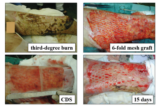
Kashiwa N, Ito O, Ueda T, et al. (2004) Treatment of full-thickness skin defect with concomitant grafting of 6-fold extended mesh auto-skin and allogeneic cultured dermal substitute. Artif Organs 28: 444–450. doi: 10.1111/j.1525-1594.2004.00009.x
    [24] Ohtani T, Okamoto K, Kaminaka C, et al. (2004) Digital gangrene associated with idiopathic hypereosinophilia: treatment with allogeneic cultured dermal substitute (CDS). Eur J Dermatol 14: 168–171.
    [25] Moroi Y, Fujita S, Fukagawa S, et al. (2004) Clinical evaluation of allogeneic cultured dermal substitutes for intractable skin ulcers after tumor resection. Eur J Dermatol 14: 172–176.
    [26] Yamada N, Uchinuma E, Kuroyanagi Y (2008) Clinical trial of allogeneic cultured dermal substitute for intractable skin ulcers of the lower leg. J Artif Organs 11: 100–103. doi: 10.1007/s10047-008-0406-7
    [27] Chen WYJ, Abatangelo G (1999) Functions of hyaluronan in wound repair. Wound Repair Regen 7: 79–89. doi: 10.1046/j.1524-475X.1999.00079.x
    [28] West DC, Hampson IN, Arnold F, et al. (1985) Angiogenesis induced by degradation products of hyaluronic acid. Science 228: 1324–1326. doi: 10.1126/science.2408340
    [29] Sattar A, Rooney P, Kumar S, et al. (1994) Application of angiogenic oligosaccharides of hyaluronan increases blood vessel numbers in rat skin. J Invest Dermatol 103: 576–579. doi: 10.1111/1523-1747.ep12396880
    [30] Lees VC, Fan TP, West DC (1995) Angiogenesis in a delayed revascularization model is accelerated by angiogenic oligosaccharides of hyaluronan. Lab Invest 73: 259–266.
    [31] Pardue EL, Ibrahim S, Ramamurthi A (2008) Role of hyaluronan in angiogenesis and its utility to angiogenic tissue engineering. Organogenesis 4: 203–214. doi: 10.4161/org.4.4.6926
    [32] Postlethwaite AE, Seyer JM, Kang AH (1978) Chemotactic attraction of human fibroblasts to type I, II, and III collagens and collagen-derived peptides. P Natl Acad Sci USA 78: 871–875.
    [33] Carpenter G, Cohen S (1976) Human epidermal growth factor and the proliferation of human fibroblasts. J Cell Physiol 88: 227–237. doi: 10.1002/jcp.1040880212
    [34] Carpenter G, Cohen S (1979) Epidermal growth factor. Annu Rev Biochem 48: 193–216. doi: 10.1146/annurev.bi.48.070179.001205
    [35] Park JS, Kim JY, Cho JY, et al. (2000) Epidermal growth factor (EGF) antagonizes transforming growth factor (TGF)-beta 1-induced collagen lattice contraction by human skin fibroblasts. Biol Pharm Bull 23: 1521–1523. doi: 10.1248/bpb.23.1521
    [36] Xin X, Yang S, Ingle G, et al. (2001) Hepatocyte growth factor enhances vascular endothelial growth factor-induce angiogenesis in vitro and in vivo. Am J Pathol 158: 1111–1120. doi: 10.1016/S0002-9440(10)64058-8
    [37] Conway K, Price P, Harding KG, et al. (2006) The molecular and clinical impact of hepatocyte growth factor, its receptor, activators, and inhibitors in wound healing. Wound Repair Regen 14: 2–10. doi: 10.1111/j.1524-475X.2005.00081.x
    [38] Kondo S, Kuroyanagi Y (2012) Development of wound dressing composed of hyaluronic acid and collagen sponge with epidermal growth factor. J Biomat Sci-Ploym E 23: 629–643. doi: 10.1163/092050611X555687
    [39] Kondo S, Niiyama H, Yu A, et al. (2012) Evaluation of a wound dressing composed of hyaluronic acid and collagen sponge containing epidermal growth factor in diabetic mice. J Biomat Sci-Ploym E 23: 1729–1740.
    [40] Yu A, Takeda A, Kumazawa K, et al. (2015) Preliminary clinical study using a novel wound dressing composed of hyaluronic acid and collagen containing EGF. Open J Regen Med 4: 6–13. doi: 10.4236/ojrm.2015.41002
    [41] Wolfort FG, Dalton WE, Hoopes JE (1972) Chemical peel with trichloroacetic acid. Brit J Plast Surg 25: 333–334.
    [42] Baker TJ, Gordon HL, Mosienko P, et al. (1974) Longterm histological study of skin after chemical peeling. Plast Reconstr Surg 53: 522–525. doi: 10.1097/00006534-197405000-00003
    [43] Coleman WP, Brody HJ (1997) Advances in chemical peeling. Dermatol Clin 15: 19–26. doi: 10.1016/S0733-8635(05)70411-3
    [44] Brody HJ, Monheit GD, Resnik SS, et al. (2000) A history of chemical peeling. Dermatol Surg 26: 405–409. doi: 10.1046/j.1524-4725.2000.00505.x
    [45] Anitha B (2010) Prevention of complications in chemical peeling. J Cutan AesthetSurg 3: 186–188. doi: 10.4103/0974-2077.74500
    [46] Cui XL, Iwasa M, Iwasa Y, et al. (2000) Arginine-supplemented diet decreases expression of inflammatory cytokines and improves survival in burned rats. JPEN 24: 89–96. doi: 10.1177/014860710002400289
    [47] Ochoa JB, Strange J, Kearney P, et al. (2001) Effects of L-arginine on the proliferation of T lymphocyte subpopulations. JPEN 25: 23–29. doi: 10.1177/014860710102500123
    [48] Kirs SJ, Barbul A (1990) Role of arginine in trauma, sepsis, and immunity. JPEN 14: 226–229. doi: 10.1177/014860719001400514
    [49] Shi HP, Efron DT, Most D, et al. (2000) Supplemental dietary arginine enhances wound healing in normal but not inducible nitric oxide synthase knockout mice. Surgery 128: 374–378. doi: 10.1067/msy.2000.107372
    [50] Zhu H, Ka B, Murad F (2007) Nitric oxide accelerates the recovery from burn wounds. World J Surg 31: 624–631. doi: 10.1007/s00268-007-0727-3
    [51] Witte MB, Barbul A (2003) Arginine physiology and its implication for wound healing. Wound Repair Regen 11: 419–423. doi: 10.1046/j.1524-475X.2003.11605.x
    [52] Matsumoto Y, Arai K, Momose H, et al. (2009) Development of wound dressing composed of hyaluronic acid sponge containing arginine. J Biomat Sci-Polym E 20: 993–1004. doi: 10.1163/156856209X444394
    [53] Marionnet C, Vioux-chagnoleau C, Pierrard C, et al. (2006) Morphogenesis of dermal-epidermal junction in a model of reconstracted skin: beneficial effects of vitamin C. Exp Dermatol 15: 625–633.
    [54] Lima CC, Pereira APC, Silva JRF, et al. (2009) Ascorbic acid for the healing of skin wounds in rats. Braz J Biol 69: 1195–1201. doi: 10.1590/S1519-69842009000500026
    [55] Wu YL, Gohda E, Iwao M, et al. (1998) Stimulation of hepatocyte growth factor production by ascorbic acid and its stable 2-glucoside. Growth Horm IGF Res 8: 421–428. doi: 10.1016/S1096-6374(98)80313-4
    [56] Niiyama H, Kuroyanagi Y (2014) Development of novel wound dressing composed of hyaluronic acid and collagen sponge containing epidermal growth factor and vitamin C derivative. J Artif Organs 17: 81–87.
    [57] Yamamoto A, Ohno H, Kuroyanagi Y (2016) Evaluation of epidermal growth factor-incorporating skin care product in culture experiment using human fibroblasts. Open J Regen Med 5: 44–54. doi: 10.4236/ojrm.2016.52004
    [58] Mitchell JD, Lee R, Hodakowski GT, et al. (1994) Prevention of postoperative pericardial adhesions with a hyaluronic acid coating solution: Experimental safety and efficacy studies. J Thorac Cardiov Sur 107: 1481–1488.
    [59] Harris ES, Morgan RF, Rodeheaver GT (1995) Analysis of the kinetics of peritoneal adhesion formation in the rat and evaluation of potential antiadhesive agents. Surgery 117: 663–669. doi: 10.1016/S0039-6060(95)80010-7
    [60] Risberg B (1997) Adhesions: preventive strategies. Eur J Surg 577: 32–39.
    [61] Tsai SW, Fang JF, Yang CL, et al. (2005) Preparation and evaluation of a hyaluronate-collagen film for preventing post-surgical adhesion. Int Med Res 33: 68–76. doi: 10.1177/147323000503300106
    [62] Park SN, Jang HJ, Choi YS, et al. (2007) Preparation and characterization of biodegradable anti-adhesive membrane for peritoneal wound healing. J Mater Sci-Mater M 18: 475–482.
    [63] Temiz A, Ozturk C, Bakunov A, et al. (2008) A new material for prevention of peritendinous fibrotic adhesions after tendon repair: oxidized regenerated cellulose (Interceed), an absorbable adheison barrier. Int Orthop 32: 389–394. doi: 10.1007/s00264-007-0335-8
    [64] Liu Y, Skardal A, Shu XZ, et al. (2008) Prevention of peritendinous adhesions using a hyaluronan-derived hydrogel film following partial-thickness flexor tendon injury. J Orthop Res 26: 562–569. doi: 10.1002/jor.20499
    [65] Rajab TK, Wallwiener M, Talukdar S, et al. (2009) Adhesion-related complications are common, but rarely discussed in preoperative consent; a multicenter study. World J Surg 33: 748–750. doi: 10.1007/s00268-008-9917-x
    [66] Cheung J, Tsang H, Cheung J, et al. (2009) Adjuvant therapy for the reduction of postoperative intra-abdominal adhesion formation. Asian J Surg 32: 180–186. doi: 10.1016/S1015-9584(09)60392-4
    [67] Brochhausen C, Schmitt V, Rajab T, et al. (2011) Intraperitoneal adhesions-an ongoing challenge betwenn biomedical engineering and the life sciences. J Biomed Mater Res A 98: 143–156.
    [68] Kuroyanagi M, Yamamoto A, Shimizu N, et al. (2014) Development of anti-adhesive spongy sheet composed of hyaluronic acid and collagen containing epidermal growth factor. J Biomat Sci-Polym E 25: 1253–1265. doi: 10.1080/09205063.2014.926579
  • This article has been cited by:

    1. Stefano Picotti, Luca Forte, Jo Serrentino, A pre-market interventional, single-arm clinical investigation of a new topical lotion based on hyaluronic acid and peptides, EGYFILTM, for the treatment of pain and stiffness in soft tissues, 2023, 24, 1471-2474, 10.1186/s12891-023-06903-y
    2. John Kechagias, Stephanos Zaoutsos, Effects of 3D-printing processing parameters on FFF parts’ porosity: outlook and trends, 2024, 1042-6914, 1, 10.1080/10426914.2024.2304843
  • Reader Comments
  • © 2017 the Author(s), licensee AIMS Press. This is an open access article distributed under the terms of the Creative Commons Attribution License (http://creativecommons.org/licenses/by/4.0)
通讯作者: 陈斌, bchen63@163.com
  • 1. 

    沈阳化工大学材料科学与工程学院 沈阳 110142

  1. 本站搜索
  2. 百度学术搜索
  3. 万方数据库搜索
  4. CNKI搜索

Metrics

Article views(9963) PDF downloads(1569) Cited by(2)

Article outline

Figures and Tables

Figures(14)  /  Tables(1)

Other Articles By Authors

/

DownLoad:  Full-Size Img  PowerPoint
Return
Return

Catalog